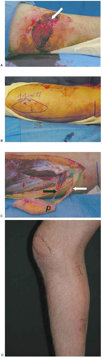
Fig. 1: (A) left knee wound with quadriceps tendon
repair exposed laterally
[arrow], (B) design of LSAP
flap ["x" mark the site of "perforators" identified
using an audible Doppler, vertical line (right) coincides
with the defect at level
of knee joint, margins of
lateral head of gastrocnemius muscle also outlined],
(C) elevated island LSAP flap [black arrow = branch
of the lateral sural artery
(note the path of its intramuscular
dissection above), white arrow = branch of
lateral sural
cutaneous nerve, p = perforator], (D) healed flap inset
at lateral
left knee, with calf scar noted after primary closure
of donor site.