


Mi SciELO
 Servicios personalizados
Servicios Personalizados
Revista
Articulo
 Inglés (pdf)
 Articulo en XML
 Referencias del artículo
 Enviar articulo por email
Indicadores
-
 Citado por SciELO
 -
 Accesos
Links relacionados
-
 Citado por Google
 -
 Similares en
SciELO
Similares en
SciELO -
 Similares en Google
Compartir

 Permalink
Medicina Oral, Patología Oral y Cirugía Bucal (Internet)
versión On-line ISSN 1698-6946
Med. oral patol. oral cir.bucal (Internet) vol.12 no.1 ene. 2007
Oral leiomyoma in retromolar trigone. A case report
Ramón Luaces Rey 1, Fernanda Lorenzo Franco 2, Guillermo Gómez Oliveira 1,
Beatriz Patiño Seijas 1, Dolores Guitián 3, José Luis López-Cedrún Cembranos 4
(1) MD. Resident. Department of Oral and Maxillofacial Surgery
(2) MD. DDS. PhD. Staff. Department of Oral and Maxillofacial Surgery
(3) MD. Staff. Department of Pathological Anatomy. Department of Oral and Maxillofacial Surgery
(4) MD. DDS. PhD. Chief of Department of Oral and Maxillofacial Surgery. Juan Canalejo Hospital. A Coruña. Spain
ABSTRACT
Leiomyomas are bening tumours originated on smooth muscle. The most frequent site of appearance are uterine myometrium, gastrointestinal tract and skin. The highest incidence ocurs between 40 and 49 years of age. Its presentation is unusual in children or in older patients. Leiomyomas are unfrequent in the oral cavity, but in this location are usually localized on tongue, lips and palate.
Leiomyomas use to appear as well-defined masses, with slow growth and totally asymptomatic. Pain is present just in rare cases. The treatment is surgical escision. Recurrences are extremely unfrequent.
The diagnosis is mainly determined by histological studies due to its unspecific clinical appearance. Histopathologicaly proliferation of smooth muscle cells is observad without necrotic areas. A low number of mitotic figures can be seen.
We present the case report of a 25-year old male patient, with a leiomioma on his right retromolar trigone.The low incidence of this pathology, the age of the patient and the inusual location, make the report of the case worthy.
Key words: Leiomyoma, oral cavity, oral tumour.
RESUMEN
Los leiomiomas son tumores benignos originados en el músculo liso. Su localización más frecuente es el útero, el tracto gastro-intestinal y la piel. Se presenta habitualmente entre los 40 y 49 años de edad, siendo muy rara su aparición en la infancia y en la senectud. Son muy infrecuentes a nivel de la cavidad oral, pero cuando se dan en esa localización, asientan principalmente en la lengua, los labios o el paladar.
Inicialmente suelen presentarse como una masa muy bien definida, de lento crecimiento y totalmente asintomática. En raras ocasiones producen dolor. Su tratamiento es casi siempre quirúrgico, siendo las recurrencias excepcionales.
Dada su clínica inespecífica, su diagnóstico es principalmente histológico, observándose en las muestras una proliferación de células musculares lisas, sin focos de necrosis y con escasas mitosis.
Presentamos el caso clínico de un paciente de 25 años de edad con un leiomioma en trígono retromolar derecho. Dada la escasa incidencia de este tipo de patología, la edad del paciente y su inusual ubicación, se justifica la presentación de este caso.
Palabras clave: Leiomioma, cavidad bucal, tumor oral.
Introduction
Oral leiomyoma has a low incidence. When it presents on this location, it appears as a slow-growth mass, symptomatic just in exceptional cases (1). Main symptoms are pain, teeth mobility, or even difficulty in chewing, usually when the tumour is located in tongue, lips or palate (2).
Due to its unspecific clinical presentation, diagnosis is made after histological study (3), where the typical small smooth muscle cells are seen, with uniform-size and with no malignity criteria. In order to achieve more specific analysis, and more precise differential diagnosis, it´s posible to make immunohistochemical studies.
Surgery is the only succesful treatment at this moment. Its important to obtain a complete resection in order to avoid recurrences (2) (usually easily due to its characteristics as a well-circumscribed tumour).
Case report
A 25-year-old male patient, without remarkable medical history or toxic habits, came to our clinic with a 1 x 1,5 cm tumor placed on right retromolar trigone. The mass had appeared three weeks before. It was painless, no adhered and well-defined with normal mucosa overlying (Figure 1).

Fine Needle Aspiration was negative for malignant cells. The result was unspecific with a possible glandular origin. Semi-inclusion of theeth 4.8 was observed, with no apparent relation with the lesion. No palpable cervical nodes were found. CT scan of the neck showed bilateral 1 cm. lymph nodes on level II, and subcentimetric ones on right level III and on submaxilar area. No masses or pathological contrast captation were observed.
The mass was excised under general anaesthesia with a 1cm free-margin. The diagnosis suspected was a lesion originated on minor salivary glands of retromolar trigone.
The patient had a correct evolution without any post-operative incident.
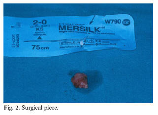
Macroscopically the tumor had greyish colour, elastic consistence, rough appearance and it was encapsulated (Figure 2)
The histological report informed that the tumour was constituted of small spindle cells, with eosinophilic cytoplasm and central and basophilic nucleus. The cells appeared in a typical whirling pattern. The lesion showed prominent vascular spaces between the bands of smooth muscle cells. No mitotic figure, nuclear atypia or necrotic areas were identified. Histological diagnosis was leiomyoma, with a clear origin on the smooth muscle placed under oral mucous (Figure 3).

The patient is disease free one year after surgical excision
Discussion
Leiomyoma is a benign tumour of smooth muscle. It can appear in any location where this kind of tissue is present.
This lesion occurs most often in female tract. This represents approximately 95% of the total (4). The remaining 5% use to be placed in gastro-intestinal tract and skin. Oral leiomyomas are considered uncommon neoplasms, around 0,065 %.
The origin of leiomyomas of oral cavity is restricted to three areas with smooth muscle in their histological analysis: tunica media of the blood vessels (5), ductus lingualis and circumvallate papila (6).
Since 1884(7), when oral leiomyoma was first reported, less than 150 cases have been published (8).
The most common sites of occurrence of the oral leiomyoma are tongue, lips, palate and cheek (2). Other less frequent locations are the flour of the mouth and the gingiva. Retromolar trigone is an unusual place, according to the reviewed literature.
Clinically, the leiomyomas use to be unspecific masses, with several aspects from normal to more congestive mucosa, according to their vascularity (9). These use to be well-defined, slow growing, asymptomatic masses. Some symptoms are induced (1) due to local growth. Deglution difficulty, toothache, loose teeth, or pain refered in TMJ (10) have been reported. Shortness of breath can be produced by huge tumours (11). Pain is suspected to be prompted by local ischemia due to tumoral vessel contraction (12) and due to neural irritation near the tumour (13).
Oral leiomyoma is more frequent in men than in women, with a 1,43: 1 ratio (9). Peak age incidence is 40-49 years old (2).
There have not been risk factors related with this kind of tumour. In our case, there is not suspected relation between semi-erupted teeth 4.8 and local tissue damage and the leiomyoma.
Several oral tumours should be included in the diferencial diagnosis, benigns lesions such as fibroma, neurofibroma, lipoma or mucocele or malignant ones such as leiomyosarcoma. Histological studies are necessary to achieve diferencial diagnosis.
The definitive diagnosis in leiomyoma is the histological one. Typical smooth muscle cell proliferation (small and spindle) is observed without necrosis areas or mitotic figures. The malignant counterpart: the leiomyosarcoma should be taken into account when the number of mitotic figures per field is over ten (14). Difference between leiomyoma and low grade leiomyosarcoma is not always easy (15).
Histologically three types of leiomyomas can be differentiated (3): solid leiomyoma (25 %), vascular leiomyoma or angiomyoma (74 %) and leioblastoma or ephiteoid leiomyoma (1 %).
Hematoxylin and eosin stains can be used in this kind of tumour. Special stains such as Masson´s trichrome, Van Gieson´s stain, or Mallory´s phosphotungstic acid (PTAH) are specific for muscle cells and collagen fibres, Immmunohistochemical detection of actine (smooth muscle marker) can also be useful.
Wide surgical resection is the only reported treatment in reviewed literature with successful results. Recurrence rate is very low if complete resection is achieved (2). It has been reported a recurrence case two weeks after surgery (16), probably due to incomplete resection.
References
1. Cherrick HM, Dunlap CL, King OH. Leiomyomas of the oral cavity. Review of the literature and clinicopathologic study of seven new cases. Oral Surg Oral Med Oral Pathol 1973;35:54-66. [ Links ]
2. Wertheimer-Hatch L, Hatch GF, Hatch KF, Davis GB, Blanchard DK, Foster RS et al. Tumors of the oral cavity and pharynx. World J Surg 2000;24:395-400.
3. Ezinger F, Lattes R,Torloni H. Histological typping of Soft Tissue and Tummors. Geneva: World Health Organization; 1969. p. 30-1.
4. Farman AG. Benign smooth muscle tumors. S Afr Med J 1975;49:1333-45.
5. Stout AP. Leiomyoma of the oral cavity. Am J Cancer 1938;34:31-6.
6. Glas E. Beitrage Zur Pathologie der Zungengrudtumoren. Wein Klin Wochenschr 1905;18:747.
7. Blanc E.Travaux originaux. Gaz Hebd Med Chir 1884;21:611.
8. Lloria Benet M, Bagan JV, Lloria de Miguel E, Borja Morant A, Alonso S. Leiomioma oral: a proposito de un caso. Med Oral 2003;8:215-9.
9. Brooks JK, Nikitakis Ng, Goodman NJ, Levy BA. Clinicopathologic Characterization of oral angioleiomiomas. Oral Surg Oral Med Oral Pathol Oral Radiol Endod 2002;94:221-7.
10. Grippaudo G,Becelli R. Leiomioma del muscolo massetere:descrizione di un caso clinico. Minerva Estomatol 1996;45:277-80.
11. Kotler HS, Gould NS, Gruber B. Leiomyoma of the tongue presenting as a congenital airway obstruction. Int J Pediatr Otorhinolaryngol 1994; 29:139-45.
12. Duhing JT, Ayer JP. Vascular leyomioma: A study of sixty-one cases. Arch Pathol 1959;68:424-30.
13. Toida M, Koizumi H, Shimokawa K. Painful angiomyoma of the Oral Cavity:Report of a case and Review of the literature. J Oral Maxilofac Surg 2000;58:450-3.
14. Robins SL, Corten RI. Pathologic Basis of Diseases (ed.2). Philadelpia: PA, Saunders; 1979. p. 209-10.
15. Leung K, Wong DY, Li W. Oral leiomyoma:report of a case. Maxillofac Surg 1990;48:735-8.
16. Svane TJ, Smith BR, Consentino BJ, Cundiff EJ, Ceravolo JJ. Oral leiomiomas: review of the literature and report of a case of palatal angioleiomyoma. J Periodontol 1986;57:433-5.
![]() Correspondence:
Correspondence:
Dr Ramón Luaces Rey
Juan Canalejo Hospital
Department of Oral and Maxillofacial Surgery
Xubias de Arriba, 84
15006 A Coruña. Spain
E-mail: rluarey@canalejo.org
Received: 3-07-2005
Accepted: 23-10-2006

