Introduction
Emotions play a fundamental role in our lives since they intervene in our way of thinking and acting, and are accompanied by a physiological response in our organism. One of the pioneering researchers in this area of study was Paul Ekman who, as early as 1969, participated in a study that attempted to identify a series of emotions of universal nature for all human beings, regardless of their culture (Ekman et al., 1969). As a result of this line of work, Ekman & Friesen (1971) identified joy, fear, disgust, sadness, surprise and anger as the six basic emotions. Since then, much research has focused on describing how information expressed in faces is processed. The scientific study of emotions has changed, in part due to the use of technology and advances in neuroscience. In the recent years, imaging of brain activity and identification of the biological basis of emotions have led to a greater understanding of processes underlying emotional recognition (Park et al., 2017; Spunt & Adolphs, 2019).
Memory and other cognitive abilities have been extensively evaluated in the aging process (Craik & Salthouse, 2011; Tucker-Drob et al., 2019). However, changes in emotional processing and recognition, as well as the implications of deficits in these functions, have received less attention (Cotter et al., 2018; Manenti et al., 2019). The recognition of facial expression of emotions is considered one of the most relevant capabilities of human beings, intervening in communication and interpersonal interaction and providing valuable information aimed at responding appropriately to different situations (Hutchings et al., 2017). Moreover, this ability is of great importance from a phylogenetic and ontogenetic point of view, since it plays an important role in survival (Hsieh et al., 2012; Hutchings et al., 2017). The study of facial emotion recognition (FER) has traditionally been done through static visual stimuli (photographs), being the stimuli developed by Ekman & Friesen (1976) the most used. These images compile a set of faces that reflect the expression of emotions identified as universal.
This ability to recognize emotions is progressively impaired both in normative (Virtanen et al., 2017; Williams et al., 2009) and pathological aging (Henry et al., 2008; Hot el at., 2013; Maki et al., 2013; Snowden et al., 2008). However, impaired recognition does not seem to affect all emotions in the same way. Different research suggests that positive emotions such as joy could be more preserved and better identified than negative emotions (Chiu et al., 2016; Guaita et al., 2009; Hot et al., 2013; Kumfor et al., 2014a). In this sense, negative emotions are recognized more poorly in neurodegenerative pathologies such as Alzheimer's Disease (AD), Corticobasal Syndrome, Frontotemporal Dementia, Lewy Body Dementia or Semantic Dementia, as well as in normal aging (Chiu et al., 2016; Fernandez-Duque & Black, 2005; Guaita et al., 2009; Heitz et al., 2016; Hsieh et al., 2012; Kipps et al., 2009; Kumfor et al., 2014c; Maki et al., 2013; Park et al., 2017). In AD, emotion recognition is impaired throughout the course of the disease (Weiss et al., 2008). Several studies suggest that deficits in FER at different stages of AD would not be attributable to alterations in visuospatial or attentional abilities, since recognition of neutral expressions is generally preserved (Fernandez-Duque & Black, 2005; Narme et al., 2013; Sapey-Triomphe et al., 2015). Similarly, inappropriate behaviors or indifference displayed by patients could be attributed to deterioration in the ability to recognize and understand emotions rather than to cognitive aging (Shimokawa et al., 2001). The correlation between impaired emotional recognition and the patient's own empathic capacity shows that the neural substrates involved seem to be the same for the experience of emotion at intra- and interindividual levels (Kipps et al., 2009). The difficulty in the interpretation of emotions, a feature shared by different pathologies such as AD or schizophrenia, could partly justify the social isolation manifested by these patients, influencing their lifestyle (Danjou et al., 2019; Mohamed et al., 2010).
The analysis of the emotion recognition ability and its possible relationship with different variables can contribute to a more complete understanding of the aging process. Therefore, the general objective of the present review was the analysis of the main variables involved in FER in people with AD. Similarly, and with the aim of providing a novel approach compared to previous reviews in this field of study (Torres Mendonça de Melo Fadel et al., 2019), we have analyzed in detail the sex and age of the participants in prior published studies, as well as possible biomarkers, neuroimaging tests and/or biological or physiological markers identified or analyzed in the literature.
Method
Criteria for the selection of studies
In order to minimize possible biases in the selection of articles, strict inclusion and exclusion criteria were applied in the present review. The inclusion criteria established were: (a) those articles in which the ability of facial emotion recognition in human subjects is explored, and (b) articles in which at least one of the experimental groups are made up of people with AD. Exclusion criteria were as follows: (a) theoretical or systematic reviews, meta-analyses, book chapters, theses, and communications to conferences (b) those papers aimed at the validation and/or reliability of instruments; and (c) papers based on a recognition or rehabilitation program. The period analyzed included 20 years (1999-2019). In addition, the PICO strategy was applied as a tool to improve the search and retrieval of information from the scientific literature (Liberati et al., 2009):
(P)- Participants: Participants were required to be older adults diagnosed with AD. However, if the study sample included both participants with AD and with other types of dementia for comparison (e.g., Vascular Dementia, Frontotemporal Dementia...), the paper was not excluded if the information presented in it was sufficient to address the object of the study.
(I)- Intervention: The assessment carried out in the investigation had to be based on the ability of facial recognition of emotions by means of different instruments.
(C)- Comparison: Primary studies taking into account comparisons of facial emotion recognition ability between different groups, with at least one of the groups consisting of people with AD.
(O) - Result: Should refer to the different study variables and their results, in regard to facial recognition of emotions.
Search procedure and strategy
For the present review, a literature search was conducted on the Web of Science (WoS) databases of Clarivate Analytics (which includes all the journals indexed on Medline) and on PsycINFO of the American Psychological Association, according to the PRISMA (Preferred Reporting Items for Systematic Reviews and Meta-Analyses) guidelines (Liberati et al., 2009; Moher et al., 2009). In addition, the general recommendations for conducting systematic reviews were followed (Siddaway et al., 2019). For the literature research strategy, the MeSH Thesaurus and PsycINFO Thesaurus descriptors related to the thematic area studied were used as a reference, with the following strategy: ("emotion recognition" OR "emotional cognition" OR "facial affect recognition") AND ("Alzheimer" OR "dementia"). The initial search generated 299 results on WoS and 195 on PsycINFO, obtaining a total of 385 different documents, since 109 were retrieved in both databases. Figure 1 shows the flow chart applied in the document selection process. After applying the established inclusion and exclusion criteria, 28 articles were obtained. The articles comprising the present study were selected after review and critical reading of their titles and abstracts. In case of doubtful articles, a critical reading of the full text was carried out after scanning the abstract. A consensus was reached by two experts in the field on the inclusion of all the preselected articles. The articles included in the review are marked with an asterisk (*) in the list of references.
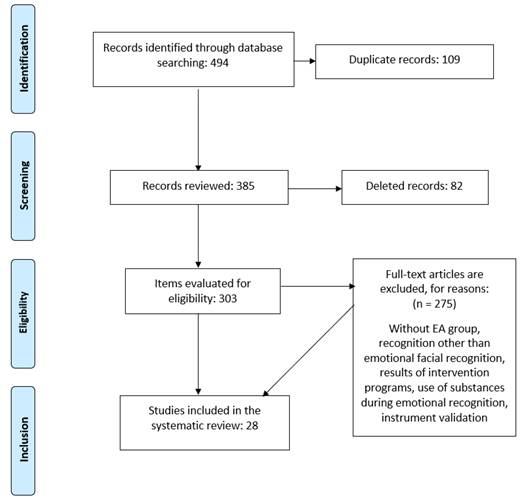
Figure 1 Flow diagram, phases of the systematic review for the selection of articles. Adapted from “Preferred reporting items for systematic reviews and meta-analyses”. Moher et al., 2009.
Codification of variables
For each of the selected articles, the following variables were determined: type of emotion evaluated (joy, sadness, anger, disgust, fear, surprise), specifically identifying the articles that evaluated the six basic emotions mentioned above and those that used neutral faces in their evaluation; description of the sample. The instruments used to evaluate the FER were also identified, in addition to the identification of those studies that used neuroimaging tests and/or biological and/or physiological markers.
Methodological quality assessment
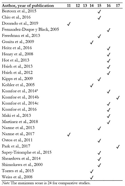
The methodological quality of primary studies is a potential source of bias. Therefore, the risk of bias in the studies selected independently by two reviewers, was assessed using the Methodological Index for Non-randomized Studies (MINORS) (Silva Junior et al., 2020; Slim et al., 2003). This tool consists of 12 items to be answered based on the information available in the articles. The items are scored as follows: 0 (not reported), 1 (reported but inadequate), 2 (reported and adequate). The ideal score is 16 for non-comparative studies and 24 for comparative studies. Discrepancies in the ratings were jointly reviewed and pooled to reach consensus. The highest score obtained in the MINORS tool when reviewing the selected articles was 17 (Kumfor et al., 2014b; Park et al., 2017) while the lowest score achieved was 11 (Dourado et al., 2019; Narme et al., 2017) (see Table 1). Giving the nature of the studies included, we can point out some limitations that may influence the methodological quality of the studies: in general, the study protocol is considered as a guarantee of methodological quality, however, not all the studies detailed the protocol that indicated that prior to the study they had the approval of the Ethics Committee for research in humans. Informant bias is another possible methodological bias. In addition, the articles reviewed generally do not report information about blinded evaluations of the criteria and interpretation of the results.
Results
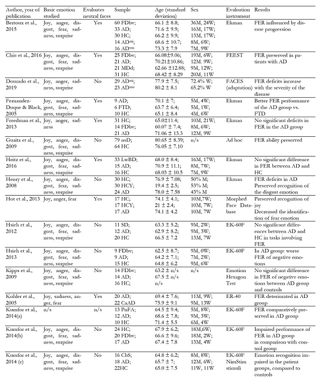
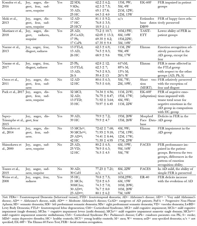
The result of the final selection was made up of a total of 28 articles, with the main results obtained summarized in Table 2, which contains the following information: authors and year of publication, types of emotion studied, emotions assessed, sample, sex, assessment instrument, results. The most relevant aspects of these sections will be discussed below.
Types of emotions evaluated
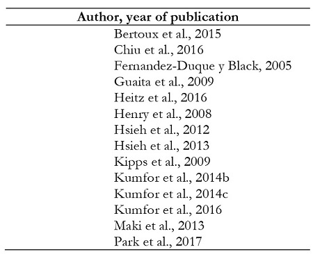
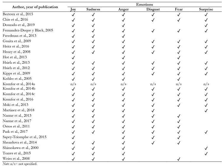
In most articles, the assessment of FER was based on the subject's identification of the basic emotions: anger, disgust, fear, joy, sadness, and surprise. It is important to highlight that 50% of the reviewed publications assessed all basic emotions (Bertoux et al., 2015; Chiu et al., 2016; Fernandez-Duque & Black, 2005; Guaita et al., 2009; Heitz et al., 2016; Henry et al., 2008; Hsieh et al., 2012; Hsieh et al., 2013; Kipps et al., 2009; Kumfor et al., 2014b; 2014c; 2016; Maki et al., 2013; Park et al., 2017) (see Table 3). The remaining 50% of the articles assessed only some of the emotions (Dourado et al., 2019; Freedman et al., 2013; Hot et al., 2013; Kholer et al., 2005; Kumfor et al., 2014a; Martinez et al., 2018; Narme et al., 2013; Narme et al., 2017; Ostos et al., 2011; Sapey-Triomphe et al., 2015; Sherardova et al., 2014; Shimowaka et al., 2000; Torres et al., 2015; Weiss et al., 2008).
The main emotions not assessed in these articles were: surprise, disgust, fear and sadness. Specifically, 29% of the articles did not use the surprise emotion for its assessment (Hot et al., 2013; Kholer et al., 2005; Narme et al., 2013; 2017; Ostos et al., 2011; Sapey-Triomphe et al., 2015; Sherardova et al., 2014; Weiss et al., 2008); 25% of the articles did not include disgust (Dourado et al., 2019; Freedman et al., 2013; Hot et al., 2013; Kholer et al., 2005; Shimowaka et al., 2000; Torres et al., 2015; Weiss et al., 2008); fear was not assessed in 14% of the articles (Dourado et al., 2019; Freedman et al., 2013; Shimowaka et al., 2000; Torres et al., 2015); and sadness was not assessed in 7% of the papers (Hot et al., 2013; Sapey-Triomphe et al., 2015) (see Table 4).
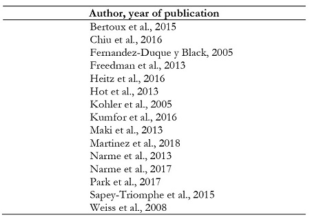
Fifty-four percent of the reviewed publications also included neutral stimuli faces (showing no emotion) that are generally considered masking stimuli (Bertoux et al., 2015; Chiu et al., 2016; Fernandez-Duque & Black, 2005; Freedman et al., 2013; Heitz et al., 2016; Hot et al., 2013; Kholer et al., 2005; Kumfor et al., 2016; Maki et al., 2013; Martinez et al., 2018; Narme et al., 2013; 2017; Park et al., 2017; Sapey-Triomphe et al., 2015; Weiss et al., 2008) (see Table 5).
Sample description
A large number of reviewed articles compared the AD group with other neurodegenerative diseases, mainly Frontotemporal Dementia (Bertoux et al., 2015; Chiu et al., 2016; Fernandez-Duque & Black, 2005; Freedman et al., 2013; Hsieh et al., 2013; Kipps et al., 2009; Kumfor et al., 2014b; Narme et al., 2013; 2017; Park et al., 2017), Vascular Dementia (Shimokawa et al., 2000), Semantic Dementia (Hsieh et al., 2012), Progressive Non-Fluent Aphasia (Kumfor et al., 2014a; Kumfor et al., 2016), Parkinson's Disease (Narme et al., 2017; Martinez et al., 2018), and Lewy Body Dementia (Heitz et al., 2016). Out of all the articles analyzed, three of them incorporated groups with Mild Cognitive Impairment (Park et al., 2017; Sheardova et al., 2014; Weiss et al., 2008). Comparisons with groups at different stages of AD were made in only one article (Dourado et al., 2019). Similarly, 23 articles incorporated a control group consisting of age-matched healthy participants for comparison (Bertoux et al., 2015; Chiu et al., 2016; Fernandez-Duque & Black, 2005; Freedman et al., 2013; Guaita et al., 2009; Heitz et al., 2016; Henry et al., 2008; Hot et al., 2013; Hsieh et al., 2012; 2013; Kipps et al., 2009; Kumfor et al., 2014a; 2014b; 2014c; 2016; Maki et al., 2013; Narme et al., 2013; Ostos et al., 2011; Park et al., 2017; Sapey-Triomphe et al., 2015; Shimokawa et al., 2000; Sheardova et al., 2014; Weiss et al., 2008). From the three articles that included healthy caregivers, in two of them these participants were used as controls (Kohler et al., 2005; Martinez et al., 2018); in the third study this sample of caregivers was only used for data collection (Torres et al., 2015). Regarding the remaining articles, three others incorporated control groups of different ages (young and old) (Henry et al., 2008; Hot et al., 2013; Maki et al., 2013). Finally, only two articles had no control group (Dourado et al., 2019; Narme et al., 2017).
Instruments used for the assessment of facial recognition of emotions
The instruments integrated by photographs were the most used for the evaluation of emotions, especially the "Pictures of Facial Affect" (POFA) by Ekman and Freisen (1976) that represents the six basic emotions, also including neutral expressions (Bertoux et al., 2015; Fernandez- Duque & Black, 2005; Freedman, et al., 2013; Heitz et al., 2016; Narme et al., 2013; 2017; Sheardova et al., 2014). In some articles the "Facial Expressions of Emotions- Stimuli and Test" (FEEST was also used) (Young et al., 2002). FEEST is a test a test that integrates two subtests: 1) “The Ekman 60 Faces Test", and 2) "The Emotion Hexagon Test" (Chiu et al., 2016; Henry et al., 2008; Hsiesh et al., 2012; 2013; Kipps et al., 2009; Kumfor et al., 2014a; 2014b; 2014c; Park et al., 2017).
Another sets of facial expressions used for emotion assessment on faces were NimStim stimuli ("The Research Network on Early Experience and Brain Development" (Kumfor et al., 2014c; 2016), DB99 (Advanced Telecommunications Research Institute International, Inc. Nara, Japan) database stimuli (Maki et al., 2013), "The Awareness of Social Inference Test-Emotion Evaluation Test" (TASIT-EET) (Martinez et al., 2018), stimuli belonging to "The Multimodal Emotion Recognition Test" (MERT) (Ostos et al., 2011), as well as the "Penn Emotion Recognition Test" (ER40) (Kohler et al., 2005; Weiss et al., 2008). Finally, several studies used the FACES battery adaptation as a test of emotional recognition (Dourado et al., 2019; Torres et al., 2015; Shimokawa et al., 2000). In addition to the studies that showed photographs in paper format, we found other works in which photographs were shown in digital format (Bertoux et al., 2015; Chiu et al., 2016; Fernandez-Duque & Black, 2005; Henry et al., 2008; Hot et al., 2013; Hsieh et al., 2012; 2013; Kipps et al., 2009; Kholer et al., 2005; Kumfor et al., 2014a; 2014b; 2014c; 2016; Maki et al., 2013; Narme et al., 2013; 2017; Park et al., 2017; Sapey-Triomphe et al., 2015; Weiss et al., 2008). Instruments were also used in digital format in which images are presented in a continuum of different intensities and/or between expressions that can be confusing (Chiu et al., 2016; Kipps et al., 2009; Kholer et al., 2005; Maki et al., 2013; Sapey-Triomphe et al., 2015) (see Table 6).
Another aspect which we find repeatedly in the reviewed articles is the use of labels to identify different emotions (Bertoux et al., 2015; Chiu et al., 2016; Dourado et al., 2019; Fernandez-Duque & Black, 2005; Freedman, et al., 2013; Henry et al., 2008; Hot et al., 2013; Hsieh et al., 2012; 2013; Kipps et al., 2009; Kumfor et al., 2014b; 2014c; 2016; Martinez et al., 2018; Narme et al., 2013; 2017; Ostos et al., 2011; Park et al., 2017; Sapey-Triomphe et al., 2015; Sheardova et al., 2014; Shimokawa et al., 2000; Torres et al., 2015; Weiss et al., 2008). Among the different stimuli used for the assessment of emotional recognition we found dynamic demonstrations of emotions in the format of video vignettes (Freedman, et al., 2013; Kumfor et al., 2014b, 2014c; Martinez et al., 2018; Ostos et al., 2011; Sapey-Triomphe et al., 2015), vocalizations (Hsiesh et al., 2013), musical excerpts (Hsiesh et al., 2012), and drawings (Dourado et al., 2019; Shimokawa et al., 2000; Torres et al., 2015).
Use of neuroimaging tests and/or biological and/or physiological markers
In the process of reviewing the articles, we found that 11 of them used neuroimaging tests and/or biological or physiological markers to support the results in FER testing. Specifically, they used neuroimaging tests (Heitz et al., 2016; Hsieh et al., 2012; Kumfor et al., 2014b, 2014c; 2016; Ostos et al., 2011; Park et al., 2017; Sapey-Triomphe et al., 2015; Sheardova et al., 2014; Shimokawa et al., 2000) biomarkers in cerebrospinal fluid (Bertoux et al., 2015) and electrodermal conductance response (Ostos et al., 2011).
Main results in the AD groups
Different articles suggest the existence of deficits in FER in the AD group (Bertoux et al., 2015; Dourado et al., 2019; Henry et al., 2008; Kohler et al., 2005; Kumfor et al., 2014b; 2014c; Martinez et al., 2018; Park et al., 2017; Sapey-Triomphe et al., 2015; Sheardova et al., 2014; Shimokawa et al., 2000; Weiss et al., 2008) and this decline appears to be related to the disease progression itself (Bertoux et al., 2015; Dourado et al., 2019; Sheardova et al., 2014; Torres et al., 2015; Weiss et al., 2008). The comparison of people with AD with healthy younger and older control groups indicated that a decline in performance is observed in people with AD when compared to younger but not with elderly people (Hot et al., 2013; Henry et al., 2008; Maki et al., 2013). However, other research suggests that in people with AD this ability seems to be relatively preserved (Fernandez-Duque & Black, 2005; Freedman et al., 2013; Guaita et al., 2009; Heitz et al., 2016; Henry et al., 2008; Hot et al., 2013; Hsieh et al., 2012; 2013; Kipps et al., 2009; Kumfor et al., 2014a; Maki et al.,2013; Narme et al., 2013; Ostos et al., 2011; Torres et al., 2015). Another study confirmed that the AD group showed worse emotional recognition than their caregivers (Kohler et al., 2005; Martinez et al., 2018). In the comparison of FER performance between people with AD and people with Frontotemporal Dementia, it is observed that the AD group maintains this ability more preserved (Bertoux et al., 2015; Chiu et al., 2016; Fernandez-Duque & Black, 2005; Freedman et al., 2013; Hsieh et al., 2013; Kipps et al., 2009; Narme et al., 2013; 2017; Park et al., 2017).
Analysis of joy as the emotion best identified in face recognition
If we perform an individual analysis of the emotions, we observe that joy is one of the best identified emotions in the different experimental groups (Chiu et al., 2016; Guaita et al., 2009; Hot et al., 2013; Maki et al., 2013; Martinez et al., 2018; Narme et al., 2013; Ostos et al., 2011). Valence (classification of the emotion as positive, negative or neutral) is another aspect assessed with positive emotions, being the best recognized in most studies (Fernandez- Duque & Black, 2005; Guaita et al., 2009; Heitz et al., 2016; Hsieh et al., 2012; 2013; Kipps et al., 2009; Kumfor et al., 2014c; Park et al., 2017; Maki et al., 2013).
Outcomes obtained using other instruments for the assessment of emotional recognition
It is also interesting to compare the emotional recognition capacity when the stimuli applied are not photographs but other types of materials (vignettes or video clips, narrations, music, vocalizations...). It is confirmed that with the use of video vignettes or video clips no significant differences in recognition are observed between groups with AD and Frontotemporal Dementia (behavioral variant) (Freedman et al., 2013) or between AD patients and healthy elderly people (Henry et al., 2008). On the opposite side, in several prior studies those groups including patients with different pathologies displayed worse performance in this modality, in comparison with controls (Kumfor et al., 2014b; 2014c; Martinez et al., 2018). Interestingly, a study developed by Ostos et al. (2011) compared performance between dynamic versus static presentation and observed differences between people with AD and a control group. In another research developed by Hsieh et al. (2012) in which musical fragments were used for emotion recognition, it was observed that patients with AD and Semantic dementia performed worse in this condition than in facial recognition.
Discussion
It is unquestionable that the ability to recognize and understand emotions is a skill that we use in our daily lives and that can condition the correct functioning at a social and a motivational level, increasing adaptation to a continuously changing environment. An inadequate identification of emotions can contribute to impairments in social relationships and/or behavioral disorders (Brown et al., 2018; Miller et al.,2013; Narme et al., 2017). In both normative and pathological aging, joy seems to be the best identified emotion (Chiu et al., 2016; Guaita et al., 2009; Hot et al., 2013; Maki et al., 2013; Virtanen, et al., 2017) even in stimuli such as musical excerpts (Hsieh et al., 2012). This advantage can be considered a bias in the recognition of positive emotions, since these are represented in minority compared to negative ones (positive: joy and surprise, although the valence of surprise remains controversial; negative: anger, sadness, fear, disgust). This bias may be related to changes that may favor the processing of positive emotions in structures such as the amygdala or ventromedial prefrontal cortex (Mather, 2016). In the analysis of the results, we have been able to verify the presence of a bias in the recognition of positive emotions (Guaita et al., 2009; Park et al., 2017; Maki et al., 2013) both in the individual study of emotions and in the analysis of their valence (positive or negative value of the emotion). This positivity bias appears to be present in older adults compared to younger adults (Carstensen, 2006; Comblain, D'Argembeau, & Van der Linden, 2004; Gallo et al., 2011; Mather & Carstensen, 2005; Meléndez et al., 2018; Meléndez et al., 2020) in both recall and recognition (Mather & Carstensen, 2003; Ziaei & Fischer, 2016). Furthermore, this trend has been related to changes at the neuroanatomical level involving lateralization in emotional processing (Gainotti, 2019; Lichtenstein-Vidne et al., 2017) linking gray matter in the frontal and temporal regions, amygdala and insula, to the recognition of positive and negative emotions respectively (Hsieh et al., 2013; Park et al., 2017). In this line, changes in temporal and frontal neural networks related to selective impairment of facial features recognition have also been described (Sapey-Triomphe et al., 2015).
In the aging process, deficits in FER (Sarabia-Cobo et al., 2016) have sometimes been related to the excessive cognitive demand involved in these tasks in patients with neurodegenerative diseases. However, the use of "labels" that identify emotions could improve performance by serving as a visual support to identify emotional expression. The introduction of these labels is intended to justify that deficits in recognition are not due to excessive cognitive demand, language difficulties or visuoperceptual deficits, but would be attributable to specific deficits in the FER ability (Chiu et al., 2016; Freedman, et al., 2013; Henry et al., 2008; Hot et al., 2013; Hsieh et al., 2012; 2013; Kipps et al., 2009; Kumfor et al., 2014a; 2014b, 2014c; 2016; Martinez et al., 2018; Narme et al., 2013; 2017; Ostos et al., 2011; Park et al., 2017; Sapey-Triomphe et al., 2015; Sheardova et al., 2014; Shimokawa et al., 2000; Torres et al., 2015; Weiss et al., 2008). Another aspect that could justify deficits observed would be possible alterations in face recognition. For this reason, several investigations include among the assessment instruments such as the "Benton Facial Recognition Test" that evaluates the ability to recognize unfamiliar human faces (Fernandez-Duque & Black, 2005; Freedman et al., 2013; Henry et al., 2008; Hot et al., 2013; Ostos et al., 2011).
It has been suggested that the intensity of the stimuli could be related to an excessive cognitive demand for correct face identification. Sapey-Triomphe et al. (2015) indicate the possibility that subtle images could be considered more complex than those presented with high intensity. In this study, they compared images with different intensities regarding emotion expression (0%, 20%, 40%, 60%, 80% and 100%), suggesting that lower intensity correlated with more difficulty in emotion recognition, probably attributable to a greater cognitive effort. Therefore, stimulus intensity would be an element that favors correct emotional recognition (Sarabia-Cobo et al., 2016). Similarly, the correct identification of faces in printed images compared to other modalities (e.g., when using vocalizations, or musical fragments) could be related to the relevance of face recognition and correct emotion identification for survival (Hsieh et al 2012; 2013). When other types of stimuli (such as video vignettes) are used, the results are not uniform (Freedman et al., 2013; Henry et al., 2008; Martínez et al., 2018). However, some studies point at an improvement in recognition (Calvo et al., 2010), which could be attributed to the contextual and nonverbal information that these types of dynamic presentations of the emotions may contain.
In relation to the age variable, our review suggests that the older the age, the worse the emotional recognition (both in normative and pathological aging) (Abbruzzese et al., 2019; Ostos et al., 2011; Sapey-Triomphe et al., 2015; Sarabia-Cobo et al., 2016), especially in those studies comparing performance with young people (Henry et al., 2008; Hot et al., 2013; Maki et al., 2013). These differences have been related to changes in brain structures such as the amygdala, cingulate cortex and orbitofrontal cortex involved in the recognition process (De Winter et al., 2016; Hot et al., 2013; Porcelli et al., 2019). We could speak about an alteration in recognition ability and identification time in relation to age and sex (Demenescu et al., 2014). In the few articles comparing groups of different age, a relationship between this variable and emotion identification is observed in both normative and pathological aging. Since age may be a factor of great importance in emotional recognition, we consider that incorporating a control group of young people in research conducted in older age groups would allow us to analyze results more correctly, taking into account an evolutionary perspective. Future longitudinal studies should address the issue of emotional recognition and expression in greater depth, considering the age of the participants.
If we focus on the sex variable, in most of the studies analyzed, this factor has only been analyzed as a sociodemographic variable (Bertoux et al., 2015; Freedman et al., 2013; Guatia et al., 2009; Heitz et al., 2016; Henry et al., 2008; Hsieh et al., 2013; Ostos et al., 2011; Park et al., 2017; Sapey-Triomphe et al., 2015; Sheardova et al., 2014). One of the difficulties we found for the assessment of this variable is the use in the different articles reviewed of the terms sex and gender, which can sometimes make it difficult to identify the publications related to this variable, as well as its interpretation. Other studies evaluated the ability to correctly identify the sex of photographs in order to justify those deficits in FER are due to a more specific deficit related to emotional information since sex is an invariant feature of the face (Fernandez-Duque & Black, 2005; Narme et al., 2013; Sapey-Triomphe et al., 2015). Only three investigations performed an analysis taking into account the sex variable of the participants (Guaita et al., 2009; Martinez et al., 2018; Narme et al., 2013). Recent studies point to mood and sex, among other variables, as modulating factors of emotional processing (Demenescu et al., 2014; Lichtenstein-Vidne et al., 2017). Based on these results, it is suggested that it would be important for the sex of the participants to be studied as a variable that can modulate the results in FER.
Based on the results of this review, which reflects a scarce approach to the subject of the study from a perspective that takes into account the sex of the participants, the need for an in-depth analysis from this approach is confirmed (Guatia et al., 2009; Koch et al., 2007; Ruiz & Moreno, 2017). The analysis of inequalities depending on sex aims to enrich the analysis and generates a more rigorous knowledge of reality. Already classic studies point to the need of contemplating the sex variable as an important modulating factor in both the encoding and decoding of emotions (Zuckerman et al., 1979) and, currently, this approach is identified as a growing field of scientific study (Abbruzzese et al., 2019; Luchsinger et al., 2020; Sullivan et al., 2017). Attending to brain dimorphism, it would be convenient to perform analyses of performance in experimental tasks including the variable “sex” (Pletzer, 2019). In addition, the need for research assessing emotion processing over the life cycle arises (Pierguidi & Righi, 2016). This type of experimental approach may prove to be of great value as a tool for understanding the aging process itself, as well as for determining whether there are changes in FER in both normative and pathological aging. Therefore, future research should consider analyzing gender and age as variables to test the results in facial emotion recognition tasks.
In recent years, with advances in the field of neuroscience and applied techniques, it has become possible for the results in experimental studies to be supported by biomarkers, neuroimaging tests or other neurobiological measures (Bertoux et al., 2015; Heitz et al., 2016; Kumfor et al., 2016; Park et al., 2017; Sapey-Triomphe et al., 2015). This type of research may help to deepen the neurobiological mechanisms of emotional recognition from a gender and evolutionary perspective, contributing to clarify some controversies in this field of study. Longitudinal studies that evaluate facial emotional recognition in relation to neurobiological data that are currently being used in the diagnosis and classification of AD, would also be necessary. Furthermore, these studies should evaluate sex differences, both in the pathogenesis of AD and in the ability of facial recognition of emotions in people affected by this neurodegenerative pathology. Likewise, it would be important to analyze the role of sex hormones in emotion recognition (Osório et al., 2018).
The present systematic review aims to provide a novel approach compared to previous review studies, since we analyzed the possible modulating role of the aforementioned variables (age and sex) in emotional recognition. The methodology used in this review followed quality standards; nevertheless, the present review has limitations derived in part from the inherent nature of the studies included, since the objective of the studies (to evaluate the FER in persons with AD) makes it impossible to randomize the participants. Often the articles do not report information about the possibility of blinding the research staff in charge of analyzing the data, a technique that would avoid informant biases. The size of the sample and the variety of instruments and methodology employed is another limitation that we have been able to detect in the selected articles.
In the present review we consulted the bibliographic databases Web of Science and PsycINFO, which tend to index papers published in English, so that the indexing of papers published in a different language may be considered a limitation. For this reason, it was established as a prior criterion to include only works published in English, although this criterion was only applied when eliminating a single article written in French. In addition, publications such as conference proceedings, research reports and others that fall into the so-called "gray literature", and often reflect partial results that are later transferred to published articles, were not included. On the other hand, it is possible that from the search performed it could be difficult to detect articles that do not confirm significant differences in FER between people with AD and control groups, or with other pathologies, since these manuscripts with negative results tend not to be submitted or are sometimes rejected for publication (Jáuregui-Lobera, 2016). Although there is ample literature about FER, the results in this regard are controversial. The detailed analysis of the selected articles, highlights the existence of great heterogeneity in terms of material and methods in the investigations. This lack of systematization in the study of FER is a source of variation between studies that may be at the basis of the heterogeneity of the results, and that should be taken into account when interpreting and generalizing the results obtained.
Also, it is important to note that sex and age differences are widely considered in research on different psychological constructs (Chen et al., 2020; Garcia et al., 2020; Garcia et al., 2019). However, results of much research on aging processes do not consider variations between men and women (Baltes et al., 1986; Parkin & Java, 1999), as is observed in the literature on emotion recognition in people with AD. We believe that it would be of interest for future studies on emotion recognition in AD to examine in more detail age-associated changes, considering similarities and differences between men and women, given that a growing body of research supports the idea that sex may be a key factor in AD pathogenesis, since most people with AD are women (Frozza et al., 2020). Furthermore, when sex and age differences are not controlled in statistical analyses, spurious relationships between the variables analyzed can be inferred (Fuentes et al., 2020). Similarly, it would be of interest to estimate the effect sizes associated with variations in sex and age observed in emotional recognition in patients with AD by providing meta-analytic evidence (Gryngberg et al., 2012). Even if factors have small or moderate effect sizes, it is important that they are considered in evaluations (Gorostiaga et al., 2019). However, beyond the contribution (larger or smaller) of each of the multiple factors involved in emotion recognition in AD (e.g., gender and age), in the first place it is crucial to identify whether the pattern of age-associated changes is the same or different in men and women, or whether they could alternatively vary as a function of the type of emotional stimulus being recognized. Some of these preliminary questions on which no clear and systematic knowledge is observed in the literature have been addressed in the present work.
Conclusions
The number of people affected by AD is increasing significantly due to the aging of the population. Given that we do not have effective pharmacological treatments yet, it is necessary to identify other approaches that allow the earliest possible diagnosis and interventions that help to delay the progression of the disease. FER may be altered in prodromal or very early stages of AD so it could be a possible marker for both diagnosis and differentiation with other types of dementia such as Frontotemporal Dementia (Park et al., 2017).
One of the main conclusions after reviewing the articles selected for the present review is that, in general, people with AD present deficits in FER, although they seem to maintain the ability to identify faces that reflect positive emotions such as joy (Guaita et al., 2009; Maki et al., 2013). This preservation has been related to changes at a neuroanatomical level in areas involved in emotional recognition. The emotions worst identified by AD patients are the negative ones (fear, anger, disgust and sadness) which could contribute to the differentiation between AD and other types of dementia (Park et al., 2017). The emotion surprise (which can be qualified as both positive and negative) is the least used in the studies analyzed. On the other hand, 54% of the studies reviewed in the present work included neutral faces as masking stimuli. Regarding the type of instrument applied for FER assessment, we concluded that static images were the most used, especially photographs developed by Paul Ekman and colleagues (Ekman & Friesen, 1976; Young et al., 2002).
In most of the papers reviewed, we found that the sex of the subjects was analyzed as a sociodemographic data, without being considered as a modulating variable that could explain individual differences. The few articles that include this variable in the analyses performed suggest the possible interaction of sex with the recognition of some emotions such as fear and anger (Guaita et al., 2009; Martinez et al., 2018; Narme et al., 2013). From an evolutionary perspective we can conclude that, in general, it has been observed that the older the age, the worse the emotion recognition through the face. This influence of age has been confirmed both in normative aging and in pathologies such as AD. Only three of the articles reviewed in the present work included groups of different ages (young and old) in the sample (Henry et al., 2008; Hot et al., 2013; Maki et al., 2013), which makes it difficult to perform analyses from an evolutionary perspective. Approaching FER research from this perspective, and complementing it with the use of different neuroimaging techniques, may contribute to the clarification of the controversies that arise in this field of research. We believe that future research should incorporate a developmental and gender perspective in addition to deepening the knowledge of the deficit in the ability of facial recognition of emotions in different types of dementia, as this could contribute to both diagnosis and new forms of intervention in people affected by these pathologies.


 texto en

