Mi SciELO
Servicios Personalizados
Revista
Articulo
Indicadores
-
 Citado por SciELO
 -
 Accesos
Links relacionados
-
 Citado por Google
 -
 Similares en
SciELO
 -
 Similares en Google
Compartir
Revista Española de Enfermedades Digestivas
versión impresa ISSN 1130-0108
Rev. esp. enferm. dig. vol.105 no.6 Madrid jun. 2013
https://dx.doi.org/10.4321/S1130-01082013000600012
LETTERS TO THE EDITOR
Giant hepatic hydatid cyst with mediastinal extension
Quiste hidatídico hepático gigante con extensión al mediastino
Key words: Hepatic hydatid cyst. Echinococcosis. Hydatidosis.
Palabras clave: Quiste hidatídico hepático. Equinococosis. Hidatidosis.
Dear Editor,
Echinococcosis is a zoonosis caused by Echinococcus granulosus tapeworm larvae. It is endemic in the Mediterranean countries, Middle East and South America (1). The liver is the organ most frequently affected (50-70%), followed by the lungs (25-30%) (2). Hepatic hydatidosis is frequently characterized by insidious symptoms and diagnosis is often difficult (3).
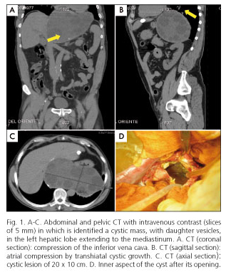
We report an unusual case of giant hydatid cyst (HC), which grew exophytically from the left hepatic lobe to the mediastinum, where it caused compression of various anatomical structures.
Case report
A 60-year-old male with no past medical history presented with few days of epigastric discomfort, jaundice, dark urine and acholic stools. Physical examination revealed only jaundice. Laboratory values showed AST 57 IU/L, ALT 172 IU/L, GGT 564 IU/L, ALP 272 IU/L, total bilirubin 8.15 mg/dL, direct bilirubin 5.72 mg/dL. Positive serology for hydatidosis. CT scan (Fig. 1 A, B and C) showed a large cystic mass of 20 x 10 x 12 cm in the left hepatic lobe, with vesicles inside, compressing the inferior vena cava and extending through the hiatus into the mediastinum, displacing descending aorta, esophagus, and right atrium. He was treated with albendazole for four weeks and subsequently underwent surgery (Fig. 1D). Through a midline laparotomy, a puncture-aspiration of the cyst, sterilization with hypertonic saline solution and partial pericystectomy with residual cavity drainage were performed. Biopsy confirmed the hydatid nature of the cyst. The patient had an uncomplicated recovery and was discharged on the seventh postoperative day on a therapeutic regimen of albendazol.
Discussion
The diagnosis of hepatic hydatidosis is usually casual or may be made after intra and extrahepatic complications (4). Clinical manifestations depend on the topography and the size of the lesion. Giants HC are extremely rare and symptoms are noted when their diameter reaches 10 cm (5).
The combination of imaging and serology usually enables diagnosis. Ultrasonography has a high diagnostic sensitivity (90-95%), and it is important to follow-up (6). CT scan is the imaging modality of choice to determine the presence of complications. This technique is useful for detection of extra-abdominal locations and for surgical planning (4).
Currently, therapeutic options in hydatid liver disease include medical treatment, percutaneous aspiration and drainage, and open or laparoscopic surgery (7). Chemotherapy is the preferred treatment when the disease is inoperable or when the cysts are too numerous. This treatment is also applied in combination with surgery as prophylaxis against the spread of cystic content and to minimize the recurrence of cysts. Surgery still remains as the standard for liver HC treatment because completely eliminates the parasite, treat complications associated and prevent recurrences (8). Radical procedures (pericystectomy and hepatectomy) are associated with lower rates of morbidity, hospital stay, and recurrence rates. However, its application must be made on an individual basis (9). In the particular case reported, it is remarkable the large size of the cyst and that it was not originated in the right lobe, as usual (10), but it was located in the left lobe, involving the mediastinum. Therefore, it should be noted that in the presence of a mediastinal mass of possible abdominal origin, liver hydatidosis should be included in the differential diagnosis.
Daniel Fernández-Martínez, José Antonio Álvarez-Pérez, Pablo Granero-Castro,
Raquel Rodríguez-Uría, Jimy Harold Jara-Quezada, Antonio Rodríguez-Infante and Germán Mínguez-Ruiz
Department of General and Digestive Surgery. Hospital Universitario Central de Asturias. Oviedo, Spain
References
1. Ozturk G, Aydinli B, Yildirgan MI, Basoglu M, Atamanalp SS, Polat KY, et al. Posttraumatic free intraperitoneal rupture of liver cystic echinococcosis: A case series and review of literature. Am J Surg 2007;194:313-6. [ Links ]
2. Khuroo MS, Wani NA, Javid G, Khan BA, Yattoo GN, Shah AH, et al. Percutaneous drainage compared with surgery for hepatic hydatid cysts. New Engl J Med 1997;337:881-7. [ Links ]
3. Turgut AT, Altin L, Topçu S, Kiliçoglu B, Aliinok T, Kaptanoglu E, et al. Unusual imaging characteristics of complicated hydatid disease. Eur J Radiol 2007;63:84-93. [ Links ]
4. Adán Merino L, Alonso Gamarra E, Gómez Senent S, Froilán Torres C, Martín Arranz E, Segura Cabral JM. Hidatidosis hepática: manejo actual de una entidad aún presente. Rev Esp Enferm Dig 2008;100:1130-48. [ Links ]
5. Suárez Grau JM, Gómez Bravo MA, Álamo Martínez JM, Rubio Cháves C, Marín Gómez LM, Suárez Artacho G, et al. Giant hydatid cyst involving the right hepatic lobe. Rev Esp Enferm Dig 2009;101:133-5. [ Links ]
6. Safioleas M, Misiakos E, Manti C, Katsikas D, Skalkeas G. Diagnosis and treatment of hepatic hydatid disease of the liver. World J Surg 1994;18:859-63. [ Links ]
7. Salemis NS. Giant hydatid liver cyst. Management of residual cavity: A case report. Ann Hepatol 2008;7:174-6. [ Links ]
8. Alonso Casado O, Moreno González E, Loinaz Segurola C, Gimeno Calvo A, González Pinto I, Pérez Saborido B, et al. Results of 22 years experience in radical surgical treatment of hepatic hydatid cysts. Hepatogastroenterology 2001;48:235-43. [ Links ]
9. Priego P, Nuño J, López Hervás P, López Buenadicha A, Peromingo R, Die J, et al. Hidatidosis hepática. Cirugía radical vs. no radical: 22 años de experiencia. Rev Esp Enferm Dig 2008;100:82-5. [ Links ]
10. Losada Morales H, Burgos San Juan L, Silva Abarca J, Muñoz Castro C. Experience with the surgical treatment of hepatic hydatidosis: Case series with follow-up. World J Gastroenterol 2010;16:3305-9. [ Links ]


 texto en


