Mi SciELO
Servicios Personalizados
Revista
Articulo
Indicadores
-
 Citado por SciELO
 -
 Accesos
Links relacionados
-
 Citado por Google
 -
 Similares en
SciELO
 -
 Similares en Google
Compartir
Revista Española de Enfermedades Digestivas
versión impresa ISSN 1130-0108
Rev. esp. enferm. dig. vol.106 no.5 Madrid may. 2014
PICTURES IN DIGESTIVE PATHOLOGY
Gastric and duodenal pseudomelanosis: A propos of two cases
Pseudomelanosis gástrica y duodenal: a propósito de dos casos
Alejandra Ochoa-Palominos, Fernando Díaz-Fontenla, Cecilia González-Asanza, Beatriz Merino-Rodríguez, Óscar Nogales-Rincón and Pedro Menchén Fernández-Pacheco
Department of Digestive Diseases. Hospital General Universitario Gregorio Marañón. Madrid, Spain
Introduction
Gastric and duodenal pseudomelanosis is an uncommon condition characterized by pigment deposition within macrophages in the lamina propria of the mucosa. It is usually associated with oral iron ingestion, but also with antihypertensive drugs and diseases such as blood hypertension, chronic kidney failure, gastrointestinal bleeding, chronic heart failure, and diabetes mellitus.
Case reports
Case report 1
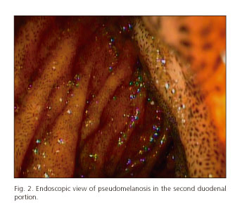
A 60-year-old male with blood hypertension, ischemic heart disease, idiopathic retroperitoneal fibrosis with chronic splenic thrombosis, chronic kidney failure, and iron deficiency anemia. He was on treatment with doxazosin, atenolol, hydralazine, ferrous sulfate, furosemide, and acenocoumarol, among others. An upper digestive endoscopy (UDE) was ordered to rule out esophageal-gastric varices, which identified multiple millimetre coffee-colored lesions in the gastric body, duodenal bulb, and second duodenal portion (Figs. 1 and 2). Histopathology revealed the presence of blackish-brown deposits within lamina propria macrophages (Fig. 3).
Case report 2
A 78-year-old male with blood hypertension, diabetes mellitus, ischemic heart disease, heart failure, peripheral vascular disease, iron deficiency anemia, and chronic kidney failure. He was on treatment with furosemide, hydralazine, ferrous sulfate, acetylsalicylic acid, lisinopril, carvedilol, doxazosin, and insulin. He underwent UDE for anemia, which identified blackish longitudinal stripes in the gastric antrum and multiple dark point-like spots in the duodenal bulb, and second duodenal portion. Histopathology confirmed the presence of iron deposits using Perls' technique (Fig. 4).
Discussion
Duodenal pseudomelanosis is more common than gastric pseudomelanosis - We found around 50 case reports of the former in the literature versus only 5 of the gastric variant (1-5). Differential diagnosis includes melanoma, Peutz-Jeghers syndrome, severe ischemic lesions in the gastric mucosa, and heavy metal toxicity. Both conditions are benign, and no association with malignant or inflammatory degeneration has been reported.
References
1. Mitty RD, Wolfe GR, Cosman M. Initial description of gastric melanosis in a laxative-abusing patient. Am J Gastroenterol 1997;92:707-8. [ Links ]
2. Weinstock LB, Katzman D, Wang HL. Pseudomelanosis of stomach, duodenum, and jejunum. Gastrointestinal Endoscopy 2003;58:578. [ Links ]
3. Rinesmith SE, Marsh WL. Gastric pseudomelanosis. Gastroenterology & Hepatology 2006;2:806-7. [ Links ]
4. Kibria R BC. Pseudomelanosis of the stomach. Endoscopy 2010;42(Supl. 2):E243-4. [ Links ]
5. Alraies MC, Alraiyes AH, Baibars M, Shaheen K. Pseudomelanosis of the stomach. QJM 2014;107:83-4. [ Links ]


 texto en


