Mi SciELO
Servicios Personalizados
Revista
Articulo
Indicadores
-
 Citado por SciELO
 -
 Accesos
Links relacionados
-
 Citado por Google
 -
 Similares en
SciELO
 -
 Similares en Google
Compartir
Revista Española de Enfermedades Digestivas
versión impresa ISSN 1130-0108
Rev. esp. enferm. dig. vol.107 no.11 Madrid nov. 2015
Ectopic pancreas in gallbladder. Clinical significance, diagnostic and therapeutic implications
Páncreas ectópico en vesícula biliar. Significado clínico e implicaciones diagnósticas y terapéuticas
Elena M. Sanchiz-Cárdenas1, Rocío Soler-Humanes1, Ana I. Lavado-Fernández2, Rafael Díaz-Nieto1 and Miguel A. Suárez-Muñoz1
1 General and Digestive Surgery Department, and 2 Pathology Department. Hospital Clínico Universitario Virgen de la Victoria. Málaga, Spain
ABSTRACT
Ectopic or heterotopic pancreas is defined as the presence of pancreatic tissue in an anatomical place not related to the pancreas, being it most frequent locations the stomach and small bowel. Its finding in the gallbladder is exceptional. Since the first case was reported by Otschkin in 1916, about 30 cases have been described in literature. We report the case of a 43 years-old male patient who had an urgent laparoscopic cholecystectomy with the diagnosis of acute cholecystitis, which pathological study showed the existence of chronic cholecystitis with heterotopic pancreatic tissue in the gallbladder wall.
Key words: Ectopic pancreas. Heterotopic pancreas. Gallbladder.
RESUMEN
Páncreas ectópico o heterotópico se define como la presencia de tejido pancreático en una localización anatómica que no tiene relación con el páncreas, siendo sus localizaciones más frecuentes el estómago y el intestino delgado.
Su hallazgo en la vesícula biliar es excepcional. Desde que Otschkin publicara el primer caso en 1916, alrededor de 30 más han sido descritos en la literatura.
Presentamos el caso de un paciente varón de 43 años al que se le realizó una colecistectomía laparoscópica urgente con diagnóstico de colecistitis aguda cuyo estudio histopatológico demostró la existencia de colecistitis crónica con tejido pancreático heterotópico en la pared de la vesícula biliar.
Palabras clave: Páncreas ectópico. Páncreas heterotópico. Vesícula biliar.
Introduction
Heterotopic pancreas, also called ectopic pancreas, is an embryologic abnormality, defined as the presence of pancreatic tissue without continuity or anatomic or vascular communication with the pancreatic gland. It can be located in the stomach, duodenum, proximal jejunum and Meckel's diverticulum (1).
It also has been described in spleen, ileum, mesentery, lung, mediastin, liver, biliary duct, gallbladder, and fallopian tube. Histologically, it is similar to the normal pancreas, with exocrine glands, ducts, and even Langerhans islets (2).
This condition has an estimated frequency of 1/500 surgical interventions in the upper gastrointestinal tract (3). In spite of its congenital origin, it is usually diagnosed in adulthood because most patients are asymptomatic.
The clinical significance of the presence of heterotopic pancreas in the gallbladder is uncertain because of its incidental finding at microscopic exploration after extirpation for cholecystopathy. As a rare entity, it is not usually considered in the initial differential diagnosis (1,4).
Case report
A 43-years-old male with hypertension and biliary colics with known cholelytiasis as clinical background is admitted in Emergency Department for abdominal pain in the right upper quadrant for 48 hours and vomits. Physical examination revealed pain and tenderness located in the right upper abdomen, with positive Murphy's sign.
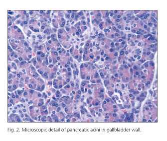
The requested blood test had a leukocyte range of 6.7x10³/µL (reference: 4-10.5 103/µL) with associated neutrophilia. Total bilirrubin of 0.6 mg/dl (reference: 0.2-1.10 mg/dl), AST 66 mg/dl (reference 8-40 mg/dl), amylase 43 (reference 25-115 mg/dl), PCR 15.84 mg/dl (reference less than 5 mg/dl). An urgent abdominal ultrasound was performed, which showed a distended gallbladder with wall edema and cholelithiasis (Fig. 1). With the diagnosis of acute cholecystitis, the patient underwent an urgent laparoscopic cholecystectomy. He had a favourable postoperative recovery without complications. The pathological specimen study revealed chronic cholecystitis and ectopic pancreatic tissue in the gallbladder wall (Fig. 2).
Discussion
Heterotopic pancreas, first described by Jean Schultz in 1727, is defined as pancreatic tissue in an anatomical place that is not related with the pancreatic gland (5).
Four types of pancreatic heterotopia are defined according with Heinrich classification in 1909, modified by Fuentes in 1973:
- Type I: Pancreatic tissue with acini, ducts, and islets like pancreatic gland.
- Type II: Canalicular variant with pancreatic ducts.
- Type III or exocrine pancreas with acinar tissue.
- Type IV or endocrine pancreas, with cellular islets (2).
Its real incidence is unknown because most patients do not manifest symptoms, it has been described in 2% of laparotomies, and 0.5-13.7% of autopsies. Upper gastrointestinal tract is the most frequent place, at stomach (25-38%), especially in submucosa (75%), duodenum (30%), and jejunum (15%) (5,6).
Symptomatic cases are nonspecific like abdominal pain, nausea, vomits, anorexia, weight loss, anemia or melena. Abdominal pain is the most frequent symptom and can be explained by inflammation and irritation of the surrounded tissue secondary to enzyme and pancreatic hormone secretion (2).
In gallbladder case, the presence of ectopic pancreas is extremely rare, with a few cases described, being most of them an incidental finding after cholecystectomy by cholescytopathy with location more frequent in neck and fundus (1,7-9).
Its diagnosis is difficult because is asymptomatic in most cases, when there are symptoms, they usually are nonspecific and similar to acute or chronic cholecystopathy, without being necessarily associated with cholelithiasis; thus the diagnosis is usually incidental during surgery or at histological study (7).
In reported cases in which the incidental finding of an asymptomatic lesion in the gallbladder led to cholecystectomy, it was for suspected malignancy, but not for the possibility of ectopic pancreas (6,10).
At histological study, ectopic pancreas in gallbladder is described as a polypoid, exofitic grown or like yellow nodules with size ranging from a few millimeters up to four centimetres. Fifty five percent of them are located in gallbladder neck (73% submucosa) (7).
Soto et al. found high amylase and lipase levels in bilis related to ectopic pancreatic tissue in gallbladder and proposed that this exocrine activity may cause pain and acute or chronic cholecystopathy with or without associated lithiasis, and malign lesions in the biliar tract due to the damage that can cause the elevation of pancreatic enzymes in the gallbladder and biliary tract mucosa (8). As well as amilasuria in the case described by Klimis et al., also have been described cases of gallbladder obstruction and perforation (6,7).
As in this case, the preoperative clinical orientation of ectopic pancreas in the gallbladder is difficult because of its infrequency, especially in the urgent context, so it is not considered in the differential diagnosis. In addition, the actual diagnostic tools like ultrasonography and CT scan can not differentiate between the presence of aberrant pancreas in gallbladder and other lesions like cholesterol polyps, adenoma or neoplasia, so its finding is usually incidental after cholecystectomy for cholecistopathy, being a challenge the preoperative diagnosis. However, despite its rarity, it should be considered in the differential diagnosis of lesions in the gallbladder wall without stones, like polyps or nodules, especially if coexists hyperamilasuria of unknown origin (7,10,11). Therefore, the attitude to this condition is controversial. In most cases surgical treatment is decided, not only by the presence of symptoms, but for diagnostic reasons and exclusion of malignancy (5).
References
1. Elhence P, Bansal R, Agrawal N. Heterotopic pancreas in gallbladder associated with chronic cholecystolithiasis. Int J Appl Basic Med Res 2012;2:142-3. DOI: 10.4103/2229-516X.106360. [ Links ]
2. Sathyanarayana SA, Deutsch GB, Bajaj J, et al. Ectopic pancreas: A disgnostic dilema. Int Journal Angiol 2012;21:177-80. DOI: 10.1055/s-0032-1325119. [ Links ]
3. Biswas A, Husain EA, Feakins RM, et al. Heterotopic pancreas mimicking cholangiocarcinoma. Case report and literature review. JOP 2007;8:28-34. [ Links ]
4. Al-Shraim M, Ezzadien M, Elhakeen H, et al. Pancreatic heterotopias in the gallbladder associated with chronic cholecystitis: A rare combination. JOP 2010;11:464-6. [ Links ]
5. Guimaraes M, Rodrigues P, Goncalves G, et al. Heterotopic pancreas in excluded stomach diagnosed after gastric bypass surgery. BMC Surg 2013;13:56. DOI: 10.1186/1471-2482-13-56. [ Links ]
6. Soto A, Hashimoto M, Sasaki K, et al. Elevation of pancreatic enzymes in gallbladder bile associated with heterotopic pancreas. A case report and review of the literature. JOP 2012;13:235-8. [ Links ]
7. Klimis T, Roukonakis N, Kafetzis I, et al. Heterotopic pancreas of the gallbladder associated with chronic cholecystitis and high levels of amylasuria. JOP 2011;12:458-60. [ Links ]
8. Gucer H, Bagcy P, Coskunoglu EZ, et al. Heterotopic pancreatic tissue located in the gallbladder wall. A case report. JOP 2011;12:152-4. [ Links ]
9. Shiwani MH, Gasling J. Heterotopic pancreas of the gallbladder associated with chronic cholecystitis. JOP 2008;9:30-2. [ Links ]
10. Foucault A, Veilleux H, Martel G, et al. Heterotopic pancreas presenting as suspicious mass in the gallbladder. JOP 2012;13:700-1. [ Links ]
11. Weppner JL, Wilson M, Ricca R, et al. Heterotopic pancreatic tissue obstructing the gallbladder neck, a case report. JOP 2009;10:532-4. [ Links ]
![]() Correspondence:
Correspondence:
Elena M. Sanchiz-Cárdenas.
General and Digestive Surgery Department.
Hospital Clínico Universitario Virgen de la Victoria.
Campus Universitario de Teatinos, s/n.
29010 Málaga, Spain
e-mail: esanchizcardenas@gmail.com
Received: 25-11-2014
Accepted: 16-02-2015


 texto en


