Mi SciELO
Servicios Personalizados
Revista
Articulo
Indicadores
-
 Citado por SciELO
 -
 Accesos
Links relacionados
-
 Citado por Google
 -
 Similares en
SciELO
 -
 Similares en Google
Compartir
Revista Española de Enfermedades Digestivas
versión impresa ISSN 1130-0108
Rev. esp. enferm. dig. vol.108 no.12 Madrid dic. 2016
https://dx.doi.org/10.17235/reed.2016.4051/2015
Segmental colitis caused by idiopathic myointimal hyperplasia of mesenteric veins
Mariana N. Costa1, Joana Saiote1, Maria José Pinheiro2, Pedro Duarte1, Teresa Bentes1, Mário Ferraz-Oliveira3 and Jaime Ramos1
Departments of 1Gastroenterology, 2Surgery and 3Pathology. Centro Hospitalar Lisboa Central. Lisbon, Portugal
ABSTRACT
Diseases causing colonic ischemia may be mistaken with other causes of segmental colitis such as inflammatory bowel disease, especially in young patients. The authors present the case of a 47-year-old male with severe proctosigmoiditis. Assessment excluded infectious causes, thrombophilia and systemic vasculitis. The initial histological specimen was suggestive of inflammatory bowel disease and therapy was initiated with intravenous steroids and, at day 5, infliximab, with no response. The patient was proposed for surgery. Pathological examination of the surgical specimen revealed an idiopathic myointimal hyperplasia of mesenteric veins, a rare entity exhibiting necrotizing phlebitis with rapid progression to segmental necrosis in the rectosigmoid colon. In this paper the authors discuss the differential diagnosis of proctosigmoiditis in young ages and the approach to this exceptionally rare ischemic entity.
Key words: Ischemic colitis. Idiopathic myointimal hyperplasia of mesenteric veins.
Introducción
Diseases causing colonic ischemia may have similar presentation symptoms to other causes of segmental colitis such as inflammatory bowel disease, especially in young patients. Idiopathic myointimal hyperplasia of mesenteric veins (IMHMV) is a rare condition causing segmental colonic ischemia that should be considered in the differential diagnosis of severe colonic inflammatory bowel disease refractory to intensive medical treatment. Preoperative diagnosis of IMHMV could be difficult as intimal thickening venules are in the submucosa and deeper layers. Even when a full-thickness biopsy is performed the diagnosis may be inconclusive. Standard treatment is surgical resection and there are no reports of postoperative disease recurrence. The following case illustrates this rare clinical condition in a young adult male patient.
Case report
A 47-year-old Caucasian male patient was admitted to our medical department on February 2012 with bloody diarrhea (more than 20 stools per day), lower abdominal cramping pain, proctalgia and malaise.
The patient reported a previous 9 month history of hypogastric cramp-like pain, 4-5 small volume stools, without blood, mucus or pus, as well as a compelling urge to defecate and fecal incontinence. One month before, the patient had been assessed on the emergency department of another hospital due to clinical deterioration with anal pain, persistent urge to defecate, tenesmus and straining at stool. There, he had undergone a colonoscopy with biopsy and a pelvic CT scan. The colonoscopy showed edema of the rectal mucosa and sigmoid colon. The biopsies did not reveal any significant alterations. The pelvic CT scan identified parietal thickening of the rectum. At that time, the patient was treated with an antispasmodic. No improvement was observed.
Patient's personal and family histories were irrelevant and epidemiological context was unremarkable. He had not been undergoing any other course of medication and denied known allergies, smoking, alcohol and history of injected or inhaled drug use.
Upon physical examination, he was afebrile with lower abdominal tenderness with no signs of peritoneal irritation.
Routine blood analysis revealed normal hemoglobin (13.8 g/dL), no leukocytosis nor neutrophilia with a slightly raised level of C-reactive protein (6.3 mg/dL; normal level: < 5 mg/dL). Infectious causes were excluded.
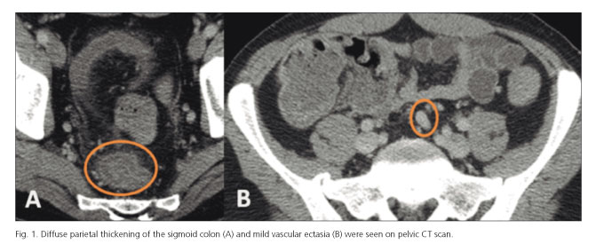
Radiologically, there was a marked diffuse parietal thickening (13 mm) of the rectum and sigmoid colon, peri-colic fat densification with some small ganglion formations and mild vascular ectasia. There was no intra peritoneal free fluid (Fig. 1).
The rectosigmoidoscopy showed diffuse edema of the rectal mucosa with sudden transition to a circumferential and continuous mucosal ulceration and luminal stenosis of the sigmoid colon. Histologic examination showed mucosal edema and vascular congestion without significant glandular lesions, with mild inflammatory infiltration and fragments of base of ulcer without pericriptitis, crypt abscesses or granulomas.
Intravenous methylprednisolone (40 mg daily) was initiated. There was no improvement in the clinical course after 5 days of intravenous steroids, so treatment with intravenous 400 mg infliximab (5 mg/kg) was started.
Colonoscopy was repeated to evaluate the disease extent and severity, revealing a granular edematous mucosa in the proximal rectum, circumferential necrotic ulceration extending from 10 to 30 cm from the anal verge and a nodular mucosa followed by a well-defined transition to normal mucosa at this level. Above the ulcerated area, the colon mucosa was normal (Fig. 2). Biopsy samples were taken. Histologic evaluation showed edema and hemorrhage of the chorion, focal ischemic type changes and several small vessels with fibrinoid necrosis and lumen thrombosis, aspects suggestive of ischemic etiology (Fig. 3).
Autoimmunity, systemic vasculitis and thrombophilia tests were negative.
The patient was proposed for surgery and a Hartmann procedure was performed. The macroscopic examination of the surgical specimen showed an extensive ulcerated area 13 cm long (Fig. 4). On histological examination of the ulcerated region granulation tissue was observed, which reached the submucosa with neovascular proliferation and extensive lesions of fibrinoid necrosis and vasculitis, thrombosis and nerve hyperplasia. On the adventitia, several vessels with intimal hyperplasia conditioning luminal stenosis were seen. The lymph nodes showed sinus histiocytosis and ectasia. The mesorectal adipose tissue had lesions with steatonecrosis. The histological aspects were consistent with IMHMV (Fig. 5).
Discussion
A significant diagnostic challenge is raised by segmental colitis because of its broad differential diagnosis. Many diseases such as diverticulitis, radiation colitis, mucosal prolapsed/solitary ulcer syndrome, infectious diseases, colon carcinoma, inflammatory bowel disease and colonic ischemia may present a segmental colonic involvement.
Colonic ischemia may result from various etiologies but it can be broadly classified as non-occlusive and occlusive. Venous occlusion is an uncommon cause of ischemic bowel disease, and the majority of such cases are caused by venous thrombosis (1,2). Non-thrombotic occlusion of the mesenteric veins is rare and has previously been described in vasculitis associated with systemic lupus erythematosus, Behçet's disease, enterocolic lymphocytic phlebitis and IMHMV (3,4).
Colonic ischemia is more common in the elderly but younger patients may also be affected. In these patients, segmental ischemic colitis is a rare differential diagnosis of inflammatory bowel disease (5) and it results of a hypoxic tissue injury with secondary necrosis and inflammation (6). It may present a spectrum of severity ranging from mild, transient mucosal erosion to fibrous scarring with stricture formation or even transmural infarction (7).
IMHMV is a rare entity exhibiting necrotizing phlebitis with rapid progression to segmental necrosis in the rectosigmoid colon. The precise incidence of IMHMV is unknown, as only 21 cases have been reported in the literature to date (Table I). This disease is known to occur predominantly in young, healthy men, with a sudden onset of crampy abdominal pain accompanying repeated episodes of diarrhea and bloody stools (4,8). The pathophysiologic mechanism seems to be an abnormal arteriovenous shunting with elevated venous pressure and consecutive congestion and ischemia of the involved bowel segment (3). IMHMV diagnosis can be based on clinical manifestations causing acute, progressive ulcers in the rectosigmoid colon and pathohistologic findings on the resected specimen. A biopsy is generally not helpful for diagnostic purposes since specimens that include concentric intimal thickening venules in the submucosa and deeper layers are often not obtained (9). Pathohistologic examination of a resected specimen reveals a striking pattern of scattered circumferential intimal thickening of vessels, with or without lymphocyte infiltration around venules and complete obliteration of venules from the submucosa to subserosa (4). Regarding treatment, complete resection of the necrotic segments is recommended. The postoperative clinical prognosis is generally good, with no recurrence (4).
Our patient is a typical case of this extremely rare condition. The results of the initial biopsy revealed nonspecific changes in the intestinal mucosa. As with other cases of IMHMV, medical therapies were ineffective. Given that subsequent biopsies suggested a possible ischemic etiology, our patient underwent a surgical resection. The histological examination of the resected specimen established the diagnosis of IMHMV. Overall, this case presents an entity that is difficult to diagnose since both the endoscopic and biopsy appearance are nonspecific and only the surgical specimen provides the diagnosis.
References
1. Grendell JH, Ockner RK. Mesenteric venous thrombosis. Gastroenterology 1982;82:358-72. [ Links ]
2. Hunter GC, Guernsey JM. Mesenteric ischemia. Med Clin North Am 1988;72:1091-115. [ Links ]
3. Abu-Alfa AK, Ayer U, West AB. Mucosal biopsy findings and venous abnormalities in idiopathic myointimal hyperplasia of the mesenteric veins. Am J Surg Pathol 1996;20:1271-8. DOI: 10.1097/00000478-199610000-00014. [ Links ]
4. Genta RM, Haggitt RC. Idiopathic myointimal hyperplasia of mesenteric veins. Gastroenterology 1991;101:533-9. [ Links ]
5. Brandt LJ, Boley SJ. AGA technical review on intestinal ischemia. American Gastrointestinal Association. Gastroenterology 2000; 118:954-68. [ Links ]
6. Feuerstadt P, Brandt LJ. Colon ischemia: Recent insights and advances. Curr Gastroenterol Rep 2010;12:383-90. DOI: 10.1007/s11894-010-0127-y. [ Links ]
7. Hwang S-S, Chung W-C, Lee K-M, et al. Ischemic colitis due to obstruction of mesenteric and splenic veins: A case report. World J Gastroenterol 2008;14:2272-6. DOI: 10.3748/wjg.14.2272. [ Links ]
8. Kao PC, Vecchio JA, Hyman NH, et al. Idiopathic myointimal hyperplasia of mesenteric veins: A rare mimic of idiopathic inflammatory bowel disease. J Clin Gastroenterol 2005;39:704-8. DOI: 10.1097/00004836-200509000-00011. [ Links ]
9. Mizoshita T, Tanida S, Joh T. A case of punched-out ulcer occurring in the rectosigmoid colon with sudden onset of bloody stools. Gastroenterology 2011;141:e9-10. DOI: 10.1053/j.gastro.2010.05.092. [ Links ]
10. Savoie LM, Abrams A V. Refractory proctosigmoiditis caused by myointimal hyperplasia of mesenteric veins: Report of a case. Dis Colon Rectum 1999;42:1093-6. DOI: 10.1007/BF02236711. [ Links ]
11. Lavu K, Minocha A. Mesenteric inflammatory veno-occlusive disorder: A rare entity mimicking inflammatory bowel disorder. Gastroenterology 2003;125:236-9. DOI: 10.1016/S0016-5085(03)00663-2. [ Links ]
12. García-Castellanos R, López R, De Vega VM, et al. Idiopathic myointimal hyperplasia of mesenteric veins and pneumatosis intestinalis: A previously unreported association. J Crohns Colitis 2011;5:239-44. DOI: 10.1016/j.crohns.2010.12.003. [ Links ]
13. Chiang C-K, Lee C-L, Huang C-S, et al. A rare cause of ischemic proctosigmoiditis: Idiopathic myointimal hyperplasia of mesenteric veins. Endoscopy 2012;44(Suppl2):E54-5. DOI: 10.1055/s-0031-1291529. [ Links ]
14. Feo L, Cheeyandira A, Schaffzin DM. Idiopathic myointimal hyperplasia of mesenteric veins in the elderly. Int J Colorectal Dis 2013;28:433-4. DOI: 10.1007/s00384-012-1480-0. [ Links ]
15. Korenblit J, Burkart A, Frankel R, et al. Refractory pancolitis: A novel presentation of idiopathic myointimal hyperplasia of mesenteric veins. Gastroenterol Hepatol 2012;8:696-700. [ Links ]
16. 1Thomas BS. Myointimal hyperplasia of the mesenteric veins mimicking infectious colitis. Int J Colorectal Dis 2013;28:727. DOI: 10.1007/s00384-012-1522-7. [ Links ]
17. Lanitis S, Kontovounisios C, Karaliotas C. An extremely rare small bowel lesion associated with refractory ascites. Idiopathic myointimal hyperplasia of mesenteric veins of the small bowel associated with appendiceal mucocoele and pseudomyxoma peritonei. Gastroenterology 2012;142:e5-7. DOI: 10.1053/j.gastro.2011.11.052. [ Links ]
18. Wangensteen KJ, Fogt F, Kann BR, et al. Idiopathic myointimal hyperplasia of the mesenteric veins diagnosed preoperatively. J Clin Gastroenterol 2015;49:491-4. DOI: 10.1097/MCG.0000000000000290. [ Links ]
19. Sahara K, Yamada R, Fujiwara T, et al. Idiopathic myointimal hyperplasia of mesenteric veins: Rare case of ischemic colitis mimicking inflammatory bowel disease. Dig Endosc 2015;27:767-70. DOI: 10.1111/den.12470. [ Links ]
20. Abbott S, Hewett P, Cooper J, et al. Idiopathic myointimal hyperplasia of the mesenteric veins: A rare differential to be considered in idiopathic colitis. ANZ J Surg 2015 Jul 3. (E-pub ahead of print) DOI: 10.1111/ans.13210. [ Links ]
21. Laskaratos F-M, Hamilton M, Novelli M, et al. A rare cause of abdominal pain, diarrhoea and GI bleeding. Gut 2015;64:214. DOI: 10.1136/gutjnl-2014-308319. [ Links ]
22. Zijlstra M, Tjhie-Wensing JW, Van Dijk M, et al. Idiopathic myointimal hyperplasia of mesenteric veins: An unusual cause of diarrhoea. Ned Tijdschr Geneeskd 2014;158:A7752. [ Links ]
![]() Correspondence:
Correspondence:
Mariana Nuno Costa.
Department of Gastroenterology.
Alameda de Sto. António dos Capuchos.
1169-050 Lisbon, Portugal
e-mail: mariananunocosta@gmail.com
Received: 24-10-2015
Accepted: 28-10-2015


