Dear Editor,
In relation to the article published in this journal by Alberto Herreros de Tejada et al. 1, we recently diagnosed a case of adenocarcinoma of the proximal anal canal with an exceptional immunohistochemistry. This was identified as an incidental finding after a hemorrhoidectomy in a patient with serrated polyposis syndrome (SPS).
Case report
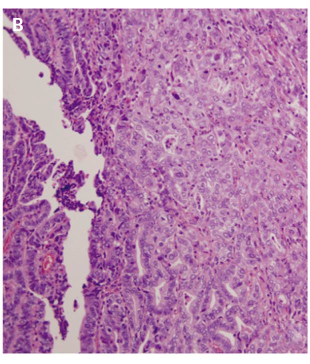
We present the case of a 48-year-old female, current smoker, with a history of endoscopically controlled SPS. The patient was assessed for grade III hemorrhoids, and a hemorrhoidectomy in accordance with the Milligan-Morgan technique was performed. Pathological analysis of the specimen revealed a poorly differentiated infiltrating adenocarcinoma that originated from a tubular-villous adenoma with high-grade dysplasia at the squamocolumnar junction (Fig. 1A and B). The superficial adenomatous lesion and the infiltrating component were identical and expressed cytokeratin 7 intensely, and were negative for cytokeratin 20 (Fig. 1C). A multidisciplinary committee decided to widen the previous resection margins after a computed tomography (CT) scan confirmed the absence of distant or lymph node disease. No residual tumor infiltration was found in the surgical specimen. PET-CT after three months follow-up showed lymphadenopathy at the sigmoid colon mesenterium. Given these findings and the history of SPS, a proctocolectomy with end ileostomy was performed. Histopathological analysis identified multiple serrated adenomas with low-grade dysplasia with metastasis of poorly differentiated carcinoma that was morphologically the same as that observed in the anal canal in 2/109 lymph nodes analyzed (pT0N1bM0). The patient started adjuvant chemotherapy.


Fig. 1 A. Microscopic image of the epithelium in the squamocolumnar transition zone (arrow); the polyp mucosa is cranial to it (hematoxylin-eosin). B. Microscopic image of infiltrating tumor foci in the submucosa, with poorly differentiated and disorganized cells with nuclear pleomorphism (hematoxylin-eosin). C. Immunohistochemistry of the polyp at the hemorrhoid. Submucosal tumor clusters in the center and on the right (arrows), both reaching the limit of the resection plane, hindering a proper assessment of the deeper margin.
Discussion
Anal adenocarcinomas account for up to 10% of anorectal tumors 2. They are more commonly located proximal to the dentate line and are histologically and immunohistochemically similar to rectal carcinomas (CK7- and CK20+). The main clinical implication is regional lymphatic dissemination 3. Intense CK7 expression makes it necessary to rule out both ductal anal carcinoma and metastatic lesions. In rare cases with a high-grade and poorly differentiated colorectal phenotypes, expression of CK20 may be replaced by CK7 4. As a T1 lesion with no evidence of lymphatic or distant disease was identified via a CT scan, we opted for close surveillance following margin extension. With regard to SPS, factors associated with malignancy are serrated adenomas with dysplasia and their multiplicity 1.


