Meu SciELO
Serviços Personalizados
Journal
Artigo
Indicadores
-
 Citado por SciELO
 -
 Acessos
Links relacionados
-
 Citado por Google
 -
 Similares em
SciELO
 -
 Similares em Google
Compartilhar
Nutrición Hospitalaria
versão On-line ISSN 1699-5198versão impressa ISSN 0212-1611
Nutr. Hosp. vol.27 no.5 Madrid Set./Out. 2012
https://dx.doi.org/10.3305/nh.2012.27.5.5933
The use of body circumferences for the prediction of intra-abdominal fat in obese women with polycystic ovary syndrome
El uso de circunferencias corporales para la predicción de la grasa intra-abdominal en mujeres obesas con el síndrome del ovario poliquístico
F. Rodrigues de Oliveira Penaforte1,2, C. Cremonezi Japur2, R.W. Díez-García2, C. Salles Macedo3 and P. García Chiarello2
1Departamento de Nutrição. Universidade Federal do Triângulo Mineiro. Uberaba. Minas Gerais. Brazil
2Laboratorio de Práticas e Comportamento Alimentares (PrátiCA). Curso de Nutrição e Metabolismo. Faculdade de Medicina de Ribeirão Preto. Universidade de São Paulo. Ribeirão Preto. São Paulo. Brasil
3Departamento de Ginecologia e Obstetricia. Faculdade de Medicina de Ribeirão Preto. Universidade de São Paulo. Ribeirão Preto. São Paulo. Brasil
ABSTRACT
Introduction: Computerizd tomography (CT) is the gold standard for the evaluation of intra- (IAF) and total (TAF) abdominal fat; however, the high cost of the procedure and exposure to radiation limit its routine use.
Objective: To develop equations that utilize anthropometric measures for the estimate of IAF and TAF in obese women with polycystic ovary syndrome (PCOS).
Methods: The weight, height, BMI, and abdominal (AC), waist (WC), chest (CC), and neck (NC) circumferences of thirty obese women with PCOS were measured, and their IAF and TAF were analyzed by CT.
Results: The anthropometric variables AC, CC, and NC were chosen for the TAF linear regression model because they were better correlated with the fat deposited in this region. The model proposed for TAF (predicted) was: 4.63725 + 0.01483 x AC - 0.00117 x NC - 0.00177 x CC (R2 = 0.78); and the model proposed for IAF was: IAF (predicted) = 1.88541 + 0.01878 x WC + 0.05687 x NC -0.01529 x CC (R2=0.51). AC was the only independent predictor of TAF (p < 0.01).
Conclusion: The equations proposed showed good correlation with the real value measured by CT, and can be used in clinical practice.
Key words: Obesity. Polycystic ovary syndrome. Abdominal fat. Anthropometry.
RESUMEN
Introducción: La tomografia computarizada (TC) es el estándar de oro para la evaluación de la grasa intra-abdominal (GIA) y abdominal total (GAT), pero los altos costos y la exposición a la radiación limitan su uso rutinario.
Objetivo: Desarrollar ecuaciones para la estimación de la GIA y la GAT en mujeres obesas con el sindrome del ovario poliquistico, utilizando medidas antropométricas.
Métodos: Se evaluó el peso, la altura, el IMC y las circunferencias abdominal (CA), cintura (CC), pecho (CP) y cuello (Ccu) de 30 mujeres obesas con SOP. La GIA y GAT fueron analizados por la TC.
Resultados: El modelo propuesto fue: GAT = 4,63725 + 0,01483 x CA - 0.00117 x CCu - 0,00177 x CP (R2 = 0,78); y para la GIA fue: GIA = 1, 88541 + 0, 01878 x CC + 0,05687 x CCu - 0,01529 x CP (R2 = 0,51). La CA fue La única variable predictora independiente de la GAT (p < 0,01).
Conclusión: Las equaciones propuestas correlacionaronse bien con el valor real, medido a través de la TC, y se puede utilizarlas en la práctica clínica.
Palabras clave: Obesidad. Síndrome del ovario poliquistico. Grasa abdominal. Antropometría.
Abbreviations
PCOS: Polycystic ovary syndrome.
CT: Computerized tomography.
IAF: Intra-abdominal fat.
TAF: Total abdominal fat.
HCFMRP-USP: Hospital das Clínicas da Faculdade de Medicina de Ribeirão Preto da Universidade de São Paulo.
BMI: Body mass index.
WC: Waist circumference.
AC: Abdominal circumference.
TC: Trunk circumference.
NC: Neck circumference.
SD: Standard deviation.
Introduction
There is a high prevalence of obesity1 and preferential androgenic distribution of body fat in women with polycystic ovary syndrome (PCOS), which contrasts with the typical profile expected for the female gender, characterized by larger concentration of fat in the gluteal-femoral region.2
The abdominal obesity phenotype is commonly associated with insulin resistance and hyperandrogenism2. The localization of adipocytes determines differences in the metabolic role of these cells. Intra-abdominal fat has the most deleterious impact on tissue insulin resistance3 because of the high plasma levels of non-esterified fatty acids originating from intra-abdominal fat tissue lipolysis.4 For this reason, a more detailed assessment of body fat, especially in the abdominal and intra-abdominal regions, is mandatory in the case of women with PCOS. Nowadays computerized tomography (CT) is the gold standard method for evaluation of intra- (IAF) and total (TAF) abdominal fat. However, its routine use is restricted by high costs, availability, and patient exposure to radiation.5 Therefore, the aim of the present study was to develop equations for the estimate of intra- and total abdominal fat in obese women with PCOS by means of inexpensive, easy-to-apply, noninvasive anthropometric measurements.
Materials and methods
Thirty women with PCOS selected at the Gynecologic Endocrinology outpatient clinics of Hospital das Clínicas, Faculdade de Medicina de Ribeiráo Preto, Universidade de Sao Paulo (HCFMRP-USP) were included in the study using the following inclusion criteria: PCOS diagnosis according to the Rotterdam criteria (2004)6 and made at least one year before the study, age between 20 and 40 years, and body mass index (BMI) ≥ 30 kg.m-2. Previous ovarian failure, thyroid dysfunction, infectious diseases, presence of symptoms of menopause, diabetes mellitus type I or II, use of hormones over the previous months, recent weight loss (> 5% weight loss over the previous three months) and regular consumption of cigaretts and/or alcohol were the exclusion criteria. All the participants were submitted to anthropometric evaluation and CT. The study was approved by the HCFMRP-USP Research Ethics Committee and was registered under process number 4675/2006. All subjects gave written informed consent to participate.
Body weight (kg) was measured on an electronic platform-type Filizola® scale with maximum capacity of 300 kg and 0.1 kg precision, with the subject standing upright in the center of the equipment, barefoot and wearing a minimum amount of clothing. Height (m) was obtained using a height rod with 0.5 cm graduation, with the subject positioned on the center of the equipment, barefoot and bareheaded, standing upright with arms extended along the body, according to standard techniques.7 Obesity was classified according to the BMI.8
Body circumferences were measured with an inextensible tape measure with 0.1 mm graduations, according to standardized techniques.
Waist circumference (WC) - The patient was asked to stand upright, and a measure of the smallest curvature between the ribs and the iliac crest was taken without compressing the tissues.9
Abdominal circumference (AC) - AC was measured at the point corresponding to the navel, since it is the same point employed for assessment of abdominal fat by CT.
Trunk circumference (TC) - TC was measured on the posterior part of the trunk, 3 cm under the armpit. The patient was asked to place her arms parallel to the body. The tape measure was placed horizontally, and the complete circumference around the thorax was measured.10
Neck circumference (NC) - NC was measured at the upper margin of the thyroid cartilage.11
All anthropometric measurements were made by the same investigator, previously trained and habilitated for this activity.
CT was performed for the determination of IAF and TAF. All measurements were performed by the same technician. Twelve-hour fasting patients were placed in the supine position and asked to stretch their arms over their head. Oral contrast agents were not administered. Patients had been instructed to remove clothing and metal objects and to wear only a hospital gown during the exam.
IAF and TAF were measured between L4 and L5. To this end, 10 mm-thick slices were determined for all measurements according to a previously standardized procedure.12 The adipose tissue determined by assessment of body segments was calculated with the aid of the Mircro software (MRIcro, Atlanta, GA, EUA), which provides results in cm3. The volume of adipose tissue obtained was converted to mass (g) by means of the following equation: density = mass per volume unit, using the value of fat specific density, which is 0.9 g.cm-3 at 37o C.13
Statistical analyses
Results are presented as the mean ± standard deviation (SD). Correlations between the studied variables were analyzed by the Spearman coefficient. The level of significance was set at 5% (p < 0. 05).
To adjust the model for the variables IAF and TAF, multiple linear regression models were proposed, and some co-variables were considered to be possible independent variables. These models assume that the difference between the values predicted by the model and the observed values have normal distribution with constant mean and variance. In the situations in which this assumption was not confirmed, transformed response variables were employed. The model was adjusted by means of the SAS software version 9®.
Results
The patients included in the study were 30.5 ± 5.0 years old and had a BMI of 36.3 ± 4.1 kg.m-2. Anthropometric and CT data are listed in table I.
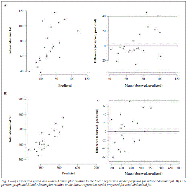
A multiple linear regression model was proposed for IAF. Waist, trunk, and neck circumferences were considered as independent variables because they correlate well with this type of fat (r = 0.74 and p < 0.0001; r = 0.62 and p < 0.0006; r = 0.70 and p < 0.0006, respectively). The proposed linear regression model was: IAF (predicted) = 1.88541 + 0.01878 x waist circumference + 0.05687 x neck circumference - 0.01529 x trunk circumference (R2 = 0.51).
A multiple linear regression model was proposed for TAF. Abdominal, trunk, and neck circumferences were considered as possible independent variables, since they correlated well with this kind of fat (r = 0.85 and p < 0.0001; r = 0.59 and p < 0.0001; r = 0.49 and p < 0.0006, respectively). Among the variables analyzed, abdominal circumference was the only independent predictor of TAF (b coefficient 0.01; p < 0.01). The proposed linear regression model was: TAF (predicted) = 4.63725 + 0.01483 x abdominal circumference - 0.00117 x neck circumference - 0.00177 x trunk circumference (R2 = 0.78). The dispersion and Bland-Altman graphs, which evaluate both equations for prediction, are displayed in figure 1.
Discussion
The predictive equation models proposed herein for the evaluation of IAF and TAF employ easy-to-perform anthropometric measurements that correlate well with the gold standard method used for such assessment (CT). Abdominal, trunk, and neck circumferences were employed for TAF prediction, whereas waist, trunk, and neck circumferences were used for IAF prediction.
There are few studies examining fat compartmentalization in PCOS, especially in the abdominal region, even though it has been documented that women with PCOS tend to accumulate fat in this area.4,14 A study conducted by Yildirim et al (2003) on the relationship between abdominal fat and metabolic disorders has found that women with PCOS have significantly larger abdominal fat accumulation compared to women without the syndrome paired for weight and age. It has also been verified that women with PCOS and abdominal distribution of body fat present a higher prevalence of metabolic disorders such as hyperinsulinemia and elevated triglyceride levels compared to control.14 Similar results have been reported by Kirchengast and Huber (2001), who described that only 30% of women with PCOS have body fat distribution of the gynoid type, whilst 100% of the women in the group without the disorder (paired for age, weight, and BMI) have the latter type of body fat distribution.3
The limitations inherent to CT for analysis of IAF and TAF justify the design of alternative and more easily accessible methods for the reliable evaluation of body fat distribution. Therefore, the regression models proposed for estimation of IAF and TAF are extremely relevant.
Previous studies aiming to develop equations for IAF and TAF estimation have already been conducted. For these models, the authors utilized demographic and anthropometric variables such as age, gender, BMI, and hip and waist circumferences,15 for example. Some of these equations were designed for populations with specific characteristics, e.g., the elderly and Indians. Nevertheless, there are no literature studies specifically involving obese women with PCOS.
In this respect, the present study was the first to develop this type of equations for application to obese women with PCOS. Our aim was to provide more easily applicable tools for the assessment of body fat distribution in this population, thereby facilitating clinical practice. It is noteworthy that there are no reference values for IAF and TAF. For this reason, it is important that analysis of these parameters by means of the proposed equations be carried out in a serial order, so that evolution of body fat distribution in patients submitted to weight loss, for instance, can be monitored.
The small number of patients evaluated and the need for future studies on the validation of the proposed equations are limitations of the present work.
We may conclude that it is possible to employ the proposed equations in clinical practice for IAF and TAF evaluation in obese women with PCOS since they related well to the real measurement obtained by the gold standard method. However, it is still necessary to validate these equations, especially to permit their use in other populations, because it is extremely important to adequately evaluate adiposity in this body segment.
References
1. Alvarez-Blasco F, Botella-Carretero JI, San Milla JL, Escobar-Morreale HF: Prevalence and Characteristics of the Polycystic Ovary Syndrome in Overweight and Obese Women. Arch Intern Med 2006; 23:2081-2086. [ Links ]
2. Cascella T, Palomba S, De Sio I, Manguso F, Giallauria F, De Simone B, Tafuri D, Lombardi G, Colao A, Orio F: Visceral fat is associated with cardiovascular risk in women with polycystic ovary syndrome. Human Reproduction 2008; 23 (1): 153-159. [ Links ]
3. Filho FFR, Mariosa LS, Ferreira SRG, Zanella MT: Gordura visceral e síndrome metabólica: mais que uma simples associação. Arq Bras Metab 2006; 50 (2): 230-238. [ Links ]
4. Wilding JP: The importance of free fatty acids in the development of type 2 diabetes. Diabet Med 2007; 24 (9): 934-945. [ Links ]
5. Rankinen T, Kim SY, Pérusse L, Després JP, Bouchard: The prediction of abdominal visceral fat level from body composition and anthropometry: ROC analysis. Inter J Obes 1999; 23: 801-809. [ Links ]
6. The Rotterdam ESHRE/ASRM-sponsored PCOS consensus workshop group. Revised 2003 consensus on diagnostic criteria and longterm health risks related to polycystic ovary syndrome (PCOS). Human Reproduction 2004; 19 (1): 41-47. [ Links ]
7. BRASIL. Vigilância alimentar e nutricional - SISVAN: orientações básicas para a coleta, processamento, análise de dados e informação em serviços de saúde. Brasilia: Ministério da Saúde, 2004. [ Links ]
8. World Health Organization. Obesity: preventing and managing the global epidemic. Report of a WHO Consulation. Geneva: World Health Organization; 1998 (Technical Report Series, No 894). [ Links ]
9. Callway CW, Chumlea WC, Bouchard C, Himes JH, Lohman TG, Martin AD, Mitchell CD, Mueller WH, Roche AF, Seefeldt VD. Circumferences. In: Lohman TG, Roche AF, Martorell R. Anthropometric standardization reference manual. Champaign, IL: Human Kinetics; 1988, pp. 39-54. [ Links ] 10. Penaforte FR, Japur CC, Diez-Garcia RW, Chiarello PG: Upper trunk fat assessment and its relationship with metabolic and biochemical variables and body fat in polycystic ovary syndrome. J Hum Nutr Diet 2011; 24 (1): 39-46. [ Links ] 11. Dixon JB, O'Brien PE. Neck circumference a good predictor of raised insulin and free androgen index in obese premenopausal women: changes with weight loss. Clinical Endocrinology 2002; 57: 769-778. [ Links ] 12. Seidell JC, Bakker CJG, van der Kooy K. Imaging techniques for measuring adipose-tissue distribution-a comparison between computed tomography and 1.5-T magnetic resonance. Am J Clin Nutr 1990; 51: 953-7. [ Links ] 13. Lukaski HC. Methods for the assessment of human body composition: traditional and new. Am J Clin Nutr 1987; 46 (4): 537-56. [ Links ] 14. Yildirim B, Sabir N, Kaleli B. Relation of intra-abdominal fat distribution to metabolic disorders in nonobese patients with polycystic ovary syndrome. Fertility and Sterility 2003; 79 (6):1358-1364. [ Links ] 15. Goel K, Gupta N, Misra A, Poddar P, Pandey RM, Vikram NK, Wasir JS: Predictive equations for body fat and abdominal fat with DXA and MRI as reference in Asian Indians. Obesity 2008; 16 (2): 451-456. [ Links ] Recibido: 3-IV-2012 ![]() Correspondence:
Correspondence:
Fernanda Rodrigues de Oliveira Penaforte
Professor of Endocrinology and Nutrition
Departamento de Clínica Médica
Faculdade de Medicina de Ribeirão Preto/USP
Avenida Bandeirantes, n.o 3900
14049-900. Ribeirão Preto. SP. Brasil
E-mail: ferpenaforte@usp.br
Aceptado: 10-V-2012


