


Meu SciELO
 Serviços customizados
Serviços Personalizados
Journal
Artigo
 texto em
  Inglês (pdf)
 Artigo em XML
 Referências do artigo
 Enviar este artigo por email
Indicadores
-
 Citado por SciELO
 -
 Acessos
Links relacionados
-
 Citado por Google
 -
 Similares em
SciELO
Similares em
SciELO -
 Similares em Google
Compartilhar

 Permalink
Archivos Españoles de Urología (Ed. impresa)
versão impressa ISSN 0004-0614
Arch. Esp. Urol. vol.62 no.1 Jan./Fev. 2009
Percutaneous treatment of stone-containing calyceal diverticulum
Tratamiento percutáneo de divertículo calicial asociado a litiasis
Pablo Garrido Abad, Inmaculada Fernández González, Almudena Coloma Del Peso, Luis Miguel Herranz Fernández, Milagros Jiménez Gálvez, Manuel Fernández Arjona, Gloria Bocardo Fajardo, Lorenzo Herrero Torres and Ignacio Pereira Sanz
Urology Department. University Hospital La Princesa. Madrid. Spain.
SUMMARY
Objectives: To report the case of a 47 years old woman with several small stones located inside a calyceal diverticulum of the right kidney and to highlight the importance of minimally invasive endourological treatment in these cases.
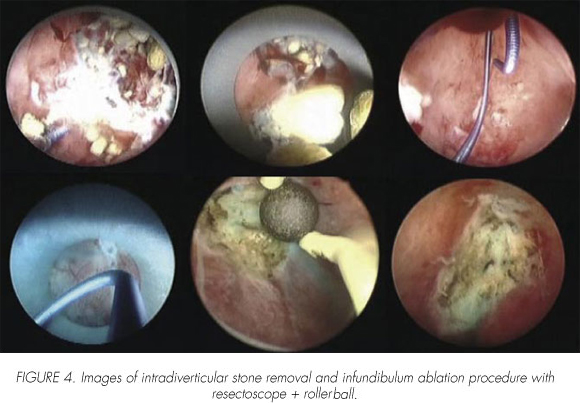
Methods: Owing to the presence of diverticular calculi and clinical symptoms of recurrent infection, we decided to perform percutaneous nephrolithotomy (PNL). After Holmium-YAG laser calculi fragmentation and removal of all stone material, we fulgurated the diverticular lining and infundibulum with a resectoscope and a rollerball electrode.
Results: The patient is free of symptoms after 6 months follow up. The disappearance of the calculi and diverticulum is confirmed with excretory urogram.
Conclusions: Endourological approach for diverticular calculi, such as percutaneous nephrolithotomy (PNL), is a minimally invasive treatment with excellent results and low morbidity. Using this procedure we are able to perform stone removal and cavity fulguration. According to this, we think that endourological techniques, and specially PNL could be the first option for treatment in selected cases of this pathology.
Key words: Calyceal diverticulum. Lithiasis. Percutaneous nephrolitotomy.
RESUMEN
Objetivo: Presentar el tratamiento percutáneo del divertículo calicial asociado a litiasis de localización posterior y resaltar la importancia que ha cobrado en los últimos tiempos el tratamiento endourológico, mínimamente invasivo, para este tipo de patología.
Métodos: Debido a la presencia de litiasis múltiple intradiverticular y sintomatología infecciosa recurrente asociada, se decide realización de nefrolitotomía percutánea (NLPC). Tras extracción de las litiaisis, se procede a la obliteración de infundíbulo calicial mediante electrocoagulación con resector y electrodo "rollerball".
Resultados: La paciente se encuentra libre de sintomatología a los 6 meses de seguimiento. Se confirma desaparición de divertículo calicial y litiasis mediante urografía intravenosa (UIV).
Conclusiones: El manejo endourológico de la litiasis alojada en un divertículo calicial de localización posterior mediante nefrolitotomía percutánea es un tratamiento mínimamente invasivo con excelentes resultados y escasa morbilidad. Permite la resolución no sólo de la patología litiásica, sino además la obliteración de la cavidad diverticular. Todo ello confirma que las técnicas endourológicas, y más específicamente la NLPC, son el tratamiento de elección en gran parte de los pacientes con esta patología.
Palabras clave: Divertículo calicial. Litiasis. Nefrolitotomía percutánea.
Introduction
The calyceal diverticula concept has been frequently modified during urology history. In 1841 Rayer (1) described intra-renal cavities, containing urine, located near of the cortical and communicated with the calyx by a thin conduct. He named urinary cysts. In 1941 Prather uses for the first time the name of calyceal diverticulum (2). Since then exists several denominations with high confusion: Cortical congenital cyst, cystic calyceal distend, pyelogenic cyst, pyelo-renal cyst, hydrocalyciosis, yuxtacalyceal cavity, cystic calyceal diverticulum, etc.
In 1965 Puigvert (3) tried to put in order all of these denominations, admitting two kinds of congenital cavities:
a) congenital dilation of Bellinis ducts.
b) ectopic calyx.
Steg (4) in 1975 defined the modern concept of calyceal diverticulum with higher precision.
Pyelocaliceal is a more appropiated and descriptive term, because includes the two types of anatomical varieties:
a) Type 1: The most frequent one. It depends on minor calyx and often near of the top, especially in the upper pole of the kidney. Usually small sized and asymptomatic.
b) Type 2: Presents direct communication with renal pelvis or major calyx. They normally grows and are associated with symptoms. Located more frequently in central portion of the kidney. The pyelogenic cyst is a type 2 diverticulum. (5).
Páramo y Resel (6) named them ductus calicoide when are center-papilar located, and calyceal diverticula when they have parafornicial origin, using the term pyelogenic cyst for those who depend from pelvis or major calyx.
The ultimate definition of calyceal diverticula consists in cystic cavities from congenital or acquired origin, covered with a non-secretor transitional epithelium, that communicates with the calyx by a very thin duct that allows retrograde urine filling of the cavity. Separated of the kidney parenchyma by a smooth sheet of connective tissue containing muscular fibers. By the presence of lithiasis inside the diverticulum, this epithelium may convert to squamous one and the nearest parenchyma may present intersticial nephritic injures. All of those cavities with different features must not be considered like a calyceal diverticulum.
Following this, we report the endo-surgical technique for the percutaneous treatment of several small stones inside posterior calyceal diverticulum.
Material y methods
We report the case of a 47 years old female complaining about recurrent urinary tract infections and right flank pain. In the abdominal x-ray are described several images of lithiasis in the theoretical location of the right kidney, that are confirmed and much better located (inside a calyceal diverticulum) in the intravenous urography (IVU). Diverticular size was 3 cms, located in the lower pole of the right kidney. In computerized tomography (CT) is described narrow communication between diverticulum and surrounding calyceal system with cortical notches in right kidney (Figure 1a and 1b). In the CT is confirmed its posterior location (Figure 2). According to this, we decided to perform a percutaneous nephrolithotomy (PNL) under general anesthesia. With the patient on lithotomy position we introduced a 6 Fr catheter to right renal pelvis. Later, patient was moved into a prone position in order to perform selective diverticular percoutaneous punction under x-ray control (Figure 3). Subsequently we introduced a Lun-derquist guide wire to distend the percutaneous trajectory until 30 Fr. Through Amplatz sheath we introduced a rigid 24 Fr nephroscope, observing many small lithiasis that were easily removed with a nitinol basket. After complete removal of the lithiasis we began retrograde infusion of blue methylene with uretheral stent to locate the infundibulum. Electrofulguration of diverticular infundibulum was performed by means of rollerball electrode in the 24 Fr. resectoscope. Finally we proceeded putting a nephrostomy catheter 20 Fr. inside the diverticulum. Through this nephrostomy catheter we carried out an anterograde pielography with no pass of contrast to excretory system.

Results
Operating time was approximately 2 hours, and no intraoperative complications were observed. The nephrostomy catheter was firstly closed and later removed 3 days after surgery. The Foley catheter was removed after 48 hours to keep low pressure in urinary tract. The hospital stay was of 4 days and antibiotic treatment lasted 10 days. The IVU after 3 months showed diverticular disappearance (Figure 5). After 6 months, patient was free of symptoms.

Discusion
Pyelocalyceal system embryologically grows from repeated ampoule ureteral yolk divisions and is surrounded by the methanephric blastema. The 3-5 first divisions generation will form renal pelvis and major calyx. Next divisions will form about 20 minor calyx. The reduction in the number of calyx is due to the absortion of several branches in the pelvis. The failure in disappearance of one of this calyx may originate a pouch connected with the collector system. The urine pressure dilates the pouch producing a definitely diverticulum (7).
Classically congenital and adquired causes have been quoted to explain pyelocaliceal diverticula origin. Nowadays, arguments for congenital origin are stronger, like the similar incidence in childhood and adult age. However, other arguments for adquired origin are the breakage of a cyst/abscess to a calyx, the presence of vesicoureteral reflux, the functional obstruction of a calyceal infundibulum, or a infundibulum-pyelic disgenesia (8).
Calyceal diverticulum incidence is low, 0.2% - 0.5% of all IVUs, occurs similar in both sex and kidneys. Often located in the superior third of the calyceal (70%), but they are also present in the inferior third (18%) and in the middle (12%) (9). Bilateral affectation is only described in 3% of the patients with diverticula. However, intradiverticular incidence of lithiasis is higher, with rates of 40% (9.5-39%) (10,11). Incidence of symptomatology associated with stone-containing diverticulum is estimated between 10% and 50% (7). The contribution of metabolic factors versus stasis in the pathogenesis of intradiverticular lithiasis formation is nowadays far to be clear. The studies of Matlaga et al., with one of the largest number of patients with stone-containing calyceal diverticulum, suggests that both (urinary stasis and metabolic factors), are involved like independent causal factors in the pathogenesis (12). Every patient with calyceal diverticulum examined by Auge et al (13) have at least one metabolic disorder: hypercalciuria, hyperuricosuria, hypercitraturia or hyperoxaluria. The most commonly metabolic disorder association described is low urinary flow rate (less than 2000 ml/day). Calyceal diverticula are frequently assymptomatic and incidentally identified during image studies done by another purpose (13). Patients usually became symptomatic when urinary stasis inside the diverticulum leads to infection or stone formation. Clinical manifestations include: haematuria, infection signs (especially chronic or recurrent urinary infections) abdominal, flank or renal pain (14). Recurrent infections may be due to diverticulum in 25% of all cases. Diverticular infundibulum obstruction may also lead to sepsis, abscess formation or arterial hypertension. Fatal cases of haemorrhage or rupture (primary or associated with IVU) are described (9).
Some authors describe mixed composition of intra-diverticular lithiasis, monohydrated calcium oxalate, monohydrate + hydroxiapatite. However, other studies cited higher incidence of struvite composition (9).
Image study most frequently used in these patients is the abdominal ultrasound (US). In US, diverticulum appears like a cystic image, often indistinguishable from another cystic lesion. In the majority of cases this study is insufficient for a precise diagnosis.
Abdominal x-ray increases the sensibility if milk of lime or stones are present. In IVU the calyceal diverticulum often is opaque due to the connection with the adjacent collector system, but the filling is retrograde and slow. If we suspect this kind of pathology, a retrograde pyelography is highly recommended to confirm the diagnosis and to establish a more precise anatomical description (11).
Computerized tomography (CT) and magnetic resonance (MRI) can be useful to determinate more exactly size and location (anterior/posterior) of the diverticulum. If image studies are not conclusive, we could perform endoscopic technics to identifícate diverticular neck.
Differential diagnosis must always include: hydrocalyx, renal cyst, medular sponge kidney, papylar necrosis, renal tuberculosis and Fraley syndrome (8).
Indications for surgery treatment include: persistent flanck pain, recurrent urinary tract infections, symptomatic diverticular lithiasis, progressive renal damage o severe hematuria (13). The number of patients who present symptomatic diverticular lithiasis is small. Moreover, symptomatology may due to another concomitant process. Thereby, this group of patients entails a challenge for urologists, with the complicated decision of choosing the subgroup of patients for surgery treatment. It is suggested that almost 83% of all diverticular lithiasis will require surgery intervention in 5 years, and this percentage increases to 89% at 10 years after diagnosis (15,16).
First line therapeutic options have highly developed in last 20 years. Previously to introduction of endourological technics and appearance of ESWL, open surgery was the gold standard treatment, in their different modalities (Partial/radical nephrectomy, open nephrostomy with diverticulectomy associated (deroofing or marsupialization).
ESWL have been widely used in the treatment of intradiverticular lithiasis due to their low morbidity and easily management. However, stone-free rate of patients treated exclusively with ESWL is between 0% and 58%, due to difficulties of stone fragments exit through thin diverticular neck. On the other hand, many patients became asymptomatic although stone fragments are present (9).
PNL, although is an invasive treatment, shows a larger number of advantages than ESWL. Percutaneous access may be performed directly into the diverticulum or indirectly into the adjacent collector system. Direct punction into the diverticulum allows the use of rigid nephroscope and ablation/fulguration of diverticular neck. Some authors prefer infundibulum dilatation, instead of closure. The stone free rate for different modalities of PNL, previously described, ranged between 70% and 100%, with a very high free of symptoms patients rate. Minor complications that occur during the procedure include: haemorrage, pneumothorax, urinary extravasation and/or slight irrigation liquid extravasation. Major complications include renal pelvis perforation with urinoma formation, pneumothorax /haemothorax that requires thoracostomy tube placement or massive haemorrage that requires balloon tamponage (9).
Some authors recommend percutaneous management like the first line treatment option when small calyceal diverticula are present. However prefer open surgery for patients with larger calyceal diverticula (with parenchymal damage associated).
Laparoscopic technics have been introduced recently for treatment of this pathology. During operating time diverticulum roof is resectioned, neck is obliterated and diverticular cavity is fulgurated. This technic also allows safe access to callyceal diverticulum in every location and complete lithiasis removal. Is not recommended in obese patients (17,18).
Appropiate treatment selection depends on diverticulum location inside the kidney and stenotic condition of diverticular neck.
The postoperative of the patients after surgical treatment is often well admitted. CT may be the best radiographic measure of success of calyceal ablation, since IVP does not permit visualization of all diverticula. Randomized trials of the various percutaneous techniques discussed with long-tem CT follow-up are ideally needed to compare efficacy and safety.
Conclusion
Endourological management of posterior calyceal diverticulum lithiasis by percutaneous nephrolithotomy is a minimally invasive treatment with excellent results and very low morbidity. Using this procedure we are able not only to perform stone removal but cavity fulguration. According to this, we think that endourological techniques, and specially PNL could be the first option treatment in selected cases with this pathology.
 Correspondence:
Pablo Garrido Abad
Santiago Bernabeu, 4 - 5º pta. 4
28036 Madrid. (Spain).
pgabad@hotmail.com
Accepted: 17th January, 2008.
References and recomended readings (*of special interest, **of outstanding interest)
1. Rayer PF. Traitement des malaides des reins. Bailliéres, Paris: 1984; 3:507. [ Links ]
2. Prather GC. Calyceal diverticulum. J Urol 1941; 45:55-8. [ Links ]
3. Puigvert A. Malformaciones de la pirámide de Malpighi. Editorial Eco AS, Barcelona. 1965. [ Links ]
4. Steg A. Les Affections kystiques du rein d´l adulte. J Urol Nephrol 1975; 91:240. [ Links ]
5. Wulfsohn MA. Pyelocaliceal diverticula. J Urol 1980; 123:1. [ Links ]
6. Páramo PG, Resel L. Patología quística renal. Ponencia del Congreso Nacional de Urología 1975; 140. [ Links ]
7. Middleton AW, Pfister RC. Stone-containing pyelocalicial diverticulum: embryogenic, anatomic, radiologic and clinical characteristics. J Urol 1974; 111:2-6. [ Links ]
**8. Lima E, La Fuente J, García Cuerpo E, Sánchez Encinas, Fernández González I, Sanz Miguelañez JL et al. Divertículos pielocaliciales. Arch Esp Urol 2000; 53:581-95. [ Links ]
9. Monga M, Smith R, Ferral H, Thomas R. Percutaneous ablation of caliceal diverticulum: Lon-term follow-up. J Urol 2000; 163:28-32. [ Links ]
10. Kim SC, Kuo RL, Tinmouth WW, Watkins S, Lingeman JE. Percutaneous nephrolithotomy for caliceal diverticular calculi: a novel single stage approach 2005; 173:1194-98. [ Links ]
*11. Gross AJ, Herrmann TRW. Management of stones in calyceal diverticulum. Curr Opin Urol 2007; 17:136-40. [ Links ]
12. Matlaga BR, Miller NL, Terry C, Kim SC, Kuo RL, Coe FL, et al. The pathogenesis of calyceal diverticular calculi. Urol Res 2007; 35:35-40. [ Links ]
13. Auge BK, Maloney ME, Mathias BJ, et al. Metabolic abnormalities associated with calyceal diverticular stones. Br J Urol 2006; 97:1053-56. [ Links ]
14. Wogan JM. Pyelocalyceal diverticulum: An unusual cause of acute renal colic. J Emerg Med 2002; 23:19-21. [ Links ]
*15. Staios D, Andrews HO, Shaik T, Bucholz NNP. Quality of life after percutaneous nephrolithotomy for caliceal diverticulum and secluded lower-pole renal stones. J Endourol 2007; 21:515-19. [ Links ]
16. Coury TA, Sonda LP, Lingeman JE, Kahnoski RJ. Treatment of painful calyceal stones. Urology 1988; 32:119-23. [ Links ]
17. Gluckman GR, Stoller ML, Irby P. Laparoscopic pyelocaliceal diverticular ablation. J Endourol 1993; 7:315-17. [ Links ]
*18. Okumura A, Murakami K, Yoshida M, Nagakawa O, Fuse H. Percutaneous endoscopic treatment for the calyceal diverticular calculi. Int Urol Nephrol 2005; 37:5-8. [ Links ]

