


Meu SciELO
 Serviços customizados
Serviços Personalizados
Journal
Artigo
 texto em
  Inglês (pdf)
 Artigo em XML
 Referências do artigo
 Enviar este artigo por email
Indicadores
-
 Citado por SciELO
 -
 Acessos
Links relacionados
-
 Citado por Google
 -
 Similares em
SciELO
Similares em
SciELO -
 Similares em Google
Compartilhar

 Permalink
Archivos Españoles de Urología (Ed. impresa)
versão impressa ISSN 0004-0614
Arch. Esp. Urol. vol.62 no.3 Abr. 2009
Current indications of open surgery for the treatment of renal lithiasis. Ureterocalycostomy as definitive treatment for lithiasis in a female with recurrent disease
Indicación actual de la cirugía abierta en el tratamiento de la litiasis renal. Ureterocalicostomía como tratamiento definitivo de litiasis en mujer con patología recidivante
Jose Luis Miján Ortiz, Francisco Valle Díaz de la Guardia, Antonio Jiménez Pacheco, Miguel Arrabal Martín, Mercedes Nogueras Ocaña and Armando Zuluaga Gómez.
Department of Urology. San Cecilio Clinic Hospital. Granada. Spain.
SUMMARY
Objective: We describe one case of recurrent lithiasis associated with anatomical alteration of the renal pelvis related to previous surgery.
Methods/Results: The patient presented a urinary tract infection episode, complicated with pyonephrosis and septicemia. In the intravenous urography, infectious radiopaque pyelocaliceal multiple and complex lithiasis can be seen, as well as kidney hydronephrosis grade III-IV. Important pyelic sclerosis secondary to previous surgery on the renal unit was seen. Nephrectomy was performed with lower pole nephrolithotomy and reconstruction of the upper urinary tract through ureterocalicostomy. Two and a half years after surgery, control urogram shows absence of urolithiasis and a slight delay of renal function.
Conclusions: Ureterocalicostomy is indicated in cases of ureteropelvic junction obstruction associated with intrarenal pelvis caused by alterations of fusion, rotation or location of kidney. It is also indicated in cases of severe peripyelic fibrosis secondary to previous pyeloplasty failure or renal surgery. In our case, in addition to the infectious component of lithiasis, an anatomical alteration, probably secondary to previous surgery, caused the chronification of lithiasis. Facing such suspicion a surgical management was undertaken to eliminate the lithiasis and get a correct derivation of the working area of the kidney, in order to prevent further recurrences.
Key words: Lithiasis. Ureterocalicostomy. Surgery.
RESUMEN
Objetivo: Presentamos un caso de litiasis recidivante asociado a alteración anatómica de la pelvis renal secundaria a cirugía.
Métodos/Resultados: La paciente presenta un episodio de infección urinaria complicada con pionefrosis y septicemia. En la urografía intravenosa se observa litiasis radiodensa infecciosa, pielolocalicial múltiple compleja, sobre riñón con hidronefrosis grado III-IV por importante esclerosis piélica secundaria a cirugía previa sobre dicha unidad renal. Se realiza nefrectomía polar inferior con nefrolitotomía y reconstrucción de la vía urinaria superior mediante uréterocalicostomía. Dos años y medio después de la cirugía la urografía de control refleja ausencia de litiasis y leve retraso de la función renal.
Conclusiones: La ureterocalicostomía está indicada en casos de obstrucción de la unión ureteropiélica asociada a una pelvis intrarrenal por alteraciones de la fusión, rotación o localización renal, y en casos de fibrosis peripiélica severa secundaria a una pieloplastía fallida o cirugía renal previa. En el caso presentado además del componente infeccioso de las litiasis, una alteración anatómica, probablemente secundaria a la cirugía previa, provocaba una perpetuación de la clínica litiásica. Ante tal sospecha se impuso una solución de tipo quirúrgico que solucionara en un tiempo tanto la eliminación de la litiasis como una correcta derivación de la zona funcionante del riñón para evitar recidivas posteriores.
Palabras clave: Litiasis. Ureterocalicostomía. Cirugía.
Introduction
In recent years, indications for performing open surgery have changed. As a result of the advance of endourological techniques and the development of extracorporeal shock wave lithotripsy (ESWL), surgery is not longer the habitual management of lithiasis, but it is seen as the last option in the face of failures of other treatments, and very complex lithiasis processes, or functional arrest. However, indications for surgery are still present in current protocols and seem to be better defined, so surgery techniques have become a sound option for some patients
Case report
We describe here the case of a 35 year-old woman with a history of lithiasic surgery in both kidneys performed in some other medical center. She reported right pyelolithotomy 15 years earlier. She underwent a left pyelolithotomy two years later, and a subsequent nephrolithotomy in left kidney, due to complex renal lithiasis, 10 months before admittance to our urology department in 2005.
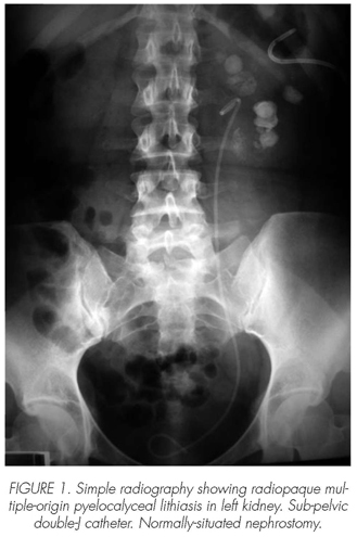
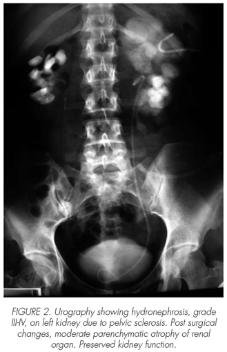
In that center, the patient presented with a urinary tract infection complicated with pyonephrosis and septicemia after placement of DJ catheter, wrongly positioned on sub-pelvic region, that did not resolve septic symptomatology, so it was decided to perform left percutaneous nephrostomy to allow renal drainage, so faced with this situation, the patient was referred to our center. Intravenous urography provided by the patient reveals an infectious radiopaque pyelocalyceal, multiple-origin and complex lithiasis that affects grade III-IV hydronephrotic kidney, as a result of a significant renal pelvis sclerosis that is secondary to previous surgery interventions on the aforementioned organ. The patient also shows a relatively preserved renal function. We also note post-surgical changes accompanied by a moderate parenchymatous atrophy on right renal organ (Figures 1 and 2). Blood test exhibits normal values for creatine and urea.
In view of these urography findings, as well as the contralateral kidney status and patient's age, we decided to perform an exploratory lumbar incision in an attempt to spare the organ by means of a lower pole nephrectomy and subsequent reconstruction of the upper urinary tract through ureterocalycostomy.
Over the post-operative period, the patient required the transfusion of 2 units of red corpuscle concentrate on account of her persistent anemia. Subsequent evolution did not report major complications, and she was discharge on the seventh day. The pathoanatomical study of the organ showed changes compatible with pyelonephritis, while the study of the calculus revealed ammonium-magnesium phosphate in the composition.
Two years and a half after the operation, the patients only refers some slight discomfort on left flank. Control urography reveals absence of lithiasis, and some nephrography delay on left region and on right secretory pathway, with no changes regarding previous status. (Image 3). Renogram shows a 30% of left renal function, and a compensatory right kidney function, 70%. Patient's renal function remains within normal values, and has only shown a single UTI that resolved with ciprofloxacin. Imaging revealed the presence of small asymptomatic bilateral remaining fragments.
Discussion
As for the management of lithiasis, open surgery not only includes the classic pyelolithotomy or nephrolithotomy procedures, but also reconstructive surgery techniques for those cases that may require them. Now, the use of open surgery for such indications has been restricted thanks to the advent of extracorporeal lithotripsy, percutaneous techniques and ureteroscopy. However, the unquestionable results of this technique, although shadowed by the current literature, make this therapy a valuable instrument against lithiasis. The number of publications on open surgery has declined significantly, although this does not mean that the various interventions included in this technique may have been abandoned (1)
Paik (2) describes his experience on 780 patients with lithiasis, out of which 42 underwent open surgery through distinct techniques: pyelolithotomy, nephrolithotomy, ureterolithotomy, among them. Indications for open surgery were as follows: complex lithiasic mass (55%); previous treatment failure (ESWL or ureteroscopy) (29%); and anatomical changes of urinary pathways, such as infundibular stenosis, among them (24%); morbid obesity (10%), or medical comorbidity (7%), which indicated that overall surgery should be performed in a single process.
In 2003, Ather (3) reports a series of 1195 patients examines the differences among the three main types of treatments, wherein a 20% of surgical procedures were performed for distinct reasons: anatomical alterations in special, failure of other procedures, but less frequently, patient's preference, management of great impacted lithiasis, or open surgery concomitant with another process (for instance, a cesarean delivery). The series also report a dramatic decline of these interventions, which now only represent an 8%, thanks to the inclusion of endoscopic pneumatic lithotripsy techniques to his center.
The Guidelines of the European Association of Urology (updated in June 2005) (4) make a review of the surgery indications aforementioned and also suggests surgery as the treatment of choice in big-sized coralliform lithiasis, and in those cases requiring correction of anatomical alterations, as the case described before. Although many authors champion combination treatments in coralliform lithiasis through percutaneous surgery and ESWL, some series yield better results with open surgery techniques, as expressed by Snyder (5), 0% of residual fragments versus percutaneous, 13%; or Esen (6) who reports better results than those seen in lithotripsy or in combination treatments.
Ureterocalycostomy is indicated in ureteropelvic junction obstruction (UJO) associated with intrarenal pelvis that result from anomalies of kidney fusion, rotation and position, as well as severe peripelvic fibrosis secondary to failed pyeloplasty or previous renal surgery. As for the case described previously, and in addition to the infectious component found in it, an anatomical change attributable to previous surgery made the lithiasis process a chronic condition. Faced with this suspicion, it was necessary to resort to surgery to manage in a single process both the lithiasis and the correct deviation of the functional region of the kidney to prevent subsequent relapses.
In general, the results of this technique are satisfactory and have already been described by diverse authors in the literature. In 2005, Matlaga (7) describes a series of 11 patients who underwent ureterocalycostomy, as a first indication in pyeloureteral junction stenosis or after failure of endourological treatments, and reports good outcomes on the kidneys that were treated. Haouas, in 2005, (8) refers other series with longer follow-ups that showed a good renal function in 12 out of 16 patients, although he also mentions some surgery failures that required subsequent nephrectomy in 2 patients, 4 and 10 years, respectively, after operation.
This technique is also used in relapses of stenosis of the pyeloureteral junction after pyeloplasty; however, this would be a restricted technique and other authors would resort to a second pyeloplasty, or an endo-pyelolithotomy. Stenosis of the new junction is the most frequent complication observed in ureterocalycostomy, and can give rise to recurring obstructions; however, the incidence of this problem is low, so it can generally be regarded as a safe technique. Today, laparoscopic surgery is an option for such cases, but it requires a good management of the technique, especially with regard to intra-corporeal suturing and knot tying. The literature reports highly successful reconstruction series performed by experienced surgeons on upper urinary tracts, Gill, 2004 (9); some cases even include ureterocalycostomy through robotic surgery.
Conclusion
An adequate indication for open surgery, as well as the anatomical correction of an altered urinary pathway, may achieve, in a single procedure, good results both for lithiasis removal and diminution of relapses.
 Correspondence:
Francisco Valle Díaz de la Guardia
Melchor Almagro, 8 4oB
18002. Granada. (Spain).
pacovd@hotmail.com
Accepted for publication: Februay, 14th 2008.
References and recommended readings (*of special interest, **of outstanding interest)
*1. Boronat Tormo F, Pontones Moreno JL, Broseta Rico E, Oliver Amoros F, Budia Alba A, Jimenez Cruz JF. Tratamiento de la litiasis renal cálcica. LEOC, NLP, cirugía abierta. Arch. Esp. Urol., 2001; 54: 909-925. [ Links ]
*2. Paik ML, Wainstein M. A current indications for open stone surgery in the treatment of renal and ureteral calculi J. Urol., 1998; 159: 374. [ Links ]
*3. Ather MH, Paryani J, Memon A, et al. A 10-year experience of managing ureteric calculi: changing trends towards endourological intervention - is there a role for open surgery? B. J. U. Int., 2001; 88: 173. [ Links ]
4. Tiselius HG, Ackermann D, Alken P, et al. Guidelines on Urolithiasis. En: EAU Guidelines. 2006. Disponible en: http://www.uroweb.org/fileadmin/user_upload/Guidelines/18%20Urolithiasis.pdf [ Links ]
5. Snyder JA, Smith AD. Staghorn calculi: percutaneous extraction versus anatrophic nephrolithotomy. J. Urol., 1986; 136: 351. [ Links ]
6. Esen AA, Kirkali Z, Guler C. Open stone surgery: is it still a preferable procedure in the management of staghorn calculi? Int. Urol. Nephrol., 1994; 26: 247. [ Links ]
**7. Matlaga BR, Shah OD, Singh D, et al. Ureterocalicostomy: a contemporary experience. Urology, 2005; 65: 42. [ Links ]
8. Haouas N, Youssef A, Sahraoui W, et al. Ureterocalicostomy: indications and results based on a series of 16 patients. Prog. Urol., 2005; 15: 641. [ Links ]
9. Gill IS, Cherullo EE, Steinberg AP, et al. Laparoscopic ureterocalicostomy: initial experience. J. Urol., 2004; 171:1227. [ Links ]
10. Mufarrij PW, Shah OD, Berger AD, et al. Robotic reconstruction of the upper urinary tract. J. Urol., 2007; 178: 2002. [ Links ]

