Meu SciELO
Serviços Personalizados
Journal
Artigo
Indicadores
-
 Citado por SciELO
 -
 Acessos
Links relacionados
-
 Citado por Google
 -
 Similares em
SciELO
 -
 Similares em Google
Compartilhar
Nutrición Hospitalaria
versão On-line ISSN 1699-5198versão impressa ISSN 0212-1611
Nutr. Hosp. vol.25 no.6 Madrid Nov./Dez. 2010
Wernicke's encephalopathy induced by total parental nutrition
Encefalopatía de Wernicke asociada a nutrición parenteral total
J. T. Sequeira Lopes da Silva1, R. Almaraz Velarde2, F. Olgado Ferrero1, M. Robles Marcos2, D. Pérez Civantos2, J. M. Ramírez Moreno3, L. M. Luengo Pérez4
1Servicio de Medicina Interna.
2Unidad de Cuidados Intensivos.
3Servicio de Neurología.
4Unidad de Nutrición Clínica y Dietética.
Hospital Infanta Cristina, Badajoz, España.
ABSTRACT
Wernicke's encephalopathy is an acute neurological syndrome due to thiamine deficiency, which is characterized by a typical triad of mental status changes, oculomotor dysfunction and ataxia. Despite the fact that Wernicke's encephalopathy, in developed countries, is frequently associated with chronic alcoholism, there have been a number of published cases associating this encephalopathy with parenteral feeding without vitamin supplementation. Diagnosis is primarily a clinical one, and can be supported by laboratory tests and imaging studies; treatment should start as soon as possible, for the morbidity and mortality (almost 20%) associated with this syndrome is high. Thiamine supplementation, along with other vitamins, is recommended for patients in risk of developing this syndrome.
Key words: Wernicke's encephalopathy. Total parenteral nutrition. Thiamine deficiency.
RESUMEN
La Encefalopatía de Wernicke es un síndrome neurológico de instauración aguda secundario a un déficit de tiamina y que se caracteriza por una típica tríada de alteración del nivel de conciencia, disfunción oculomotora y marcha atáxica. Aunque la causa más frecuente de Wernicke en nuestro medio sea el alcoholismo crónico, varios casos han sido descritos en enfermos con nutrición parenteral total sin aporte vitamínimo. El diagnóstico es principalmente clínico, apoyándose en pruebas analíticas y de neuroimagen, recomendándose empezar el tratamiento con tiamina lo más precozmente posible, dada la alta morbilidad y la alta mortalidad (de casi 20%), que se asocian a esta encefalopatía. La suplementación dietética con tiamina y otras vitaminas está indicada en todos los individuos en riesgo de desarrollar este síndrome.
Palabras clave: Encefalopatía de Wernicke. Nutrición parenteral total. Déficit de tiamina.
Introduction
Wernicke's encephalopathy (WE) is an acute neuropsychiatric syndrome that results from thiamine (vitamin B1) deficiency and that is characterized by a triad of mental-status changes, oculomotor dysfunction and ataxia.1 In recent years, although our knowledge of the predisposition factors and clinical settings associated with this syndrome has increased, it has been proven that many cases go undiagnosed. Due to its high morbi-mortality, diagnosis and treatment should be made as soon as possible. The authors report the case of a 28-year-old man that during prolonged total parenteral nutrition (TPN), due to a bleeding ulcer which required surgery in different occasions, developed a WE, with substantial improvement when intravenous thiamine was administered.
Case report
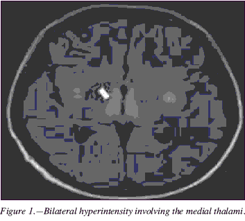
A 28-year-old man without any relevant medical history was admitted in our Intensive Care Unit with the diagnosis of hemorrhagic shock due to a bleeding ulcer in the first duodenal curve. He had required a bilateral vagotomy and pyloroplasty after failure of endoscopic treatment. At the second day after admission, parenteral nutrition with Oliclinomel N4-550® was started, being replaced after two days by Oliclinomel N8-800® with Hyperlite® and Dipeptiven®. During his stay, the patient presented a new episode of upper digestive hemorrhage requiring surgical intervention, in which a partial gastrectomy and Roux-en-Y Gastric Bypass was made. Antibiotics were prescribed due to Escherichia coli peritonitis. At the twentieth day, he had a new episode of severe hematochezia with hypotension and tachycardia, requiring urgent surgery, diagnosing pancreatic and biliary fistula; the gastroduodenal artery was sutured and a Whipple procedure was performed. In the following seven days, the patient remained sedated and intubated due to a ventilator-associated pneumonia, and a glucose 5% solution was prescribed for three days. After withdrawing the sedatives and extubating the patient, a confusional state with important mental sluggish was noticed, accompanied also by horizontal nystagmus to both sides and paralysis of the left arm. An urgent computerized tomography (CT) was requested, being informed as normal, following which a magnetic resonance imaging (MRI) was performed, revealing symmetric and bilateral hyperintense signal in the fourth ventricular floor, the periacqueductal gray matter, medial cerebral peduncles areas, medial thalami, mamillary bodies and patchy frontal cortical/subcortical lesions, topographically compatible with a WE (fig. 1). Treatment with 100 mg intravenous thiamine, three times per day, was started, with remission of the nystagmus and significant improvement of his confusional state in the three following days. Rehabilitation was requested for residual proximal palsy of the left arm, and an electroneurogram/electromyogram of the brachial plexus showed a diffuse axonal motor lesion, compatible with a critical illness polyneuropathy. The MRI performed a week after starting treatment with thiamine showed significant reduction of the size of the infra/supratentorial lesions (fig. 2).
Discussion
Thiamine is a water-soluble B-complex vitamin that participates as a coenzyme in the oxidative decarboxylation of pyruvate and alfa-ketoglutarate and also in the pentose phosphate pathway.2 In the central nervous system, thiamine is converted into thiamine pyrophosphate by the neuronal and glial cells, being responsible for the ATP synthesis, the production and maintenance of the myelin sheath, the production of aminoacids and glucose-derived neurotransmitters (e.g., glutamic acid), the acetylcholinergic and serotoninergic synaptic transmission and the axonal conduction.1
Absorption occurs in the duodenum by a rate-limited process. The recommended dose of thiamine for a healthy adult is 1,4 mg of per day or 0,5 mg per 1000 Kcal of consumed, increasing its demands in subjects with high metabolic rate (e.g., critical ill patients) or with high carbohydrate intake. Body´s stores of thiamine are approximately 30 mg, lasting between 18 to 20 days in patients with a strict free-thiamine diet,3 as was the case of our patient.
Thiamine deficiency can lead to a cardiovascular disease, known as "wet beriberi", a high-output cardiac failure with orthopnea and pulmonary and peripheral edema, and/or a neurologic syndrome, known as "dry beriberi". As in this case, severe, short-term thiamine deficiency usually leads to a WE, whereas a milder/moderate, more prolonged deficiency tends to originate a polyneuropathy, preferably involving myelin and worse distally than proximally, secondary to lesions in the peripheral nerves.1
WE is an acute syndrome that requires an emergent treatment due to its high morbi-mortality and that is characterized by the typical triad of mental confusion, oculomotor dysfunction and gait ataxia. The prevalence of this encephalopathy in men is higher than in women (1,7:1), although women appear to be more susceptible to developing a WE.4 Autopsy studies have continuously revealed a higher prevalence of typical WE brain lesions (0,8-2,8%) than expected by clinical studies (0,04-0,13%), proving that in our clinical practice a considerable number of cases are undiagnosed.
In developed countries, most cases of WE are associated with chronic alcoholism, which due to their inadequate dietary intake, reduced gastrointestinal absorption, decreased hepatic storage and impaired utilization, seem to be more susceptible in developing this encephalopathy. Other predisposition factors and clinical settings associated to WE and that are commonly encountered in our clinical practice are the hyperemesis gravidarum, gastrointestinal surgery5 (including bariatric surgery), systemic diseases such as cancer and related conditions (malignancy is the most common disorder that precipitates a WE in children), severe infections (AIDS, for example), endocrinological disorders as thyrotoxicosis and also hemo and peritoneal dialysis. Several cases associating WE with TPN without thiamine6 supplementation have been described. In his two years study, Francini-Pesenti observed a high prevalence of WE in this type of patients,3 a conclusion similar to that of Hahn, that reported an increase of this syndrome in patients with TPN, that due to a shortage of multivitamin infusions, were not receiving thiamine.
Only one-third of patients will have all three of the typical symptoms, being the confusional state the most frequent one, followed by ataxia and ocular dysfunction. 19% of patients will not show any of the typical symptoms. Mental changes range from apathy, profound indifference and mental sluggishness to, when left untreated, stupor and coma. Nystagmus is the most common oculomotor dysfunction, and usually is evoked by horizontal gaze to both sides. As the WE progresses, one can encounter bilateral lateral rectus palsy and, in advanced cases, complete ophthalmoplegia with nonreactive, miotic pupils.1-7 Ataxia, that can precede the other symptoms by a few days or weeks, is commonly due to the combination of vestibular dysfunction, polyneuropathy and the involvement of the anterior and superior vermis. Other less frequent symptoms are hypothermia, tachycardia, hearing loss and epileptic seizures. Overt of a "wet beriberi" and a WE is rare.
Diagnosis is primarily a clinical one, and the high rate of undiagnosed WE cases can be explained by the non-specific clinical presentation in many patients. Although no single test has sufficient diagnostic accuracy, the presumptive diagnosis can be confirmed by laboratory studies, like measurement of erythrocyte thiamine transketolase activity or thiamine/thiamine pyrophosphate concentration in serum, which will be decreased.8 CT is an insensitive test, and a normal result cannot rule out a WE, as was the case of our patient. MRI is currently considered the most valuable imaging study available. MRI has a sensitivity of only 53%, but a specificity of 93%, which means that it can be used to confirm the diagnosis. A typical finding is the bilateral symmetrical T2 abnormal hyperintense signal affecting the periacqueductal gray matter, around the third ventricle and the medial thalamus and the mamillary bodies,8 which can be found atrophic in a WE that has evolved for more than a week.4 The improvement of the neurological signs when parenteral thiamine is administered also confirms the diagnosis.
WE is a medical emergency and treatment should be started as soon as one considers this diagnosis. Diagnostic testing should not delay treatment. Though no randomized study exists to support a particular dosing regimen, it is recommended that patients should be treated with a minimum of 500 mg thiamine intravenously (dissolved in 100 ml of normal saline and infused over 30 minutes), three times daily for two to three days, followed by 250 mg intravenously for three to five more days, or until the end of the clinical improvement1. Magnesium and other vitamins should be replaced as well. Daily oral administration of 100 mg thiamine should be continued after completion of parenteral treatment until patients are considered no longer at risk of developing a WE.
The authors would like to point out the absolute necessity of thiamine and other multivitamin supplementation in patients with TPN, in order to prevent a WE, and to remark the importance of its clinical suspicion in undernourished patients that present any of the typical symptoms. Since the infusion of intravenous glucose solutions can precipitate a WE, these should be preceded or accompanied by the administration of thiamine.
References
1. Sechi G, Serra A. Wernicke's encephalopathy: new clinical settings and recent advances in diagnosis and management. Lancet Neurol 2007; 6: 442-55. [ Links ]
2. Fernández Suárez FE, Hernández Bujedo M, Varela Rodríguez L, Fernández Miranda A, García Arango B, Miyar Villarica MC. Beriberi tras una esofaguectomía. Rev Esp Anestesiol Reanim 2002; 49: 541-544. [ Links ]
3. Francini-Pesenti F, Brocadello F, Manara R, Santelli L, Laroni A, Caregaro L. Wernicke's syndrome during parenteral feeding: not an unusual complication. Nutrition 2009; 25: 142-146. [ Links ]
4. Charness ME, So YT. Wernicke's encephalopathy. UpToDate 2009. [ Links ]
5. Singh S, Kumar A. Wernicke encephalopathy after obesity surgery: a systematic review. Neurology 2007; 68: 807-811. [ Links ]
6. Hahn JS, Berquist W, Alcorn DM, Chamberlain L, Bass D. Wernicke encephalopathy and beriberi during total parenteral nutrition attributable to multivitamin infusion shortage. Pediatrics 1998; 101: 1-4. [ Links ]
7. Francini-Pesenti F, Brocadello F, Famnego S, Nardi M, Caregaro L. Wernicke's encephalopathy during parenteral nutrition. Journal of Parenteral and Enteral Nutrition 2007; 31: 69-70. [ Links ]
8. Attard O, Dietemann JL, Diemunsch P, Pottecher T, Meyer A, Calon BL. Wernicke encephalopathy: a complication of parenteral nutrition diagnosed by magnetic resonance imaging. Anesthesiology 2006; 105: 847-8. [ Links ]
![]() Correspondence:
Correspondence:
José Tiago Sequeira Lopes da Silva
Servicio de Medicina Interna
Hospital Infanta Cristina
Avenida de Elvas, s/n.
06006 Badajoz, España
E-mail: j.tiago.silva@hotmail.com
Recibido: 3-VIII-2010.
1a Revisión: 2-IX-2010.
Aceptado: 20-IX-2010.


