Mi SciELO
Servicios Personalizados
Revista
Articulo
Indicadores
-
 Citado por SciELO
 -
 Accesos
Links relacionados
-
 Citado por Google
 -
 Similares en
SciELO
 -
 Similares en Google
Compartir
Nutrición Hospitalaria
versión On-line ISSN 1699-5198versión impresa ISSN 0212-1611
Nutr. Hosp. vol.28 no.4 Madrid jul./ago. 2013
https://dx.doi.org/10.3305/nh.2013.28.4.6474
ORIGINAL / OBESIDAD
Bariatric surgery decreases carotid intima-media thickness in obese subjects
La cirugía bariátrica reduce el grosor íntima media carotídeo en pacientes obesos
Gonzalo García1, Daniel Bunout2, Javiera Mella3, Erik Quiroga4, María Pía de la Maza5, Gabriel Cavada3 and Sandra Hirsch5
1Internist Physician. Nutrition Department of Clínica Dávila
2Internist Physician. INTA. Universidad de Chile
3Universidad de Los Andes
4Medical Technologist. Radiology Deparment. Clínica Dávila
5Nutriology Magister. INTA. Universidad de Chile
ABSTRACT
Background: Obesity has long been associated with an increased risk of cardiovascular disease (CVD). The aim of this study was to evaluate the impact of substantial weight loss induced by bariatric surgery on carotid intima media thickness (C-IMT) (surrogate marker of early atherosclerosis) and classic factors of cardiovascular risk (CVRFs).
Methods: thirty-one obesity patients were evaluated for bariatric surgery. Twenty-seven were undergone surgery, 14 Roux-en-Y gastric bypass surgery (GBS) and 13 sleeve gastrectomy. The four obese patients who did not undergo surgery, were performed the same evaluations. Measurements: Body weight, BMI, blood pressure, total cholesterol, TC levels, LDL-C, HDL-C, TG, fasting plasma glucose and insulin, HOMA IR, and US B-mode C-IMT was measured.
Results: After 354 ± 92 days follow up, 27 patients that underwent bariatric surgery evidenced a mean body mass index decrease from 38 to 27 k/m2 (p < 0.001), simultaneously was observed improvement in CVRFs, 10 years Framingham risk and a significant reduction of therapeutic requirements. C-IMT diminished from a mean of 0.58 ± 0.14 mm to 0.49 ± 0.09 mm (p = 0.0001). Four patients that did not undergo surgery increased C-IMT from 0.52 ± 0.12 to 0.58 ± 0.13 mm (p = 0.03) with no significant changes in CVRFs.
Conclusion: Weight loss, one year after bariatric surgery, GBS and sleeve gastrectomy, decreases C-IMT; improve CVRFs and 10 years Framingham risk.
Key words: Obesity. Cardiovascular risk. Weight loss. Bariatric surgery. Carotid intima media thichkness.
RESUMEN
Introducción: La obesidad se ha asociado a un aumento del riesgo cardiovascular. El objetivo de este estudio fue evaluar el impacto de la baja de peso a través de la cirugía bariátrica en el grosor íntima media carotídea (GIMc, marcador subrogado de aterosclerosis subclínica) y los factores de riesgo cardiovascular clásicos.
Métodos: Un total de 31 pacientes obesos fueron evaluados para cirugía bariátrica, 27 fueron intervenidos quirúrgicamente, 14 sometidos a un bypass gástrico en Y de Roux y 13 a gastrectomía en manga. En los 4 pacientes que no fueron sometidos a cirugía bariátrica se realizó las mismas evaluaciones. Parámetros: peso, índice de masa corporal (IMC), presión arterial, colesterol total, LDL, HDL, triglicéridos, glicemia e insulina de ayunas, HOMA IR y medición del GIMc mediante ultrasonido.
Resultados: Luego de 354 + 92 días de seguimiento, en los 27 pacientes intervenidos se evidenció una disminución del IMC promedio de 38 a 27 k/m2 (p < 0,001), al mismo tiempo se observó una reducción en los marcadores de riesgo cardiovascular, en el riesgo de Framingham a 10 años, y una significativa reducción de la terapia farmacológica. El promedio del GIMc se redujo de 0,58 ± 0,14 mm a 0,49 ± 0,09 mm (p = 0,0001). Los cuatro pacientes que no fueron intervenidos presentaron un aumento del GIMc 0,52 ± 0,12 a 0,58 ± 0,13 mm (p = 0,03) sin cambios significativos en los marcadores de riesgo cardiovascular durante el período observado.
Conclusión: La pérdida de peso inducida por la cirugía bariátrica, tanto bypass gástrico como gastrectomía en manga, a un año de seguimiento disminuye el GIMc, mejora los factores de riesgo cardiovascular y el riesgo de Framingham a 10 años.
Palabras clave: Obesidad. Riesgo cardiovascular. Baja de peso. Cirugía bariátrica. Intima media carotídea.
Abbreviations
C-IMT: Carotid intima media thickness.
GBS: Gastric bypass surgery.
CVD: Cardiovascular disease.
CVRFs: Cardiovascular risk factors.
TC: Total cholesterol.
LDL-C: Low density lipoprotein cholesterol.
HDL-C: High density lipoprotein.
TG: Triglycerides.
HOMA-IR: Homeostatic model of assessment of insulin resistance.
US: Ultrasound.
Introduction
Obesity is one of the leading causes of increased morbidity and mortality, and its prevalence is increasing worldwide. In Chile the frequency of obesity defined as body mass index (BMI) > 30 kg/m2, increased between 2003 and 2010 from 21.9 to 25.1% among people aged over 15 years old. In the same period, the frequency of morbid obesity increased from 1.3% to 2.3%. These figures represent an increase estimated in morbidly obese from 148.000 to 300.000 subjects.1
The association between obesity and cardiovascular risk is well known.2,4 In fact, The American Heart Association identified obesity as an independent risk factor for cardiovascular disease.5,6
There are a multiple physiological and metabolic changes, such as insulin resistance, type 2 diabetes mellitus, lipid abnormalities, systolic and diastolic hypertension, left ventricular hypertrophy, sympathetic nervous system dysfunction, endothelial dysfunction, inflammatory state and obstructive sleep apnea,7 that are associated with obesity and contribute to increase the risk of cardiovascular disease.
A number of multivariate risk models have been developed to assess CVR, being the Framingham risk score, the most known and used.8 However, this method has limitations: risk assessment that stratify patients according to the number of defined risk factor can identify high-risk persons, but they tend to falsely reassure persons deemed to be who may have multiple marginal abnormalities. On the other hand, this model only gives 10 years risk estimation, patients with less 10% likelihood are considered at low risk. Moreover, this approach does not consider lifetime risk, which in the case of diseases with very slow progression rates, such atherosclerosis, could discourage their early treatment.9-11
The US measurement of carotid intima-media thickness is a widely validated method for diagnosing subclinic arteriosclerosis, and may identify individuals with low or moderate cardiovascular risk.12-16 The American Heart Association currently recommends measuring C-IMT to assess risk for atherosclerosis and CVD.17
C-IMT is a continuous variable, it is thicker in males and blacks, intermediate in whites, and thinners in Hispanics. It increases with the age at a normal progression of 0.01 to 0.02 mm/year. In Chile, the CARMELA study included the measurement of C-IMT in individuals without classic risk factors for CVD, living in Santiago which allowed the definition of normal C-IMT value in our population.18
Non-surgical weight loss programs for morbid obesity, based on some combination of diet, exercise, and behavior modification, are commonly ineffective in the long term. On the contrary, bariatric surgery results in sustained weight loss and improvements or even resolution of co-morbid conditions.19-24 Obesity surgery increased dramatically during the last decade.25 However, despite the attenuation of risk factors such as diabetes and dyslipidemia, evidence for an effect of these weight control approaches on reducing cardiovascular disease or mortality is still lacking.26
The aim of this investigation was to evaluate, in a prospective study, the effects of weight loss obtained through bariatric surgery (GBS and sleeve gastrectomy) on CVRFs and C-IMT measured by means B mode ultrasound.
Methods
A prospective study was performed, that included 31 obese subjects, of both sexes, which admitted to the bariatric surgery program of Clínica Dávila, between April 2010 and August 2011, after failing medical treatment for weight loss. Inclusion criteria were: grade 1 obesity and type 2 diabetes mellitus; grade 2 obesity plus at least one important co-morbidity (high blood pressure, dyslipidemia, glucose intolerance, type 2 diabetes mellitus); and grade 3 obesity. All participants were evaluated by the multidisciplinary team and signed an informed consent. The study was approved by the Ethics Committee of the Clinic. Patients with a history of cardiovascular disease, secondary causes of obesity, pregnancy, decompensated diabetes, non-compensated high blood pressure, eating behavior disorder and a history of alcohol consumption or other substances, were excluded from the study.
Patients were evaluated by nutriologist, psychologist and anesthetist, and when needed, by psychiatrists, cardiologists or bronchopulmonary specialists. Each case was presented in the Bariatric Surgery Committee where pertinence of surgical treatment and type of surgery, GBPS or Sleeve gastrectomy, was discussed. We opted for the GBPS in type 2 diabetics patients or whose obesity was associated with higher metabolic derangements.
The first interview two months before surgery included a complete medical history, obesity data, previous treatments, comorbidities, use of medications and interrogation about habits specially tobacco and alcohol consumption. Weight and height were measured with the patients wearing light clothes and no shoes. BMI was calculated as weight (kg) divided by height squared (m).
C-IMT measurement
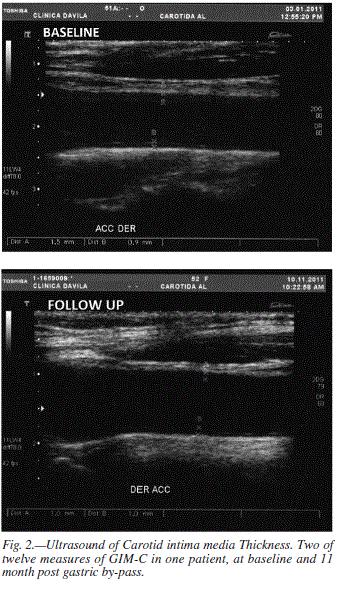
Baseline and postoperative measurements were taken to each patient, with a mean time interval of 354 ± 92.1 days, using B-mode US, by a commercially available ultrasound system (Toshiba APLIO XV) with a 7.5 MHz linear transducer (PLT 704 BT). Images were taken from the carotid wall with the patient in a supine position and a slight hyperextension and rotation of the neck to the contralateral side. Measures of the intima-media thickness (IMT), defined as the distance between the junction of the lumen and the intima, and that of the media and adventitia were recorded from the common carotid artery near 2 cm near from bulb. Three equidistant measures in the anterior and posterior walls of both carotids arteries were taken. In total, 12 measurements were taken from each patient, and an average for the final CIMT was obtained. Measurements were done by a radiologist and a medical technologist specialized in US. Both were blinded for the cardiovascular risk factors and weight loss status.
Laboratory
At baseline and 1 year postoperative laboratory measurements were carried out concomitant with carotid US measurements. A twelve hours fasting venous blood sample was obtained to measure TG, TC and HDL-C using routine laboratory methods, LDL-C was calculated using the Fridewald formula. Plasma glucose was measured by the hexokinase method. Insulin was measured by RIA. In the first evaluation a sample for glucose and insulin was taken two hours after oral 75 g de glucose, except in diabetic patients. The homeostatic model of assessment of insulin resistance (HOMA-IR) was calculated by the following formula: fasting serum insulin concentration (uIU/ml) x fasting blood glucose concentration (mg/dl)/405.27
Comorbidities: hypertension was defined as a blood pressure > 140/90 mmHg and/or use of antihypertensive medications. Patients were defined as diabetic or glucose intolerant when they had a previous diagnosis of this condition or when they met the diagnosis criteria of the American Diabetes Association28. Insulin resistance was defined if HOMA-IR > 2.6. Hypercholesterolemia was defined if total cholesterol were > 200 mg/dl. Hypertriglyceridemia if fasting triglycerides were > 150mg/dl. Mixed dyslipidemia when fasting total cholesterol > 200 mg/dl and triglycerides > 150 mg/dl. Those patients who admitted smoked in the last month were considered smokers.
Cardiovascular risk to 10-y was estimated in all patients using the 2008 gender-specific Framingham risk score.8
Statistical analysis
Baseline characteristics were expressed as mean ± SD or count with proportions, according to the type of data.
Paired-sample t-test was used to determine significance of changes before and after surgery. The significance of differences in the Framingham risk score was tested using the Wilcoxon Signed Ranks Test due to the ordinal character of the variable. Pearson correlations coefficients were calculated to assess association between risk factors and ultrasound variables C-IMT and C-IMT change.
Results
Thirty one obese subjects were included in the study (24 women/7 men). Four patients did not undergo surgery despite meeting clinical requirements, due to economic reasons. They were contacted for a second evaluation that included US C-IMT and the same laboratory tests. Gastric bypass was performed in 14 patients and Sleeve gastrectomy in 13.
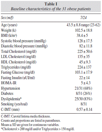
Baseline clinical and biochemical characteristics are shown in table I. Mean age of the study population was 43.6 ± 8.1 years. Mean BMI were 38.3 ± 4.3 kg/m2. At baseline we found: 18 of 29 (62%) had high levels of total cholesterol and 21 of 30 (70%) had high levels of triglycerides. Twelve of 30 (40%) had mixed dyslipidemia, 21 of 31 (68%) had hypertension and 8 of 31 (26%) were diabetic.
At baseline mean C-IMT was 0.57 ± 0.14 mm and correlated with age (r = 0.43, p = 0.03)
Table II shows baseline and follow up parameters of patients undergoing surgery. As expected, BMI showed a decrease during the observation period (366 ± 92.8 days), from 38.1 ± 4.3 to 27.3 ± 3.7 k/m2 (p < 0.001).
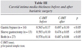
There was a significant decrease in C-IMT after both surgical procedures, from 0.578 ± 0.14 mm at baseline to 0.487 ± 0.08 mm (p < 0.0001) at the end of the follow up period (Table II). In the sleeve group rom 0.593 ± 0.1 mm to 0.476 ± 0.058 mm (p < 0.05), and from 0.565 ± 0.17 to 0.497 ± 0.10 mm (p < 0.05) in the GBPS group (table III).
All cardiovascular risk factor measured significantly improved after the observation period. The mean 10 years estimated cardiovascular risk by Framingham method decreased from 6.6% to 3.4% (p < 0.0005).
The use of prescription drugs also decreased during the follow up period. Diabetic medication from 11 to 2 participants, lipid lowering drugs from five to none, and antihypertensive from 13 to four patients (table III).
At baseline 9 of 27 patients underwent surgery showed C-IMT > 75th percentile and 15 of 27 > 50th percentile of reference population18. After follow up period only three patients persisted over > 75th percentile (fig. 1). A reduction in C-IMT was observed in 25 of 27 patients (92.6%). Only 2 of 27 patients undergoing surgery increased C-IMT, during observation period: a 0.03 and 0.1 mm increase in C-IMT during the observation period was observed in only two of 27 patients undergoing surgery. Both patients underwent GBPS.
After surgery 21 patients reached C-IMT under 5th percentile of reference population, 6 patients not achieved this end point or increase C-IMT. Parameters of the first group compared to the second group are as follow: mean age was 42.3 ± 8 years, compared 48.8 ± 4.37 years (p = 0.07), mean BMI was 37.1 vs 41.3 k/m2 (p = 0.03), mean systolic blood pressure was 141 vs 126 mmHg (p = 0.06), mean fasting insulin was 36.6 vs 20.4 uUI/ml (p = 0.02) and mean HOMA was 9.9 vs 5.4 (p = 0.04).
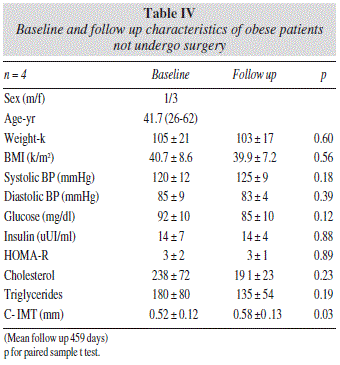
Outcomes of four patients non-undergoing surgery are shown in table IV. After a mean follow up of 459 days, the average weight, BMI and cardiovascular risk factors did not change significantly. C-IMT increased from 0.52 ± 0.12 to 0.58 ± 0.13 mm (p = 0.03), a progression rate of 0.047 mm/y.

Discussion
In this study we demonstrate that weight loss achieved after bariatric surgery is associated with a significant reduction in C-IMT.
C-IMT, measured noninvasively with the use of carotidartery ultrasonography is an independent predictor of new cardiovascular events in patients without a history of cardiovascular disease.13
Only four studies relate C-IMT and bariatric surgery. Karason et al. 1999, in a four years observational study, showed a slower rate of CIMT progression of 0.024 mm/year, among 20 morbid obese patients treated with weight-reducing gastroplasty compared with a rate of 0.068 mm/year progression in the obese group treated with dietary recommendations29. Sturm reported 37 morbid obese patients who underwent an adjustable gastric band, who experienced a CIM-T reduction from 0.56 ± 0.09 to 0.53 ± 0.08 mm (p = 0.004), and a weight loss of 21.6%, 18 months post-surgery.30 Sarmento et al., showed a reduction of CIMT among women who underwent a BPGS, from 0.73 ± 0.12 mm to 0.60 ± 0.12 mm during the first year after surgery (p < 0.05). These women experienced a 38% reduction in BMI during the same lapse.31 In a two years prospective study of 50 obese patients undergoing BPGS, Habib et al. observed a BMI reduction from 47 to 29.5 k/m2, along with a C-IMT change from 0.84 to 0.50 mm (40.5%).32 Karason and Sturm showed a smaller decrease in BMI of 17% and 21% and an annual variation of the C-IMT of +7.4% and -5.5% each. Reduction rate of C-IMT relates to the magnitude of weight loss achieved and the latter is related to the surgical technique. Gastroplasty and adjustable gastric band cause less weight loss than BPGS and sleeve gastrectomy.
Our trial and Sturm study showed the lower mean baseline C-IMT (0.58 mm, and 0.55 mm respectively) compared to the figures reported by Habib, Karason and Sarmento (0.86, 0.81 and 0.73 mm respectively) studies. This discrepancy may be due to two reasons. First the participants of our study and that of Sturm, showed had lower age. Secondly an ethnic factor may be involved, considering that Carmela study performed in Santiago and Barquisimeto population had the smallest C-IMT of all Latin American capitals studied.
Several surgical approaches to induce weight loss have been developed. This study considered the two most common techniques used in our country at this time, sleeve gastrectomy, a purely restrictive procedure, and the Roux-en-Y GBPS a mixed technique (restrictive and malabsorptive) that is probably the most common weight loss surgery procedure performed. It is beyond the scope of this study to compare the two surgical procedures. In fact there is a bias in patient selection, all diabetic patients and those with higher metabolic derangements were assigned to bypass. However it is noteworthy that while the weight loss in patients undergoing bypass was greater, the decrease in C-IMT was less. This could indicate that artery wall lesions are less reversible among subjets with worst metabolic parameters. During follow up C-IMT decreased, even taking into account that most of our patients (67%) did not have an abnormal C-IMT at baseline. Moreover, 88% of them achieved values under 5th percentile of the reference population at follow up. Since, the mean age of patients that reached < 5th percentile was less, age could be another factor that could affect C-IMT reversibility.
An increasing number of trials are using C-IMT as a surrogate end point for cardiovascular events, considering the doubtful association between C-IMT and CVR. Most of those trials that modified a single cardiovascular risk factor such as dyslipidaemia showed modest changes in C-IMT. The Pravastatin, Lipids, and Atherosclerosis in the Carotid Arteries II (PLAC-II) study demonstrated a lesser progression of C-IMT with pravastatin compared with placebo, 0.0295 and 0.0456 mm/y, respectively.33 Meteor trial demonstrated that rosuvastatin therapy resulted in a reduction of the progression of maximum C-IMT compared to placebo in middle age adults with low Framingham scores and subclinical atherosclerosis, with changes of -0.0014 and 0.0131 mm/y, respectively.34 ENHANCE trial evaluated the change in C-IMT with the combination of simvastatin and ezetimibe compared with simvastatin monotherapy. There was no significant difference between the groups.35 In hypertension, the MORE study (Multicenter Olmesartan atherosclerosis Regression Evaluation) performed in 78 hypertense patients under olmesartan therapy followed for two years, showed a reduction on C-IMT of 0.09 mm, that is similar to that obtained in our study (0.091 mm). However, the reduction in the MORE study was smaller in terms of percentage (9.2 and 15.7 respectively).36
The significant 0.047 mm/year C-IMT increase, seen in non-operated obese patients, must be analyzed taking into account the study of Hodis et al who showed that for each 0.03 mm increase per year in CIMT, there was a concomitant increase in the relative risk for non-fatal myocardial infarction or coronary death of 2.2 (95% IC 1.4-3.6) and in the relative risk for any coronary event of 3.1 (95% IC 2.1-4.5).15
Limitations of this study include the small sample size specially the number of patients not undergoing surgery.
Conclusion
Obese patients, who underwent successful Bariatric surgery in terms of weight loss, achieve a significant reduction in C-IMT, a surrogate factor of subclinical atherosclerosis.
Most operated patients reached C-IMT values less than 5 percentile of reference population. The reduction in C-IMT and cardiovascular risk factors were obtained after lowering or withdrawing pharmacologic therapy, underscoring the relevance of these surgical procedures in terms of CV risk reduction.
Reversibility of carotid wall thickness appears to inversely associate with age and metabolic involvement.
New research is needed to assess the rate of progression of the atherosclerotic process in patients with severe obesity undergoing medical therapy, to compare the impact of different surgical techniques in markers of cardiovascular risk, and to assess the long term evolution of patients underwent bariatric surgery.
Acknowledgements
We want to thank the department of radiology of Davila clinic and INTA that belongs to University of Chile.
References
1. Encuesta Nacional de Salud Chile 2009-2010. Informe Final. Santiago, Enero de 2010. Pontificia Universidad Católica de Chile. Facultad de Medicina/Departamento de Salud Pública. Disponible en: http://www.redsalud.gov.cl/portal/url/item/99bbf09a908d3eb8e04001011f014b49.pdf. [ Links ]
2. Mora S, Yanek L, Moy TF, Fallin MD, Becker LC, Becker MDl. Interaction of body mass index and Framingham risk score in prediction incident coronary disease in families. Circulation 2005; 111 (15): 1871-6. [ Links ]
3. Nguyen NT, Nguyen XM, Wooldridge JB, Slone JA, Lane JS. Association of obesity with risk of coronary heart disease: findings from the National Health and Nutrition Examination Survey, 1999-2006. Surg Obes Relat Dis 2010; 6 (5): 465-9. [ Links ]
4. Bogers RP, Bemelmans WJ, Hoogenveen RT, Boshuizen HC, Woodward M, Knekt P, et al. Association of overweight with increased risk of coronary heart disease partly independent of blood pressure and cholesterol levels: a meta-analysis of 21 cohort studies including more than 300.000 persons. Arch Intern Med 2007; 167 (16): 1720-8. [ Links ]
5. Eckel RH. Obesity and Heart disease: a statement for healthcare professionals from the Nutrition Committee, American Heart Association. Circulation 1997; 96: 3248-50. [ Links ]
6. Poirier P, Giles TD, Bray GA, Hong Y, Stern JS, Pi-Sunyer XF, et al. Obesity and cardiovascular disease: pathophysiology, evaluation and effect of weight loss: an update of the 1997 American Heart Association Scientific statement on obesity and heart disease from the Obesity Committee of the Council on Nutrition, Physical Activity, and Metabolism. Circulation 2006; 113: 898-918. [ Links ]
7. Murphy NF, MacIntyre K, Stewart S, Hart CL, Hole D, McMurray JJ. Long-term cardiovascular consequences of obesity: 20-year follow up of more than 15 000 middle-aged men and women (the Renfrew-Paisley study). Eur Heart J 2006; 27 (1): 96-106. [ Links ]
8. D'Agostino RB, Vasan RS, Pencina MJ, Wolf PA, Cobain M, Massaro JM, et al. General cardiovascular risk profile for use in primary care: the Framingham Heart study. Circulation 2008; 117 (6): 743-53. [ Links ]
9. Lloyd-Jones DM, Leip EP, Larson MG, D'Agostino RB, Beiser A, Wilson PW, et al. Prediction of lifetime risk for cardiovascular disease by risk factor burden at 50 years of age. Circulation 2006; 113: 791-8. [ Links ]
10. Ridker PM, Cook N. Should age and time be eliminated from cardiovascular risk prediction models?: Rationale for the creation of a new national risk detection program. Circulation 2005; 111: 657-8. [ Links ]
11. National Cholesterol education Program (NCEP) Expert Panel on Detection, Evaluation and Treatment of High Blood Cholesterol in Adults (Adult Treatment Panel III). Third report of the National Cholesterol Education Program (NECP) expert panel on detection, evaluation, and treatment of high cholesterol in adults (adult treatment panel III) final report. Circulation 2002; 106 (25): 3143-421. [ Links ]
12. Stein JH, Korscars CE, Hurst RT, et al. Use of carotid ultrasound to identify subclinical vascular disease and evaluate cardiovascular risk: a consensus statement from American Society of Echocardiography carotid intima-media thickness task force: endorsed by the Society for Vascular Medicine. J Am Soc Echocardiogr 2008; 21 (2): 93-111. Erratum in J Am Soc Echocardiogr 2008; 21 (4): 376. [ Links ]
13. Hodis HN, Mack WJ, LaBree L, Selzer RH, Liu CR, Liu CH, et al. The role of carotid arterial intima-media thickness in predicting clinical coronary events. Ann Intern Med 1998; 128 (4): 262-9. [ Links ]
14. Chambless LE, Heiss G, Folsom AR, Rosamond W, Szklo M, Sharrett AR, et al. Association of coronary heart disease incidence with carotid arterial wall thickness and major risk factors: the Atherosclerosis Risk in Communities (ARIC) study. Am J Epidemiol 1997; 146 (6): 483-94. [ Links ]
15. Heiss G, Sharrett AR, Barnes R, Chambles LE, Szklo M, Alzola C. Carotid atherosclerosis measured by B-mode ultrasound in populations: associations with cardiovascular risk factors in the ARIC study. Am J Epidemiol 1991; 134 (3): 250-6. [ Links ]
16. Lorenz MW, Markus HS, Bots ML, Rosvall M, Sitzer M. Prediction of clinical cardiovascular event with carotid intima-media thickness. A sistematic review and meta-analysis. Circulation 2007; 115: 459-67. [ Links ]
17. Smith SC, Greenland P, Grundy SM. Prevention Conference V: Beyond secondary prevention: identifying the high-risk patient for primary prevention: Executive Summary. Circulation 2000; 101: 111-6. [ Links ]
18. Schargrodsky H, Hernández-Hernández R, Champagne BM, Silva Aycaguer LC, Touboul PJ, Boissonnet CP, et al. CARMELA: assessment of cardiovascular risk in seven latin american cities. Am J Med 2008; 121 (1): 58-65. [ Links ]
19. Klein S, Burke LE, Bray GA, Blair S, Allison DB, Pi-Sunyer X, et al. Clinical implications of obesity with specific focus on cardiovascular disease: a statement for professionals from the American Heart Association Council on Nutrition, Physical Activity, and Metabolism: endorsed by the American College of Cardiology Foundation. Circulation 2004; 110: 2952-67. [ Links ]
20. Maggard MA, Shugarman LR, Suttorp M, Maglione M, Sugerman HJ, Livingston EH, et al. Meta-Analysis: surgical treatment of obesity. Ann Intern Med 2005; 142 (7): 547-59. [ Links ]
21. Sjöström L, Lindroos AK, Peltonen M, Torgerson J, Bouchard C, Carlsson B, et al. Lifestyle, diabetes and cardiovascular risk factor 10 years after bariatric surgery. N Engl Med 2004; 351 (26): 2683-93. [ Links ]
22. Csendes A, Burdiles P, Papappietro K, Burgos AM. Comparación del tratamiento médico y quirúrgico en pacientes con obesidad grado III (obesidad mórbida). Rev Med Chile 2009; 137 (4): 559-66. [ Links ]
23. Sjöström L, Narbro K, Sjöström CD, Karason K, Larsson B, Wedel H, et al. Effects of bariatric surgery on mortality in Swedish obese subjets. N Engl J Med 2007; 357: 741-52. [ Links ]
24. Adams TD, Gress RE, Smith SC, Halverson RC, Simper SC, Rosamond WD, et al. Long-term mortality after gastric bypass surgery. N Engl Med 2007; 357: 753-61. [ Links ]
25. Santry HP, Gillen DL, Lauderdale DS. Trends in bariatrics procedures. JAMA 2005; 294 (15): 1909-17. [ Links ]
26. Bessesen DH. Update on obesity. J Clin Endocrinol Metab 2008; 93: 2027-34. [ Links ]
27. Matthews DR, Hosker JP, Rudensky AS, Naylor BA, Teacher DR, Turner RC. Homeostatic model assessment: insulin resistance and beta-cell function from fasting plasma glucose and insulin concentration in man. Diabetologia 1985; 28 (7): 412-9. [ Links ]
28. The experte committe on the diagnosis and classification of Diabetes Mellitus. Follow-up report on the diagnosis of Diabetes Mellitus. Diabetes Care 2003; 26 (11): 3160-7. [ Links ]
29. Karason K, Wikstrand J, Sjöström L, Wendelhag I. Weight loss and progression of early atherosclerosis in the carotid artery: a four-year controlled study of obese subjects. J Obes Relat Metab Disord 1999; 23 (9): 948-56. [ Links ]
30. Sturm W, Tschoner A, Engl J, Kaser S, Laimer M, Ciardi C, et al. Effect of bariatric surgery on both functional and structural measures of premature atherosclerosis. Eu Heart J 2009; 30: 2038-43. [ Links ]
31. Sarmento P, Plavnik F, Zanella M, Pinto P, Miranda R, Ajsen S. Association of carotid intima media thickness and cardiovascular risk factor in women pre-and post-bariatric surgery. Obes Surg 2009; 19 (3): 339-44. [ Links ]
32. Habib P, Scrocco JD, Terek M, Vanek V, Mikolich JR. Effects of bariatric surgery on inflammatory, functional and structural markers of coronary atherosclerosis. Am J Cardiol 2009; 104: 1251-5. [ Links ]
33. Crouse JR III, Byington RP, Bond MG, Espeland MA, Craven TE, Sprinkle JW, McGovern ME, Furberg CD. Pravastatin, lipids, and atherosclerosis in the carotid arteries. (PLAC-II). Am J Cardiol 1995; 75 (7): 455-9. Erratum in: Am J Cardiol 1995; 75 (12): 862. [ Links ]
34. Crouse JR III, Raichlen JS, Riley WA, Evans GW, Palmer MK, O Leary DH, et al. METEOR Study Group. Effect of Rosuvastatin on progression of carotid intima-media thickness in low-risk individuals with subclinical atherosclerosis: the METEOR trial. JAMA 2007; 297 (12): 1344-53. [ Links ]
35. Kastelein JJ, Akdim F, Stroes ES, Zwinderman AH, Bots ML, Stalenhoef AF, et al. ENHANCE Investigators. Simvastatin with or without Ezetimibe in Familial Hypercholesterolemia. N Engl J Med 2008; 358: 1431-43. [ Links ]
36. Stumpe KO, Agabiti-Rosei E, Zielinski T, Schremmer D, Scholze J, Laeis P, et al. MORE study investigators. Carotid intima-media thickness and plaque volume changes following 2-year angiotensin II-receptor blockade. The Multicentre Olmesartan atherosclerosis Regression Evaluation (MORE) study. Ther Adv in Cardiovasc Dis 2007; 1 (2): 97-106. [ Links ]
![]() Correspondence:
Correspondence:
Gonzalo García Bravo
Clínica Dávila
C/ Recoleta, 464
Chile
E-mail: ggarciabravo@vtr.net
Recibido: 2-II-2013
Aceptado: 13-IV-2013.


