


Meu SciELO
 Serviços customizados
Serviços Personalizados
Journal
Artigo
 Inglês (pdf)
 Artigo em XML
 Referências do artigo
 Enviar este artigo por email
Indicadores
-
 Citado por SciELO
 -
 Acessos
Links relacionados
-
 Citado por Google
 -
 Similares em
SciELO
Similares em
SciELO -
 Similares em Google
Compartilhar

 Permalink
Nutrición Hospitalaria
versão On-line ISSN 1699-5198versão impressa ISSN 0212-1611
Nutr. Hosp. vol.30 no.4 Madrid Out. 2014
https://dx.doi.org/10.3305/nh.2014.30.4.7644
ORIGINAL / Sindrome metabolico; Diabetes
Applicability of the visceral adiposity index (VAI) in predicting components of metabolic syndrome in young adults
Aplicabilidad del visceral adiposity index (VAI) en la predicción de componentes del síndrome metabólico en adultos jóvenes
Jéssica Schuster, Patrícia Vogel, Cristiane Eckhardt and Simone Dal Bosco Morelo
Nutrition Course, Univates University Center. Lajeado/RS - Brazil
ABSTRACT
Introduction: Metabolic syndrome (MS) is one of the main risk factors for cardiovascular diseases (CVD) and identifying its components in young adults may constitute an important prevention tool.
Objective: Analyzing applicability of the Visceral Adiposity Index (VAI) for predicting components of MS in young adults.
Methods: Cross-sectional survey with 444 individuals, mean age 25.6±6.5, 77.7% females. We obtained data on weight, height, waist circumference (WC), body fat percentage (BF%), fasting glycemia, triglycerides (TG), total cholesterol (TC), HDL cholesterol (HDL-c), LDL cholesterol (LDL-c), and blood pressure (BP), as well as information on their lifestyles. Body Mass Index (BMI), Waist-to-Height Ratio (WHR) and VAI were calculated. Adiposity measurements were compared to the MS components, and for VAI, we determined the predictive capacity of MS components occurrence and the area below the ROC curve.
Results: VAI correlated to glucose (r=0.136), HDL-c (r=-436), and TG (r=0.825) in females, and amongst men, it correlated to glucose (r=0.258), HDL-c (r=-0.550), TG (r=0.897), and DBP (r=0.290). An increased VAI was associated to an increased risk of abdominal obesity (OR=1.86), hypertriglyceridemia (OR=30.74), and low HDL-c (OR=3.95). Among obesity indicators, VAI presented a larger area below the curve for increased TG and low HDL-c. Conclusion: VAI presented an association to MS components in males and females with an increased risk of abdominal obesity, hipertriglyceridemia, and low HDL-c, proving to be a good MS components predictor even among healthy young adults.
Key words: Metabolic Syndrome, Abdominal Fat, Obesity.
RESUMEN
Introducción: El síndrome metabólico (SM) es uno de los principales factores de riesgo de enfermedades cardiovasculares (ECV), y la identificación de sus componentes en los adultos jóvenes puede constituir una importante herramienta de prevención.
Objetivo: Analizar la aplicabilidad del Visceral Adiposity Index (VAI) para la predicción de componentes del SM en adultos jóvenes.
Métodos: Estudio transversal con 444 individuos, edad 25,6 ± 6,5, 77,7% del sexo femenino. Se obtuvieron datos sobre el peso, talla, circunferencia de cintura (CC), el porcentaje de grasa corporal (%GC), glucemia, triglicéridos (TG), colesterol total (CT), colesterol HDL (HDL-c), colesterol LDL (LDL-c), y la presión arterial (PA), así como información sobre sus estilos de vida. Se calculó Índice de Masa Corporal (IMC), la proporción de cintura a la altura (PCA) y VAI. Mediciones de adiposidad se compararon con los componentes del SM, y por VAI, se determinó la capacidad predictiva de ocurrencia de los componentes de SM y el área a bajo la curva ROC.
Resultados: VAI se correlaciona a la glucosa (r= 0,136), HDL-c (r=-436) y TG (r=0,825) en las mujeres y entre los hombres, se correlaciona a la glucosa (r=0,258), HDL-c (r=-0,550), TG (r=0,897), y la PAD (r= 0,290). Un aumento de VAI se asoció a un mayor riesgo de obesidad abdominal (OR=1,86), hipertrigliceridemia (OR=30,74), y bajo HDL-c (OR=3,95). Entre los indicadores de obesidad, VAI presentó una mayor área bajo la curva de aumento de triglicéridos y bajos niveles de HDL-c. Conclusión: VAI presentó una asociación a los componentes del SM en hombres, y en mujeres con un mayor riesgo de obesidad abdominal, hipertrigliceridemia y bajos niveles de HDL-c, demostrando ser un buen predictor de componentes de SM, incluso entre adultos jóvenes sanos.
Palabras clave: Síndrome metabólico, grasa abdominal, obesidad.
Abbreviations
WC: Waist Circumference.
TC: Total Cholesterol.
CVD: Cardiovascular Diseases.
DM 2: Diabetes Mellitus type 2.
BF%: Body Fat Percentage.
BMI: Body Mass Index.
HDL-c: High Density Lipoprotein .
LDL-c: Low Density Lipoprotein.
BP: Blood Pressure.
DBP: Diastolic Blood Pressure.
SBP: Systolic Blood Pressure.
WHR: Waist-to-Height Ratio.
WHiR: Waist-to-Hip Ratio.
MS: Metabolic Syndrome.
CT: Computed Tomography.
TG: Triglycerides.
VAI: Visceral Adiposity Index.
Introduction
Metabolic syndrome (MS) is defined as a complex disease represented by a set of cardiovascular risk factors, usually related to fat buildup in the abdominal region, as well as insulin resistance1. The presence of MS is defined according to the National Cholesterol Education Programs - Adult Treatment Panel III (NCEP-ATP III) criteria, presenting a combination of at least three components: Abdominal obesity (waist circumference >102cm for males and >88cm for females); increased triglycerides (≥150mg/dL); low HDL-c (<40mg/dL for males and <50mg/dL for females); elevated blood pressure (systolic ≥130mmHg or diastolic ≥85mmHg); and increased fast glycemia (≥110mg/dL)2.
Visceral adipose tissue is a metabolically active organ and intra-abdominal obesity is an independent risk factor for metabolic alterations present in MS3,4, which is associated to the development of cardiovascular diseases (CVD) and diabetes mellitus type 2 (DM2) in children, teenagers, and adults5,6,7. Computed Tomography (CT) is still considered the golden standard for evaluating distribution of body fat and quantifying intra-abdominal fat, as it allows differentiating subcutaneous and visceral tissues. CT, however, is not easily applicable in population studies and in clinical practice, especially due to its high cost and as it exposes individuals to radiation8.
Amato and contributors3, in a study performed with a Caucasian European population, have validated a visceral obesity indicator defined as Visceral Adiposity Index (VAI). The formula takes gender into consideration and combines anthropometric measurements - Waist Circumference (WC) and Body Mass Index (BMI) - with biochemical tests - triglycerides (TG) and low density lipoprotein (LDL-c). According to the study, VAI indicates the visceral adipose tissue function and its increase is independently correlated to cardiovascular and cerebrovascular risks. Another study that evaluated the applicability of VAI in predicting MS has demonstrated that it presents a significant relation to its components9.
The objective of the present study is to analyze the applicability of VAI as a predictor of MS components comparing it to other anthropometric indicators (BMI, WC, WHR, BF%) and its isolated association to a risk of MS, in a sample of young adults who use a University Center Nutrition Ambulatory in the state of Rio Grande do Sul, Brazil.
Methods
The study was performed at the Nutrition Department of Univates University Center (UNIVATES) in the city of Lajeado(Rio Grande do Sul, Brazil) after being approved by the Research Ethics Committee (COEP/UNIVATES), accredited by the Conselho Nacional de Saúde (Brazilian National Board for Health), under protocol number 110/11. It is a cross-sectional study, with a sample composed of students and staff members from UNIVATES who use the Institution's Nutrition Ambulatory, totaling 444 individuals. The data were collected between April, 2012 and March, 2014. The participants were included by adhesion, after signing a Free and Clarified Consent Term and being referred to the Institution's Nutrition Department for a nutritional consultation. We performed an anthropometric evaluation of the patients where Blood Pressure (BP), Weight (in kilos), Height (in meters), and Waist Circumference (in centimeters) were checked to later calculate BMI (Kg/m2), and Waist-to-Height Ratio (WHR - WC divided by height, both measured in centimeters).
BP was measured with a digital aneroid appliance (Omron® brand), with individual sat with arms stretched over a steady surface, with a 5 to 10 minute rest, empty bladder, and without having exercised, smoked or ingested alcoholic beverages, coffee or food 30 minutes prior to the assessment. The rotator cuff was positioned 3 cm above the antecubital fossa, maintaining the patient's arm at heart's height and with relaxed uncrossed legs. Three measurements were taken with a 1 to 2 minute interval between them, being the final BP value obtained from an arithmetical mean of the three checks10.
The weight was measured with a platform-like anthropometric scale, mounted with a Welmy® stadiometer with a maximum capacity of 150kg and 100g divisions, with the participant wearing a light scrub and no shoes, positioned at the center of the equipment, erect, with both feet together, and arms resting along the body, according to the Brazilian Ministry of Health regulations11.
Height was measured with the stadiometer mounted to the Welmy® anthropometric scale with an extension of 2 meters, divided in centimeters, subdivided in millimeters, with the participant wearing a light scrub, barefoot, without any headgear, standing, erect, arms resting along the body, raised head, looking at a fixed point at eye level, in the Frankfurt plan, according to Brazilian Ministry of Health regulations11.
BMI was calculated with weight and height measurements according to the formula: BMI = W/H2, W being weight in kilos and H height in square meters. The BMI cut-off points adopted were the ones recommended by the World Health Organization12: underweight BMI<18.5Kg/m2, eutrophia between BMI 18.5 and 24.99Kg/m2, and overweight and obesity, BMI≥30Kg/m2.
WC was assessed with an CESCORF inextensible tape measure, with a 1 mm precision, at the trunk's narrowest portion, between the ribs and the iliac crest at the moment of exhalation, with an unclothed waist, according to regulations recommended by the Brazilian Ministry of Health11. The WC cut-off points adopted were the ones recommended by the World Health Organization12 and NCEP-ATP III2, abdominal obesity being considered WC≥102cm for males and ≥88cm for females.
WHR was obtained dividing waist circumference by height, both in centimeters, and for classification purposes, compared to the cut-off points determined by Pitanga and Lessa13 for Brazilian people, which were 0.53 for females and 0.52 for males.
In a second appointment, scheduled previously, 5ml of blood were collected, with an 8 to 12 hour fast. Collections were performed by trained researchers with the use of thin needles, to minimize discomfort. The researchers used disposable and protective material to avoid contamination. Laboratory dosing was performed at the Institution's Biochemistry Laboratory, in a Mindray BS120® Automated Biochemistry equipment, through the enzymatic kinetic method. The LDL-c concentration was determined with the use of the Friedwald Formula14: LDL-C=TC-(HDL-c+TG/5). For interpreting the results, we used the cut-off points recommended by the Brazilian Diabetes Society15 and the Brazilian Cardiology Society16 with glycemia <100mg/dL, TC <200mg/dL, LDL-c <130mg/dL, HDL-c desirable >40mg/dL for males and >50mg/dL for females and TG <150mg/dL.
The Visceral Adiposity Index (VAI) was calculated with the formula proposed by Amato and collaborators3: VAI = (WC(cm)/(39.68+(1.88*BMI)))*( TG/1.03)*(1.31/HDL) for males and VAI = (WC(cm)/ (36.58+(1.89*BMI)))*(TG/0.81)*(1.52/HDL) for females.
In order to verify body fat percentage (BF%) each individual was also subjected to a quadrupolar bioimpedance test in a model 310 BIODYNAMICS equipment. The test was performed with the subject lying supine in a non-conducting surface with arms and legs 45 degrees away from the body. Contact areas were cleaned with alcohol for positioning the electrodes, an emitter electrode was positioned close to the metacarpal phalangeal joint at the right hand dorsal surface and another electrode distal to the transversal arch of the right foot's upper surface. A detector electrode was positioned between the distal prominences of the radius and the ulna on the right wrist and another one between the right ankle's median and lateral malleoli17 BF% was compared to the median recommended values according to Pollock and Wilmore18, with the optimal value between 23 and 25% for females and 14 to 16% for males, in the age interval sampled.
The data were analyzed with IBM® SPSS Statistics software version 20.0. The significance level adopted was 5% (p<0.05). We performed descriptive univariate statistics (mean, standard deviation, and frequency) and bivariate statistics (Student's t-test for independent samples, Pearson and Spearman correlations, binary logistic regression, and ROC curves). The Kolmogorov-Smirnov test was used to assess if the variables followed a normal distribution. Variables showing a normal distribution were analyzed with a Student's t-test and Pearson Correlation, while the ones that did not follow a normal distribution were analyzed through nonparametric Mann-Whitney and Spearman Correlation tests. The Pearson and Spearman Correlation tests were conducted to correlate the analyzed anthropometric, biochemical, and pressure parameters, while the Student's t-test for independent samples was applied to compare anthropometric and biochemical profiles between genders. A binary logistic regression was performed to analyze VAI's predictive strength in regards to MS components. ROC curves (receiver operating characteristic curve) were constructed and the area below the curves calculated with a 95% confidence interval. The Z test was used to compare the areas below the curves.
Results
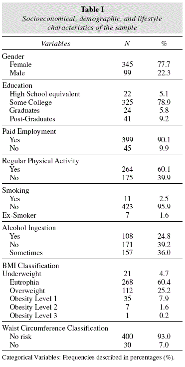
The sample's Socioeconomical, demographic, and lifestyle characteristics are described in table I.
In table II, median values for age, biochemical, anthropometric, and pressure parameters are described in the general sample, and stratified according to gender. There was a significant difference between genders for all parameters, except for LDL-c and DBP, with age, BMI, WC, WHR, and SBP being significantly higher between males; and BF%, VAI, TC, HDL-c, and TG significantly higher among females.
While analyzing the association between biochemical and pressure components of MS with anthropometric indicators of obesity, there was significant correlation of BMI with all the other components among females, of WC and WHR with glycemia, HDL-c, and BP, of BF% with glycemia, TG, and BP; and of VAI with glycemia, HDL-c, and TG. Among males, however, BMI had a significant correlation to glycemia, TG, and DBP; while WC and BF% had a significant correlation to glycemia, HDL-c, TG, and DBP; as for WHR, it presented a correlation to glycemia, TG, and DBP. Finally, VAI presented a correlation to glycemia, HDL-c, TG, and DBP. Significance values (p) and correlation coefficients (r) can be seen in table III.
When specifically analyzing VAI, it presented among females a significant association to glycemia, HDL-c, and TG in females, furthermore the increase in VAI is accompanied by an increase in glycemia and TG, and a decrease in HDL-c. Among males, VAI is correlated to glycemia, HDL-c, TG, and DBP, with an increase in VAI implying a linear increase in glycemia, TG, and DBP, but a decrease in HDL-c. While analyzing the applicability of VAI in determining the MS components occurrence relative risk, VAI was a good predictor of abdominal obesity (OR=1.86, p<0.01), hipertriglyceridemia (OR=30.74, p<0.001), and low HDL-c (OR=3,95, p<0,001). Among obesity indicators, VAI has shown a larger area under the curve for increased TG and low HDL. The results can be seen in table IV.
Discussion
The present study has demonstrated the association between VAI and other adiposity measurements with MS components. Knowles et al9, in a study with Peruvian adults, have analyzed the usage of different anthropometric indicators (BMI, WC, WHR, WHiR, and VAI) in evaluating MS risk. All the evaluated indicators have presented a significant relationship to MS components, differently from our study, where some indicators were not related to the analyzed components. The size and mean age of the sample may have determined a lower number of significant correlations in our study, for during the aforementioned investigation work a total of 1,518 individuals with a mean age of 39.3 were evaluated, while in our study 444 individuals with a mean age of 25.6 were studied showing no significant alterations in anthropometric, pressure, and biochemical parameters.
Specifically analyzing VAI, Knowles et al9 study has found an association with all MS components, with a substantial association for hipertriglyceridemia and low HDL-c in both genders. Similarly to the data found in the present study, where VAI was more highly correlated to HDL-c and TG. Considering that both these biochemical parameters compose the VAI formula, we can establish a hypothesis for this association. While evaluating the risk of MS components through adiposity indicators, VAI appears to be the best predictor of increased TG and lowered HDL-c, corroborating our findings.
In Knowles et al9 study, the anthropometric measurement that presented a more substantial correlation to fasting glycemia, both for males and females, was BMI. In our study, BMI was associated with fasting glycemia, but the indicator with the best correlation was WC, for both genders, furthermore an increase in WC is also followed by an increase in glycemia. In the aforementioned study, the pressure components of MS, SBP, and DBP were also correlated to all of the evaluated anthropometric indicators, but WC has shown higher correlation both in males and females. On the other hand, in our findings, for females, SBP and DBP were associated to all the anthropometric parameters studied except for VAI, while for males SBP was not significant for any indicator and DBP was correlated with all indicators. As previously discussed, the young age of the sample and the lack of important alterations in the anthropometric pressure and biochemical parameters may have influenced the results obtained in the present study.
In Amato et al.3 study, which suggested VAI as an indicator of the role of visceral adipose tissue, conducted with a sample of European adults, the outcomes of cardiovascular and cerebrovascular disease were correlated with BMI, WC, and VAI. VAI was independently associated with cardiovascular and cerebrovascular diseases, while WC and BMI have not shown a significant correlation. Another Amato et al.4 study, with a sample composed of Caucasian adults, has validated VAI cutoff points for cardiovascular and cerebrovascular events in individuals with MS, corroborating previous findings, which indicated the risk for cardiovascular and cerebrovascular events is higher with a higher VAI. VAI presents itself as an easy and practical tool for use in population studies and clinical practice for evaluating cardiometabolic risks associated to visceral fat4.
Salomon et al.8, in a study that has not used VAI as an adiposity measurement but compared WC and visceral fat measured by ultrasonography with MS risk factors, reported that increase in visceral fat was associated to all MS components in females, and with an increase in WC and glycemia in men. However, the predicting capacity of WC was more significant than that of visceral fat measured by ultrasonography for both genders. In our study, WC was the indicator with the best predicting power for increased glycemia in both genders.
Present study results may have been influenced by the size and mean age of the sample, as well as the reduced number of patients with no important alterations in anthropometric, pressure, and biochemical parameters, reducing the statistical strength to determine some correlations. Nevertheless, the results of this work were significant for most of the associations tested, being relevant in regards to a young and healthy sample.
Our results have demonstrated an association of VAI with MS biochemical and pressure components, in males and females, as well as an increased risk of abdominal obesity, hipertriglyceridemia, and low HDL-c. These findings allow us to conclude that VAI is a simple but efficient indicator for predicting MS components, even among young and healthy adults.
References
1. Sociedade Brasileira de Hipertensão. Sociedade Brasileira de Cardiologia. Sociedade Brasileira de Endocrinologia e Metabologia. Sociedade Brasileira de Diabetes. Associação Brasileira para Estudos da Obesidade. I Diretriz Brasileira de Diagnóstico e Tratamento da Síndrome Metabólica. Arq Bras Cardiol. 2005 Abr; 84(Suppl. I): 1-28. [ Links ]
2. Grundy SM, Becker D, Clarck LT, et al. Detection, evaluation, and treatment of high blood cholesterol in adults (Adult Treatment Panel III), Circulation. 2002; 106(25):3143-3421. [ Links ]
3. Amato MC, Giordano C, Galia M, Criscimanna A, Vitabile S, Midiri M, et al. Visceral Adiposity Index A reliable indicator of visceral fat function associated with cardiometabolic risk. Diabetes Care. 2010 Abr; 33(4): 920-2. DOI: 10.2337/dc09-1825. [ Links ]
4. Amato MC, Giordano C, Pitrone M, Galluzzo A. Cut-off points of the visceral adiposity index (VAI) identifying a visceral adipose dysfunction associated with cardiometabolic risk in a Caucasian Sicilian. Lipids Health Dis. 2011; 10(183):1-8. DOI: 10.1186/1476-511X-10-183. [ Links ]
5. Lakka HM, Laaksonen DE, Lakka TA, Niskanen LK, Kumpusalo E, Tuomilehto J, et al. The metabolic syndrome and total and cardiovascular disease mortality in middle-aged men. JAMA. 2002 Dez; 288(21):2709-16. DOI: 10.1001/jama.288.21.2709. [ Links ]
6. Codoñer-Franch P, Murria-Estal R, Tortajada-Girbés M, Castillo-Villaescusa C. del, Valls-Bellés V, Alonso-Iglesias E. New factors of cardiometabolic risk in severely obese children: influence of pubertal status. Nutr Hosp. 2010; 25(5): 845-51. DOI: 10.3305/nh.2010.25.5.4539. [ Links ]
7. Pitangueira JCD, Silva LR, Santana MLP, Silva MCM, Costa PRF, D'Almeida V, et al. Metabolic syndrome and associated factors in children and adolescents of a Brazilian municipality. Nutr Hosp. 2014; 29:865-872. DOI: 10.3305/nh.2014.29.4.7206. [ Links ]
8. Salomon EG, Hizon CE, Raboca JC. Minimum Waist Circumference and Visceral Fat Values by Ultrasonography to Identify Adult Urban Filipinos at Risk for Metabolic Syndrome. Philipp J Intern Med. 2011 Mar; 49(1): 15-21. [ Links ]
9. Knowles KM, Paiva LL, Sanchez SE, Revilla L, Lopez T, Yasuda MB, et al. Waist Circumference, Body Mass Index, and Other Measures of Adiposity in Predicting Cardiovascular Disease Risk Factors among Peruvian Adults. Int J Hyperten. 2011 Jan; 2011:1-10. DOI: 10.4061/2011/931402. [ Links ]
10. Pickering TG, Hall JE, Appel LJ, Falkner BE, Graves J, Hill MN, et al. Subcommittee of Professional and Public Education of the American Heart Association Council on High Blood Pressure Research. Recommendations for blood pressure measurement in humans and experimental animals: Part 1: blood pressure measurement in humans: a statement for professionals from the Subcommittee of Professional and Public Education of the American Heart Association Council on High Blood Pressure Research. Hyperiension. 2005; 45(5):142-61. [ Links ]
11. Brasil MdS. Aniropometria: Manual de Técnicas e Procedimenios. Ministério da Saúde: Vigilância Nutricional; 2003. [ Links ]
12. World Health Organization. Obesity: Preventing and managing the global epidemic. Geneva; 1998. [ Links ]
13. Pitanga FJG, Lessa I. Razão cintura-estatura como discriminador do risco coronariano de adultos. Rev. Assoc. Med. Bras. 2006; 52(3):157-161. [ Links ]
14. Friedewald WT, Levy RI, Fredrickson DS. Estimation of the concentration of low-density lipoprotein cholesterol in plasma, without use of the preparative ultracentrifuge. Clin Chem. 1972; 18:499-502. [ Links ]
15. Sociedade Brasileira de Diabetes. Diretrizes da Sociedade Brasileira de Diabetes 2009. Itapevi: A. Araújo Silva Farmacêutica, 2009; 3. [ Links ]
16. Sociedade Brasileira de Cardiologia. IV Diretriz Brasileira sobre Dislipidemias e Prevenção da Aterosclerose. Arq Bras Cardiol. 2007; 88(1):2-19. [ Links ]
17. Kyle UG, Bosaeus I, De Lorenzo AD, Deurenberg P, Elia M, Gomez JM, Heitmann BL, Kent-Smith L, Melchior JC, Pirlich M, Scharfetter H, Schols AMWJ, Pichard C. Bioelectrical impedance analysis-part II: utilization in clinical practice. Clinical Nutrition. 2004; 23:1430-1453. [ Links ]
18. Pollock ML, Wilmore JH. Exercícios na saúde e na doença. Avaliação e prescrição para prevenção e reabilitação. Rio de Janeiro: MEDSI, 1993; 2. [ Links ]
![]() Correspondence:
Correspondence:
Simone Dal Bosco Morelo.
Centro de Ciencias Biológicas y de la Salud,
Centro Universitário Univates.
Ru Avelino Talini, 171, Barrio Universitário -
Lajeado/RS - Brasil.
Correo postal: 95900-000.
E-mail: simonebosco@gmail.com
Recibido: 30-V-2014.
Aceptado: 08-VII-2014.
