SciELO - Scientific Electronic Library Online

 
vol.34 número3El efecto del polvo de cacao desgrasado en los cambios de colesterol inducidos de los lípidos séricos en ratasAnorexia y bulimia nerviosas: difusión virtual de la enfermedad como estilo de vida índice de autoresíndice de materiabúsqueda de artículos
Home Pagelista alfabética de revistas  

Servicios Personalizados

Revista

Articulo

Indicadores

Links relacionados

  • En proceso de indezaciónCitado por Google
  • No hay articulos similaresSimilares en SciELO
  • En proceso de indezaciónSimilares en Google

Compartir


Nutrición Hospitalaria

versión On-line ISSN 1699-5198versión impresa ISSN 0212-1611

Nutr. Hosp. vol.34 no.3 Madrid may./jun. 2017

https://dx.doi.org/10.20960/nh.463 

Original Papers

Irisin is weakly associated with usual physical activity in young overweight women

La irisina se asocia débilmente con la actividad física habitual en mujeres jóvenes con sobrepeso

Beatriz Tenorio1  , Teresa Jiménez1  , Gladys Barrera1  , Sandra Hirsch1  , María Pia De la Maza1  , Rodrigo Troncoso1  , María Belén Farias1  , Juan Manuel Rodríguez1  , Daniel Bunout1 

1Institute of Nutrition and Food Technology. University of Chile. Santiago, Chile

Abstract

Purpose:

To determine if irisin plasma levels are associated with regular physical activity, body composition and metabolic parameters in women subjected to calorie restriction.

Subjects and methods:

We studied 42 women aged 34 ± 13 years with a body mass index of 27.7 ± 1.8 kg/m2, who were subjected to a calorie restriction for three months. At baseline and at the end of the study, weight, waist and hip circumference, laboratory parameters, body composition by DEXA, resting and activity energy expenditure by indirect calorimetry and 72 hours actigraphy were measured. Fasting serum irisin was quantified using an ELISA kit.

Results:

After the intervention period, participants lost 1.5 (0.4-3.4) kg and irisin levels did not change. Irisin baseline levels were positively but weakly correlated with the level of physical activity. This association was lost at the end of the intervention. No association was found between irisin levels and body composition or insulin sensitivity or their changes after calorie restriction. No association between serum irisin levels and PGC-1α expression in peripheral blood mononuclear cells and serum irisin was observed.

Conclusions:

Fasting serum irisin was weakly associated with usual physical activity and did not change after calorie restriction.

Key words Irisin; Calorie restriction; PGC-1α

Resumen

Objetivo:

determinar si los niveles plasmáticos de irisina se asocian con la actividad física regular, composición corporal y parámetros metabólicos en mujeres sometidas a restricción calórica.

Material y métodos:

estudiamos 42 mujeres de 34 ± 13 años con un índice de masa corporal de 27,7 ± 1,8 kg/m2, quienes fueron sometidas a una restricción calórica durante tres meses. Al comienzo y final del estudio, se midieron peso, circunferencias de cintura y cadera, parámetros de laboratorio, composición corporal usando DEXA y gasto energético en reposo y en actividad mediante calorimetría indirecta y actigrafía. La irisina en ayunas se midió utilizando un kit ELISA.

Resultados:

después del periodo de intervención, las participantes bajaron 1,5 (0,4-3,4) kg y los niveles de irisina no cambiaron. La irisina basal se relacionó de forma positiva pero débil con el nivel de actividad física de las participantes. Esta asociación se perdió al final de la intervención. No se encontró una asociación entre los niveles de irisina y la composición corporal o sensibilidad a insulina o el cambio de estos parámetros después del periodo de restricción calórica. No se observó asociación entre los niveles de irisina y la expresión de PGC-1α en monocitos periféricos.

Conclusiones:

La irisina en ayunas se asoció débilmente con la actividad física habitual y no cambió después de la restricción calórica.

Palabras clave Irisina; Restricción calórica; PGC-1α

INTRODUCTION

In 2012, Boström et al. described 1 a new myokine, called irisin, which results from the cleavage of the type I membrane protein, fibronectin type III containing five domains (FNDC5). This cleavage is induced by the peroxisome proliferator-activated receptor γ (PPARy) transcriptional co-activator PGC-1α 2. In humans, preliminary evidence showed that exercise increased its plasma levels 3. In murine models, On the other hand, irisin generates beige fat cells increasing mitochondria and the expression of uncoupling protein 1 (UCP1), thereby augmenting thermogenesis and energy expenditure 4) (5) (6. In humans, its plasma concentrations increase in the presence of metabolic diseases 7; they are associated with lean body mass 8 and inversely related to visceral fat mass 9. In obese young women irisin was found to be lower than in their normal weight counterparts 10. Other authors found no association with body composition 11.

Boström postulated that physical training in humans can double the plasma concentration of irisin 1. Other authors have described a temporary increase of irisin plasma levels during acute exercise phases that is not observed during chronic exercise 12 or during high intensity workouts 13. However, the majority of these studies focused exclusively on the intensity, frequency and duration of physical activity, without describing what happens during habitual physical activity.

Pardo et al. 14 concluded that plasma irisin correlated with usual energy expenditure and fat mass, the latter being its primary predictor. Recently, Al-Daghri et al. 15 found that healthy individuals had a positive association between irisin levels and habitual physical activity.

Thus, the purpose of this work was to evaluate the association of irisin with habitual physical activity and changes in body composition in a group of women before and after a period of calorie restriction used as an intervention that modifies body composition.

MATERIAL AND METHODS

We studied 47 healthy females aged between 25 and 40 years old, with a body mass index between 27 y 30 kg/m². We excluded participants who experienced weight fluctuations over the last six months (more than 2 kg), those who were taking medications of any type other than birth control measures, those who had any underlying disease, and those who exercised vigorously or competitively.

All participants signed an informed consent form and the study was approved by the Ethics Committee of the Institute of Nutrition and Food Technology. The study was registered at clinicaltrials.gov with the number NCT01508091.

At the beginning of the study, weight and height were measured. Their body composition was determined using dual-energy X-ray absorptiometry (DEXA) in a Lunar iDXA ME+200674 equipment.

Basal and activity energy expenditure were quantified combining indirect calorimetry in a Sensor Medics Vmax Encore 29 calorimeter, actigraphy and heart rate measurement using Actiheart(r) actigraphs. First, resting energy expenditure was assessed using a ventilated hood. Then, activity energy expenditure and heart rate were measured with a breath by breath technique while cycling in a braked cycle ergometer during ten minutes (or less if the participant reported exhaustion) using a 15 watt ramp to plot energy expenditure against heart rate. Finally, participants wore the actigraphs for 72 hours during weekdays. Using resting energy expenditure values and the heart rate/energy expenditure curve, results obtained with the actigraphs were individually calibrated to measure total energy expenditure (TEE), activity energy expenditure (AEE) and physical activity level (PAL) (Actiheart software version 4.0.32, www.camtech.com.

A fasting blood sample and serial samples for two hours after a 75 g oral glucose load were obtained. Peripheral blood mononuclear cells (PBMC) were isolated from the fasting sample. Routine blood chemistry was measured in a certified clinical laboratory, including insulin and blood glucose in the fasting post prandial blood samples. Plasma irisin was measured in samples stored at -80 °C, using an ELISA kit elaborated by AdipoGen Labs, with a sensitivity of 1 ng/ml, an intra assay precision between 4.8 and 6.7% and inter assay between 8 and 9.7%. All the samples were analyzed at the Institute of Nutrition and Food Technology one month after ending the study, on the same date. The expression of PGC1α in PBMC was measured by real time polymerase chain reaction using the following primers: forward: 5' GACGTGACCACTGACAATGA 3'; reverse: 5' GGGTTTGTTCTGATCCTGTG 3'.

Participants were then instructed to restrict their calorie intake by 25% of their measured TEE with actigraphy as described above, during the ensuing three months. The proportions of the meals were 50% carbohydrates, 25% lipids, 25% proteins, and 100% of micronutrient requirements according to daily recommended intakes (DRI) 16. Hypocaloric snacks were provided to the participants and they were controlled once a week by a dietitian. Immediately after the restriction period, the same measurements made on baseline were repeated. No specific instruction was given regarding exercise during the study period.

DATA AND STATISTICAL ANALYSIS

Using serial determinations of blood glucose and insulin levels before and after a glucose load, the Matsuda index for assessment of insulin sensitivity was calculated 17. Data was analyzed comparing parameters obtained at baseline and after the restriction period. The Shapiro-Wilk test was used to determine the variable distribution. Variables with a normal distribution are reported as mean ± standard deviation, otherwise as median (interquartile range). Differences between continuous absolute values were evaluated with Student or Kruskal Wallis tests. Paired testing of data at baseline and after the intervention was performed using paired t tests or Wilcoxon signed-rank test. Correlations were assessed using Pearson correlation coefficients. Statistical significance was set at a probability of less than 0.05. STATA v.12.1. program (Statacorp, Texas, USA) was used for the statistical analysis.

RESULTS

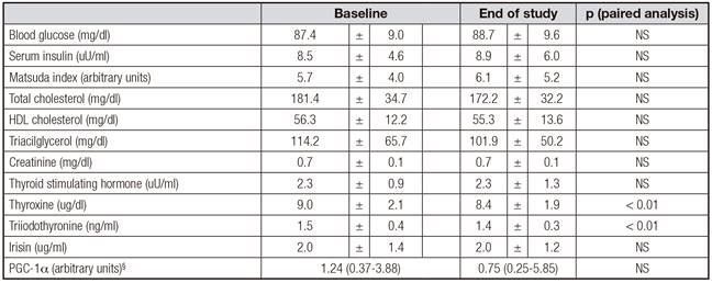
Five of the 47 women did not complete all the assessments at the end of the study, therefore data of 42 participants aged 34 ± 13 years with an initial body mass index of 27.7 ± 1.8 kg/m2 are presented. Table I shows weight, waist and hip circumferences, blood pressure, body composition, and energy expenditure of participants at baseline and at the end of the study. Significant reductions in weight, fat mass, fat free mass and resting energy expenditure were observed. Significant increases in AEE and PAL were also observed after the intervention. Table II shows laboratory values. There was no significant change in irisin levels or PGC1α expression in PBMC after the calorie restriction period. Decreases in total cholesterol, triiodothyronine and thyroxine were observed.

Table I Anthropometry, body composition and energy expenditure of participants 

Table II Laboratory values of participants 

§Expressed as median (interquartile range)

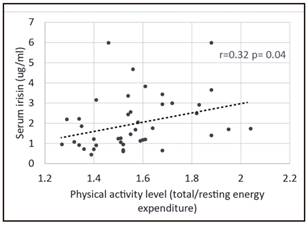
Baseline physical activity level had a positive albeit weak correlation with irisin concentration (Fig. 1). This association was not present at the end of the intervention. No association between irisin levels and other parameters was observed at baseline or at the end of the study, including body composition measures, resting energy expenditure, serum lipid levels, Matsuda index or PGC1α expression in PBMC.

Figure 1 Correlation between baseline irisin levels and physical activity level. 

DISCUSSION

After the restriction period, participants reduced their body weight and fat mass. Irisin levels were only weakly associated with the physical activity level of participants at baseline. However, we did not observe any change in irisin levels after the calorie restriction period even though subjects increased their physical activity and their body composition changed.

We measured body composition, energy expenditure and metabolic parameters using accurate and adequate methods. For body composition, we used DEXA, a method that in our hands has a measurement error of less than 3% 18. For total energy expenditure we used a combination of indirect calorimetry and actigraphy in which actigraphs were calibrated with the individual heart rate/energy expenditure curves obtained during an incremental exercise test. This method provides reliable measures of total energy expenditure when compared with wearable indirect calorimeters 19) (20. To assess insulin resistance we used the Matsuda index, which is the method with the best concordance with euglycemic insulin clamp 17.

Disappointingly, irisin was only weakly associated with baseline physical activity level, and this association was lost after the intervention. Despite the changes in fat and fat free mass achieved with calorie restriction, no change in irisin levels were observed. These results cast doubts about the real meaning of circulating irisin levels or the accuracy of the measurement methods used to determine this hormone. While Boström used Western Blot against a sequence of FNDC5 21, later studies used different ELISA kits to quantify Irisin. Albrecht and colleagues recently evaluated the three more commonly used antibodies in ELISA kits for irisin and found that they have cross-reactions with plasma proteins different from irisin. However, the antibody used by AdipoGen Labs (which we used) marked a protein that weights 25kDa, which is within the weight range of irisin. However, this group concluded that the proteins these kits identified were not irisin 22. These results have been recently challenged by other authors 23. Even more, a recent report showed that irisin is a true circulating hormone, when measured by tandem mass spectrometry 24. However, the concerns about the accuracy of ELISA kits to measure irisin still persist.

To date, only two published studies mention a possible relationship between habitual physical activity and plasma concentration of irisin 14) (15. However, the methods to measure physical activity used in these reports are less accurate than ours. One group used accelerometers without individual calibration, and the other only used questionnaires. What is usually reported is that training, specifically resistance training, increases irisin levels, and that this increase correlates with the buildup in muscle mass 25.

Unlike other reports, we did not observe a change in irisin levels after weight loss 26, with thyroid function tests 27 or insulin 28) (29. Unfortunately, we did not observe significant changes in insulin sensitivity at the end of the intervention period.

The upstream regulator of FNDC-5 cleaving and irisin liberation is PGC-1α 30) (31. Therefore, we measured the expression of PGC1α in mRNA of PBMCs by rtPCR. Again, we found no association of its expression with irisin. It must be born in mind however that we did not measure the expression of the molecule in muscle, where it should have the direct effect, and that we obtained a great dispersion of values. Again, the lack of relationship between the precursor and the hormone causes us to wonder about the real value of the hormone as a marker of PGC-1α activation 32.

In summary, we only found a weak association of irisin with usual physical activity in these women, and we seriously doubt about the real physiological role of the hormone in muscle physiology.

REFERENCES

1. Boström P, Wu J, Jedrychowski MP, Korde A, Ye L, Lo JC, et al. A PGC1-alpha-dependent myokine that drives brown-fat-like development of white fat and thermogenesis. Nature 2012;481:463-8. [ Links ]

2. Castillo-Quan JI. From white to brown fat through the PGC-1?-dependent myokine irisin: Implications for diabetes and obesity. Dis Model Mech 2012;5:293-5. [ Links ]

3. Liu J. Irisin as an exercise-stimulated hormone binding crosstalk between organs. Eur Rev Med Pharmacol Sci 2012;19:316-21. [ Links ]

4. Irving BA, Still CD, Argyropoulos G. Does IRISIN have a BRITE future as a therapeutic agent in humans? Curr Obes Rep 2014;3:235-41. [ Links ]

5. Vaughan RA, Gannon NP, Barberena MA, Garcia-Smith R, Bisoffi M, Mermier CM, et a. Characterization of the metabolic effects of irisin on skeletal muscle in vitro. Diabetes Obes Metab 2014;16:711-8. [ Links ]

6. Choi HY, Kim S, Park JW, Lee NS, Hwang SY, Huh JY, et al. Implication of circulating irisin levels with brown adipose tissue and sarcopenia in humans. J Clin Endocrinol Metab 2014;99:2778-85. [ Links ]

7. Chen JQ, Fang LJ, Song KX, Wang XC, Huang YY, Chai SY, et al. Serum irisin level is higher and related with insulin in acanthosis nigricans-related obesity. Exp Clin Endocrinol Diabetes 2015. [E-pub ahead of print] [ Links ]

8. Anastasilakis AD, Polyzos SA, Saridakis ZG, Kynigopoulos G, Skouvaklidou EC, Molyvas D, et al. Circulating irisin in healthy, young individuals: Day-night rhythm, effects of food intake and exercise, and associations with gender, physical activity, diet, and body composition. J Clin Endocrinol Metab 2014;99:3247-55. [ Links ]

9. Miyamoto-Mikami E, Sato K, Kurihara T, Hasegawa N, Fujie S, Fujita S, et al. Endurance training-induced increase in circulating irisin levels is associated with reduction of abdominal visceral fat in middle-aged and older adults. PLoS One 2015;10:e0120354. [ Links ]

10. Belviranli M, Okudan N, Çelik F. Association of circulating irisin with insulin resistance and oxidative stress in obese women. Horm Metab Res 2016. [E-pub ahead of print] [ Links ]

11. Gao S, Cheng Y, Zhao L, Chen Y, Liu Y. The relationships of irisin with bone mineral density and body composition in PCOS patients. Diabetes Metab Res Rev 2015. DOI: 10.1002/dmrr.2767. [E-pub ahead of print] [ Links ]

12. Norheim F, Langleite TM, Hjorth M, Holen T, Kielland A, Stadheim HK, et al. The effects of acute and chronic exercise on PGC-1?, irisin and browning of subcutaneous adipose tissue in humans. FEBS J 2014;281:739-49. [ Links ]

13. Löffler D, Müller U, Scheuermann K, Friebe D, Gesing J, Bielitz J, et al. Serum irisin levels are regulated by acute strenuous exercise. J Clin Endocrinol Metab 2015;100:1289-99. [ Links ]

14. Pardo M, Crujeiras AB, Amil M, Aguera Z, Jiménez-Murcia S, Baños R, et al. Association of irisin with fat mass, resting energy expenditure, and daily activity in conditions of extreme body mass index. Int J Endocrinol 2014;2014:857270. [ Links ]

15. Al-Daghri NM, Alokail MS, Rahman S, Amer OE, Al-Attas OS, Alfawaz H, et al. Habitual physical activity is associated with circulating irisin in healthy controls but not in subjects with diabetes mellitus type 2. Eur J Clin Invest 2015;45:775-81. [ Links ]

16. USDA. Dietary guidelines for Americans 2015-2020. 8th edition. 2015. Available at: https://health.gov/dietaryguidelines/2015/guidelines/Links ]

17. Matsuda M, DeFronzo RA. Insulin sensitivity indices obtained from oral glucose tolerance testing: Comparison with the euglycemic insulin clamp. Diabetes Care 1999;22:1462-70. [ Links ]

18. Gajardo H, Barrera G. Quality control of bone densitometry: Precision, reproducibility, and clinical application. Rev Med Chil 1998;126:56-62. [ Links ]

19. Crouter SE, Churilla JR, Bassett DR Jr. Accuracy of the Actiheart for the assessment of energy expenditure in adults. Eur J Clin Nutr 2008;62: 704-11. [ Links ]

20. Spierer DK, Hagins M, Rundle A, Pappas E. A comparison of energy expenditure estimates from the Actiheart and Actical physical activity monitors during low intensity activities, walking, and jogging. Eur J Appl Physiol 2011; 111:659-67. [ Links ]

21. Novelle MG, Contreras C, Romero-Picó A, López M, Diéguez C. Irisin, two years later. Int J Endocrinol 2013;2013:746281. [ Links ]

22. Albrecht E, Norheim F, Thiede B, Holen T, Ohashi T, Schering L, et al. Irisin - A myth rather than an exercise-inducible myokine. Sci Rep 2015;5:8889. DOI: 10.1038/srep08889 [ Links ]

23. Polyzos SA, Mantzoros CS. An update on the validity of irisin assays and the link between irisin and hepatic metabolism. Metabolism 2015;64:937-42. [ Links ]

24. Jedrychowski MP, Wrann CD, Paulo JA, Gerber KK, Szpyt J, Robinson MM, et al. Detection and quantitation of circulating human irisin by tandem mass spectrometry. Cell Metab 2015;22:734-40. [ Links ]

25. Kim H, Lee HJ, So B, Son JS, Yoon D, Song W. Effect of aerobic training and resistance training on circulating irisin level and their association with change of body composition in overweight/obese adults: A pilot study. Physiol Res 2015. [E-pub ahead of print] [ Links ]

26. Crujeiras AB, Pardo M, Arturo RR, Navas-Carretero S, Zulet MA, Martínez JA, et al. Longitudinal variation of circulating irisin after an energy restriction-induced weight loss and following weight regain in obese men and women. Am J Hum Biol 2014;26:198-207. [ Links ]

27. Ruchala M, Zybek A, Szczepanek-Parulska E. Serum irisin levels and thyroid function - Newly discovered association. Peptide 2014;60:51-5. [ Links ]

28. Assyov Y, Gateva A, Tsakova A, Kamenov Z. Irisin in the glucose continuum. Exp Clin Endocrinol Diabetes 2015. [E-pub ahead of print] [ Links ]

29. Duran ID, Gülçelik NE, Ünal M, Topçuoglu C, Sezer S, Tuna MM, et al. Irisin levels in the progression of diabetes in sedentary women. Clin Biochem 2015;48:1268-72. [ Links ]

30. Yang Z, Chen X, Chen Y, Zhao Q. PGC-1 mediates the regulation of metformin in muscle irisin expression and function. Am J Transl Res 2015;7:1850-9. [ Links ]

31. Huh JY, Panagiotou G, Mougios V, Brinkoetter M, Vamvini MT, Schneider BE, et al. FNDC5 and irisin in humans: I. Predictors of circulating concentrations in serum and plasma and II. mRNA expression and circulating concentrations in response to weight loss and exercise. Metabolism 2012;61:1725-38. [ Links ]

32. Pekkala S, Wiklund PK, Hulmi JJ, Ahtiainen JP, Horttanainen M, Pöllänen E, et al. Are skeletal muscle FNDC5 gene expression and irisin release regulated by exercise and related to health? J Physiol 2013;591:5393-400. [ Links ]

Received: August 12, 2016; Accepted: October 02, 2016

Correspondence: Daniel Bunout. INTA University of Chile. 138-11 Santiago, Chile e-mail: dbunout@inta.uchile.cl

Creative Commons License This is an open-access article distributed under the terms of the Creative Commons Attribution License (CC BY-NC-ND 4.0), which permits unrestricted use, distribution, and reproduction in any medium, provided the original work is properly cited.