


Mi SciELO
 Servicios personalizados
Servicios Personalizados
Revista
Articulo
 texto en
  Articulo en XML
 Referencias del artículo
 Enviar articulo por email
Indicadores
-
 Citado por SciELO
 -
 Accesos
Links relacionados
 Citado por Google
 -
 Similares en
SciELO
Similares en
SciELO  Similares en Google
Compartir

 Permalink
Archivos de la Sociedad Española de Oftalmología
versión impresa ISSN 0365-6691
Arch Soc Esp Oftalmol vol.79 no.10 oct. 2004
EDITORIAL
EMERGENCE OF SERRATIA MARCESCENS AS AN OCULAR
SURFACE PATHOGEN
APARICIÓN DE SERRATIA MARCESCENS COMO
UN PATÓGENO DE SUPERFICIE OCULAR
HUME E BH, WILLCOX M DP1
Serratia marcescens, formerly known as Bacillus prodigiosus, is an aerobic, gram negative, motile organism whose natural habitat is soil and water. S. marcescens has had an interesting history as some isolates produce prodigiosin, a red pigment. From as early as the 12th century, reports of the "blood of Christ" appearing on holy bread were documented. In the 19th century scientists discovered that this red blood-like appearance on starchy foodstuffs was due to a living organism, S. marcescens. With the development of biochemical identification techniques white type strains were identified, consequently there was an increase in the number of infections reported to be due to S. marcescens. S. marcescens is now a widely recognized opportunistic pathogen. It has caused a wide range of nosocomial infections including urinary tract infections, endocarditis, bacteremia, arthritis and osteomyelitis. The urinary tract is the most common source of isolates, consisting of 97% of all hospital isolates in one study.
Serratia marcescens has been implicated in endophthalmitis, keratoconjunctivitis, contact lens related keratitis, non contact lens related keratitis and contact lens induced acute red eye (CLARE).
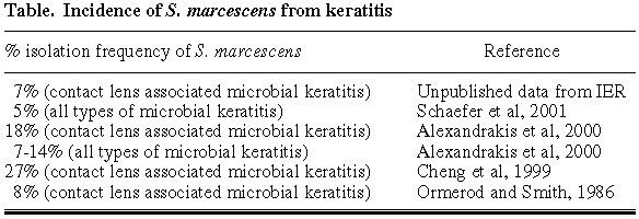
Ulcerative keratitis (also known as bacterial keratitis) is the most serious complication of contact lens wear. It is eighty times more likely to occur in contact lens wear than in non contact lens wear and is characterized by the loss of epithelium and stromal infiltration and may result in the loss of vision. Corneal rings associated with corneal ulceration have been determined to be due to an accumulation of PMNs. In contact lens associated microbial keratitis, extended wear use of lenses accounts for 79% of these cases. There is 8-15 times the likelihood of developing ulcerative keratitis in the overnight wearing of lenses as compared to the daily wear of contact lenses. The lowest incidence is associated with the use of hard or rigid lenses. Epithelial defects may be a predisposing factor for infection. P. aeruginosa has been repeatedly reported to be the most common pathogen implicated in contact lens related keratitis however a wide range of other gram negatives have been implicated as well as gram positive bacteria. Serratia marcescens has been reported as a causative organism in several studies. In one study about 8% of contact lens associated microbial keratitis incidents were due to S. marcescens. A study in 1999 reported that S. marcescens was the most common gram negative isolated from contact lens related microbial keratitis cases (table).
CLARE is only seen in extended lens wear, that is, the day and night wearing of contact lenses. It is one of the most common adverse responses associated with the extended wear of lenses. This inflammatory reaction occurs in 7% of extended lens wearers per annum, and is a major reason for lens discontinuation. CLARE typically occurs in the early hours of the morning whereby the patient wakes with acute pain, photophobia, tearing and redness of the conjunctiva, however there is no epithelial loss or ulceration. The cornea is infiltrated with white blood cells. Upon culturing of the contact lens, the lens yields confluent numbers of Gram-negative bacteria. Gram-negative carriers are 8 times as likely of having a CLARE response than a non gram negative carrier. S. marcescens has previously been implicated in CLARE and accounts for 24% of isolates in studies conducted at the Institute for Eye Research.
The source of bacterial contamination of contact lenses, in particular by Serratia marcescens has not been elucidated. The patients' hygiene regimen is a possible source of corneal contamination. Contact lens solutions (preserved and unpreserved) have been found to be contaminated with bacteria. Out of sixty-three lens soaking solutions, forty-seven were contaminated (73%), twenty-two of which were contaminated with Pseudomonas sp., twenty with Serratia sp. and fifteen with Klebsiella sp. Other contaminants included Acinetobacter sp., Proteus sp., Moraxella sp. and Neisseria sp. Contamination by S. marcescens occurred most frequently in preserved solutions and contamination by P. aeruginosa in home made saline. S. marcescens and S. liquefaciens (the latter now classified as Serratia marcescens) have been found to persist in a variety of disinfecting solutions. S. marcescens has the ability to adapt to and grow in disinfecting solutions containing chlorhexidine. Additionally, S. marcescens can survive in chlorhexidine hand washes in concentrations up to 20 mg/mL. This concentration of chlorhexidine is much higher than in lens disinfecting solutions. It has also been shown that when grown in a biofilm, S. marcescens is more resistant to lens disinfectants than both P. aeruginosa and Staphylococcus epidermidis. In a more recent study with newer disinfecting solutions, S. marcescens was found to be killed after six hours in the majority of solutions, although this sounds promising, this study was only carried out with one strain so may not be indicative of the efficacy lens solutions against S. marcescens as a whole.
Contact lens cases have also been found to be a favorable environment for bacterial contamination. S. marcescens has been found in 31% of storage cases and in high numbers where chlorhexidine was used as a disinfectant.
Although solutions and cases are known to harbor bacteria, some extended wear patients wear disposable lenses, so they do not use contact lens storage cases or solutions. Occasionally they may use saline to wet or clean lenses however, in general, these are unlikely to be sources of contamination in the disposable extended lens wearer. Body sites, in particular the fingertips, are more likely to be a source of contamination as they are often used to manipulate the lens. The ability of bacteria to persist on the hands is a concern for extended lens wearers. Finger and hand contamination in hospital infections is well known. Studies have shown that handling of contact lenses is a predominant source of lens contamination. However, these studies have not typed strains and so have not proved that the same bacteria on the fingers are the same as those on the lens.
One of the most common environmental sources that the contact lens wearer may be exposed to is domestic water (from showering or washing), which may also contribute to the contamination of contact lenses by pathogenic bacteria (such as S. marcescens, S. liquefaciens, P. aeruginosa, Aeromonas hydrophilia and Stenotrophomonas maltophilia). These strains that have been implicated more than once in adverse reactions to contact lens wear and are commonly found in water supplies.
In addition to being resistant to contact lens solutions, Serratia has been shown to be resistant to many antibiotics. This poses a severe problem considering the increasing emergence of keratitis events. S. marcescens have three outer membrane porins similar to Escherichia coli suggesting that the chromosomal beta-lactamase of S. marcescens plays a role in the intrinsic resistance to beta-lactam antibiotics.
In summary, it appears that Serratia marcescens is an important pathogen in ulcerative keratitis associated with contact lens wear. It has a relatively high incidence of resistance to disinfectants and other antimicrobial agents. This is especially worrying as it indicates that corneal infections by S. marcescens may be very difficult to control and treat effectively.
1 PhD. Director of Science, Vision CRC. Professor, School of Optometry and Vision Science. University of New South Wales. Australia.
E-mail: M.Willcox@visioncrc.org
REFERENCES
- Alexandrakis G, Alfonso EC, Miller D. Shifting trends in bacterial keratitis in south Florida and emerging resistance to fluoroquinolones. Ophthalmology 2000; 107: 1497-1502.
- Cheng KH, Leung SL, Hoekman HW, Beekhuis WH, Mulder PG, Geerards AJ et al. Incidence of contact-lens-associated microbial keratitis and its related morbidity. Lancet 1999; 354: 181-185.
- Schaefer F, Bruttin O, Zografos L, Guex-Crosier Y. Bacterial keratitis: a prospective clinical and microbiological study. Br J Ophthalmol 2001; 85: 842-847.
- Ormerod LD, Smith RE. Contact lens-associated microbial keratitis. Arch Ophthalmol 1986; 104: 79-83.

