


Mi SciELO
 Servicios personalizados
Servicios Personalizados
Revista
Articulo
 texto en
  Inglés (pdf)
 Articulo en XML
 Referencias del artículo
 Enviar articulo por email
Indicadores
-
 Citado por SciELO
 -
 Accesos
Links relacionados
 Citado por Google
 -
 Similares en
SciELO
Similares en
SciELO  Similares en Google
Compartir

 Permalink
Revista Española de Enfermedades Digestivas
versión impresa ISSN 1130-0108
Rev. esp. enferm. dig. vol.96 no.6 Madrid jun. 2004
| ORIGINAL PAPERS |
Diagnosis of Helicobacter pylori infection in patients with bleeding ulcer disease:
rapid urease test and histology
M. Castro-Fernández, D. Sánchez-Muñoz, E. García-Díaz, J. Miralles-Sanchiz1 and J. Vargas-Romero2
Services of Digestive Diseases, 1Pathology and 2Microbiology. Valme University Hospital. Seville. Spain.
ABSTRACT
Introduction: the endoscopic diagnosis of Helicobacter pylori infection in patients with bleeding peptic ulcer is limited by a decreased sensitivity in standard invasive tests, rapid urease test and histology. There is controversy about the convenience of using one, neither, or both diagnostic tests.
Aims: to evaluate the results of simultaneously performed rapid urease test and histology in the diagnosis of Helicobacter pylori infection (H. pylori) in patients with bleeding peptic ulcer.
Patients and methods: we included 173 patients, 98 male and 75 female, with an average age of 62 years (18-88), with upper gastrointestinal bleeding secondary to duodenal ulcer (115) or gastric ulcer (58), diagnosed within 24 hours after hospital admission. None of the patients had received treatment for H. pylori, proton pump inhibitors or antibiotics in the two weeks prior to the upper gastrointestinal bleeding episode. H. pylori infection was investigated in all patients by two antral biopsy samples for histological study (hematoxilin-eosin) and one or two antral biopsies for rapid urease test (Jatrox®-H.p.-test). In cases with a negative urease test and histology, a 13C urea breath test was performed. Infection was considered present when at least one invasive test or the breath test was positive, whereas both invasive tests and the breath test had to be negative to establish an absent infection.
Results: 152 patients (88%) showed H. pylori infection, 104 patients (90%) with duodenal ulcer and 48 patients (83%) with gastric ulcer. In all 119 cases (78%) were diagnosed by the urease test and 112 cases (74%) by histology. Both methods were used to diagnose 134 of 152 cases (88%) (p < 0.05), these being positive in 97 cases and negative in 39 cases. In 18 of these 39 cases, the breath test was positive.
Conclusions: histology and urease test have similar diagnostic values for the identification of H. pylori in patients with bleeding peptic ulcer. Due to its rapid results, the urease test should be the method of choice. However, additional biopsies should be performed, and, when negative, a histological study should be carried out, since a combination of both methods allows a more precise diagnosis.
Key words: Helicobacter pylori. Urease test. Histology. Diagnosis of H. pylori. Upper gastrointestinal bleeding.
Castro-Fernández M, Sánchez-Muñoz D, García-Díaz E, Miralles Sanchiz J and Vargas-Romero J. Diagnosis of Helicobacter pylori infection in patients with bleeding ulcer disease: rapid urease test and histology. Rev Esp Enferm Dig 2003; 96: 395-401.
Recibido: 15-09-03.
Aceptado: 23-12-03.
Correspondencia: Manuel Castro Fernández. Servicio de Aparato Digestivo (9ª planta). Hospital Universitario de Valme. Carretera de Cádiz, s/n. 41014 Sevilla.
e-mail: mcastrof@meditex.es
INTRODUCTION
Helicobacter pylori (H. pylori) infection is the most frequent cause of peptic ulcer disease. The prevalence of this infection in duodenal and gastric ulcers is 90-95% and 80-85%, respectively (1,2). Patients with peptic ulcer disease can develop severe complications, such as bleeding or perforation. Thus, it is necessary to reach a precise diagnosis of H. pylori infection, as its eradication reduces ulcer recurrence and complications considerably (3-5). Today, there are several diagnostic methods available to detect H. pylori infection. These include invasive methods, which require endoscopy and gastric biopsies (rapid urease test, histology, and culture) and non-invasive methods (breath test with 13C-labelled urea, stool antigen test, and serology). They all have advantages and disadvantages in terms of availability, rapidity of results, costs, diagnostic precision, etc. (6-9). Invasive methods, especially the rapid urease test, are found to have low diagnostic sensitivity in patients with bleeding peptic ulcer. It has been suggested that the way to proceed in these cases is either to obtain biopsies only for histology or to use a non-invasive diagnostic method (6,7,10-14). The causes for decreased diagnostic sensitivity of invasive methods in cases with upper gastrointestinal bleeding are still unclear, and which diagnostic method should be used in these cases has not been established either.
The aim of this study was to analyze the diagnostic value of rapid urease tests and histology in patients with bleeding peptic ulcer.
PATIENTS AND METHODS
Patients
We included 173 patients, 98 male and 75 female, aged 62 (18-88) years on average, with upper gastrointestinal bleeding secondary to duodenal ulcer (115 cases) or gastric ulcer (58 cases), as diagnosed by an endoscopy performed within the first twenty-four hours after hospital admission. None of the patients showed either active bleeding or red blood in the stomach or duodenum. All patients had received intravenous omeprazole (40-160 mg) for less than 24 hours before endoscopy. Patients had not received treatment for H. pylori, proton pump inhibitors (PPI), or antibiotics in the two weeks prior to the upper gastrointestinal bleeding.
Diagnostic methods
H. pylori infection was investigated in all patients by antral biopsy samples, two of these for histological study (hematoxilin-eosin), and one or two for rapid urease test (Jatrox®-H.p.-Test). In cases with gastric ulcer, biopsies were taken from the lesion to confirm its benign etiology. The rapid urease test was considered to be positive when a color change (from yellow to red) occurred within 24 hours after introducing the sample into a cuvette containing the reactive and 0.5 ml of distilled water. The histological study was performed without previous knowledge of the rapid urease test results. The histological diagnosis was considered to be positive when bacterial forms mor-phologically compatible with H. pylori were detected on the glycocalix of the surface epithelium, together with the standard secondary inflammatory changes.
In cases with negative results for these two invasive methods, a 13C urea breath test was performed (TAUKIT- Isomed Farmacéutica. Madrid) 6 to 8 weeks after endoscopy, following a minimum of 2 weeks without PPI consumption according to the manufacturer's instructions. Samples were analyzed using a mass spectrophotometer. Tests were considered positive when the increased value of 13C (the difference between a baseline sample and another one taken after 30 minutes) was greater than 5 delta units (>5 ‰).
Criteria for H. pylori infection
Patients were considered to have H. pylori infection when there were positive results in, at least, one of the two invasive tests performed -rapid urease test, histological examination with hematoxilin-eosin- or in the 13C urea breath test. H. pylori infection was excluded when both invasive tests and 13C urea breath test were negative.
Statistical study
Data were studied by means of an analysis of the statistical significance of percentage differences found when comparing the results of the different diagnostic methods evaluated, applying the Chi square, Fisher's exact, and McNemar tests. We considered the results to be statistically significant when p <0.05. Confidence intervals were calculated at 95%.
RESULTS
In all, 152 of 173 patients (88%) showed H. pylori infection criteria, 104 (90%) with duodenal ulcer, and 48 (83%) with gastric ulcer. The results obtained with the different diagnostic methods are detailed in table I.
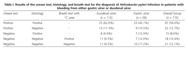
The rapid urease test was positive in 119 of 152 cases with infection criteria (78%); histology was positive in 112 cases (74%), and differences showed no statistical significance.
In addition, 134 of 152 cases (88%) were accurately diagnosed by associating the results of the two invasive diagnostic methods. We found statistically significant differences (p <0.05) when comparing this result with those of the two diagnostic techniques, separately.
DISCUSSION
The prevalence of H. pylori infection in populations with gastro-duodenal ulcer is very high. In gastric and duodenal ulcers, prevalence of 60-100% and 90-100%, respectively, have been reported (1,2). These were slightly lower or similar in bleeding ulcer disease studies when patients taking non-steroidal anti-inflammatory drugs (NSAIDs) were excluded (15). In our study there was a 90.4 and 82.7% prevalence of infection in patients with duodenal ulcer and gastric ulcer, respectively. Despite this high prevalence, it is not advisable to assume that all patients with gastro-duodenal ulcer, even when they are not taking NSAIDs, are infected by H. pylori. Due to its simplicity, precision and rapid results, the rapid urease test is considered the invasive me-thod of choice in patients requiring endoscopy (16,17), even despite the fact that, as is the case with other invasive methods, it exhibits a lower diagnostic sensitivity in cases of bleeding ulcer disease (6,11,13,18,19). Our group considered this problem in a previous report, in which patients with non-complicated gastroduodenal ulcer and patients with upper gastroduodenal bleeding were included (20). In these instances it has been recommended that only histology-rather than the urease test-should be performed as loss of sensitivity will be lower (21-23). The other alternative is to perform a non-invasive-rather than an invasive-test (i. e. a 13C urea breath test or stool antigen test) after upper gastrointestinal bleeding has ceased (7,10). Serology is not advisable (24). As for histology, the various factors which affect the accurate identification of bacteria include: type of dye used, experience of the observer, amount and quality of sample referred for examination, existence of previous treatments, density of bacterial colonies, and presence of extensive intestinal metaplasia. The causes for decreased diagnostic sensitivity of the urease test in bleeding gastro-duodenal ulcer are still controversial. Potential causes suggested for this lower sensitivity include the presence of blood in the stomach, which could lead to a transient clearance of bacterial density as a result either of a bactericide effect of serum (7,25), or bacteria proximal migration in the stomach due to intragastric pH changes, leading to a decrease in antral bacterial density; the possibility that albumin in blood serum may act as buffer on the pH indicator used in the urease test, which could prevent color change (26); or that frequent use of PPI in these patients may result in a reduction of bacterial load in the mucosa (6,27). Some of these factors could also be involved in the decreased sensitivity of histological studies. Nevertheless, the presence of both urease test and histology false negative results may share a common mechanism; our patients were under the effects of intravenous omeprazole, although for a short period of time, at the time of endoscopy, but they were not exposed to the presence of gastroduodenal blood, however.
In our study we found that no method is better than the rest in terms of their capability to detect H. pylori (78 vs 74%). However, as we expected, when both methods (urease test and histology) are simultaneously used, diagnostic value increases (88 vs 74-78%; p<0.05).
Due to its simplicity and rapidity of results, the rapid urease test is, in our opinion, the invasive method of choice. However, although this test shows a high false negative results rate when upper gastrointestinal bleeding occurs, additional biopsies should be obtained for a histological study, thus improving diagnostic yield (28). The possibility of an increase in sensitivity for both diagnostic methods when simultaneous antral and fundic biopsy samples are taken is likely. A positive result in either of these methods would be sufficient to reach a diagnosis, but when they are both negative-even considering the limitations in diagnostic sensitivity invasive methods exhibit when hemorrhagic complications are there-infection should be ruled out by using other non-invasive methods (breath test or stool antigen test) once the acute phase of the hemorrhagic process has been overcome.
Despite all this, further studies are still mandatory both to determine which diagnostic method is the most reliable for detecting H. pylori in patients with bleeding peptic ulcer, and to establish the appropriate timing in studying these patients' infections.
REFERENCES
1. Kuipers EJ, Thijs JC, Festen HP. The prevalence of Helicobacter pylori in peptic ulcer disease. Aliment Pharmacol Ther 1995; 9 (Suppl. 2): 59-69. [ Links ]
2. Gisbert JP, Boixeda D, Aller R, de la Serna C, Sanz E, Martín de Argila C, et al. Helicobacter pylori and digestive hemorrhage due to duodenal ulcer: the prevalence of the infection, the efficacy of 3 triple therapies and the role or eradication in preventing a hemorrhagic recurrence. Med Clin (Barc.) 1999; 112: 161-5. [ Links ]
3. Barhel JS. Bleeding ulcers and Helicobacter pylori. Gastrointest Endosc 1997; 46: 371-3. [ Links ]
4. Santander C, Gravalos RG, Gómez-Cedenilla A, Canter J, Pajares JM. Antimicrobial therapy for helicobacter pylori infection versus long-term maintenance antisecretion treatment in the prevention of recurrent hemorrhage from peptic ulcer: prospective randomized trial on 125 patients. Am J Gastroenterol 1996; 91: 1549-52. [ Links ]
5. Riemann JF, Schilling D, Schauwecker P, Wehlen G, Dorlars D, Kohler B. Cure with omeprazole plus amoxicillin versus long-term ranitidine therapy in Helicobacter pylori associated peptic ulcer bleeding. Gastrointest Endosc 1997; 46: 299-304. [ Links ]
6. Gisbert JP. A critical review of the diagnostic methods for Helicobacter pylori infection. Gastroenterol Hepatol 2000; 23: 135-43. [ Links ]
7. Martín de Argila C, Boixeda D. Practical considerations for the diagnosis of Helicobacter pylori infection. Med Clin (Barc) 2001; 117: 386-91. [ Links ]
8. Megraud F. Advantages and disadvantages of current diagnostic test for the detection of Helicobacter pylori. Scand J Gastroenterol 1996; 31 (Suppl. 215): 57-62. [ Links ]
9. De Boer WA, De Laat L, Megraud F. Diagnosis of Helicobacter pylori infection. Current Opinión in Gastroenterology 2000; 16 (Suppl. 1): S5-S10. [ Links ]
10. Sainz R, Borda F, Domínguez E, Gisbert JP. Grupo Conferencia Española de Consenso. Conferencia Española de Consenso sobre la infección por Helicobacter pylori. Rev Esp Enferm Digest 1999; 91: 777-84. [ Links ]
11. Colin R, Czernichow P, Baty V, Tozue I, Brazier F, Bretagne JF, et al. Low sensitivity of invasive tests for the detection of Helicobacter pylori infection in patients with bleeding ulcer. Gastroenterol Clin Biol 2000; 24: 31-5. [ Links ]
12. Howden CW, Hunt RH. Guidelines for the management of Helicobacter pylori infection. Am J Gastroenterol 1998; 93: 2330-8. [ Links ]
13. Tu T-C, Lee C-L. Wu C-H, Chen T-K, Chan C-C, Huang S-H, et al. Comparison of invasive and noninvasive tests for detecting Helicobacter pylori infection in bleeding peptic ulcers. Gastrointest Endosc 1999; 49: 302-6. [ Links ]
14. Lee JM, Breslin NP, Fallon C, O'Morain CA. Rapid urease test lack sensitivity in Helicobacter pylori diagnosis when peptic ulcer disease presents with bleeding. Am J Gastroenterol 2000; 95: 1166-70. [ Links ]
15. Gisbert JP, González L, de Pedro A, Valvuena M, Prieto B, Llorca I, et al. Helicobacter pylori and bleeding duodenal ulcer: prevalence of the infection and role of non-steroidal anti-inflammatoy drugs. Scand J Gastroenterol 2001; 36: 717-24. [ Links ]
16. Laine L, Cohen H. Helicobacter pylori: drowning in a pool of blood? Gastrointest Endosc 1999; 49: 398-402. [ Links ]
17. Lam SK, Talley NJ. Helicobacter pylori. Consensus Report of the 1997 Asia Pacific Consensus Conference on the management. J Gastroenterol Hepatol 1998; 13: 1-12. [ Links ]
18. Grino P, Pascual S, Such J, Casellas JA, Niveiro M, Andreu M et al. Comparison of diagnostic methods for Helicobacter pylori infection in patients with upper gastrointestinal bleeding. Scand J Gastroenterol 2001; 36: 1254-8. [ Links ]
19. Chung IK, Hong SJ, Kim EJ, Cho JY, Kim HS, Park SH et al. What is the best method to diagnose Helicobacter infection in bleeding peptic ulcers? A prospective trial. Korean J Intern Med 2001; 16: 147-52. [ Links ]
20. Romero-Gómez M, Vargas J, Utrilla D, Rufo MC, Otero MA, Chavez M et al. Estudio prospectivo sobre la influencia de la hemorragia por ulcus gastroduodenal en los métodos diagnósticos de infección por Helicobacter pylori. Gastroenterol Hepatol 1998; 21: 267-71. [ Links ]
21. Castillo-Rojas G, Ballesteros MA, Ponce de León S, Morales-Espinosa R, Cravioto A, López-Vidal I. Bleeding peptic ulcers and presence of Helicobacter pylori by various tests: a case-control study. Eur J Gastroenterol Hepatol 2002; 14: 1113-8. [ Links ]
22. Archimandritis A, Tzivras M, Sougioultzis S, Papaparaskevas I, Apostolopoulos P, Aviami A et al. Rapid urease test is less sensitive than histology in diagnosing Helicobacter pylori infection in patients with non-variceal upper gastrointestinal bleeding. J gastroenterol Hepatol 2000; 15: 369-73. [ Links ]
23. Calvet X,Gisbert JP. Prevención de la recidiva hemorrágica por úlcera péptica en pacientes infectados por Helicobacter pylori. GH Continuada 2002; 1: 48-51. [ Links ]
24. García-Díaz E, Castro-Fernández M, Romero-Gómez M, Vargas-Romero J. The effectiveness of (IgG-ELISA) serology as an alternative diagnostic method for detecting Helicobacter pylori infection in patients with gastro-intestinal bleeding due to gastro-duodenal ulcer. Rev Esp Enferm Dig 2002; 94: 731-6. [ Links ]
25. Houghton J, Ramamoorthy R, Pandya H, Dhirmalani R, Kim KD. Human plasma is directly bacteriocidal against Helicobacter pylori in vitro, potentially explaining the decreased detection of Helicobacter pylori during acute upper gastrointestinal bleeding. Gastrointest Endosc 2002; 55: 11-16. [ Links ]
26. Leung WK, Sung JJ, Siu KL, Chan FK, Ling TK, Cheng AF. False-negative biopsy urease test in bleeding ulcers caused by the buffering effects of blood. Am J Gastroenterol 1999; 94: 1421-2. [ Links ]
27. Graham DY, Opekun AR, Hammond F, Yamaoka Y, Reddy R, Osato El-Zimaity HM. Studies regarding the mechanism of false negative urea breath test with proton pump inhibitors. Am J Gastroenterol 2003; 98: 1005-9. [ Links ]
28. Schilling D, Demel A, Adamek HE, Nusse T, Weidmann E, Riemann JF. A negative rapid urease test is unrliable for exclusion of Helicobacter pylori infection during acute phase of ulcer bleding. A prospective case control study. Dig Liver Dis 2003; 35: 215-6. [ Links ]

