


Mi SciELO
 Servicios personalizados
Servicios Personalizados
Revista
Articulo
 Inglés (pdf)
 Articulo en XML
 Referencias del artículo
 Enviar articulo por email
Indicadores
-
 Citado por SciELO
 -
 Accesos
Links relacionados
-
 Citado por Google
 -
 Similares en
SciELO
Similares en
SciELO -
 Similares en Google
Compartir

 Permalink
Revista Española de Enfermedades Digestivas
versión impresa ISSN 1130-0108
Rev. esp. enferm. dig. vol.101 no.7 Madrid jul. 2009
CARTAS AL DIRECTOR
Incidental pseudomyxoma peritonei of the appendix diagnosed by computed tomography with sagittal reconstruction in a woman suffering from postmenopausal metrorrhagia
Seudomixoma peritoneal incidental del apéndice, diagnosticado por tomografía axial computerizada con reconstrucción sagital, en pacientes con metrorragia postmenopáusica
Key words: Computed tomography. Pseudomyxoma peritonei.
Dear Editor,
An incidental finding is something found that is unrelated to the present illness. Pseudomyxoma peritonei resulting from the appendix is a rare entity. We report an unusual case in which symptoms could not have been associated with a mucocele. Findings in the computed tomography with sagittal reconstruction were characteristics to determine its aetiology and management.
Case report and discussion
A 64-year old female patient, with no underlying diseases, was examined by a gynaecologist physician, presenting a postmenopausal metrorrhagia for one month. The oral tolerance and bowel habit remained normal in a basis daily. Neither previous traumas nor surgical or invasive studies were recorded by patient. Physical examination revealed well condition, no fever, and hemodynamic stability. His neurological, cardiological, and pulmonary examinations were normal. No lymph nodes were found. Only a suspicion of a right anexial mass was detected in abdominal evaluation. Then, an ultrasonography were ordered by Gynaecologist, showing a septated ascites mainly in pelvic cavity, in conjunction with an irregular cystic mass measuring 35 x 33 mm, and leading to suspicion of right ovary. Electrocardiogram, hemogram, white blood cell count, blood coagulation studies, blood glucose levels, ionogram, and routine kidney and liver function blood tests resulted normal. Changes in serum tumour marker level of CEA and CA 125 were not detected. The chest X-ray was normal.
Ambulatory diagnostic hysteroscopy was performed, and endometrial biopsies resulted in atrophic changes.
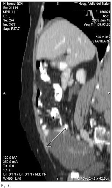
Ambulatory computed tomography (CT) of the abdomen and pelvis demonstrated the presence of non-septated ascites, associated with a cystic mass (Fig. 1), which measuring 62 x 36 mm in diameter in right pelvic compartment (Fig. 2). Appendiceal aetiology was suggested based on its location and position, its ovoid morphology, and the presence of a calcified and enhancing wall. The CT with sagittal reconstruction demonstrated that cystic mass arose from the cecum of the large colon (Fig. 3: arrow indicates the appendiceal base). All these findings together resulted to be characteristic to consider appendiceal mucocele associating pseudomyxoma peritonei (1,2). No metastases were detected.


Then, surgical approach was indicated. The specimen was microscopically studied, and a mucinous cystadenocarcinoma of the appendix associating pseudomyxoma peritonei was diagnosed, based on the amounts of mucinous ascites into which a fewer number of mucinous colonic cells with marked nuclear atypia and pleomorphism were presented. These findings are considered as a poor prognostic factor in the disease (3).
The hospital course was uneventful. The patient is free of symptoms after discharged from the hospital, waiting for chemotherapy.
We postulated that symptoms and surgical findings did not show any correlation, findings in the computed tomography were characteristics to determine its aetiology and management, so, adenocarcinoma of the appendix resulted to be an incidental finding (4,5).
The use of different investigation techniques permitted us to look at several different aspects of the same phenomena.
The authors would like to thank Department of Surgery’s members for the useful help and interesting suggestions concerning the medical and technical part of the work.
Hospital Valle del Nalón (SPH) has provided Internet services, and electronic and print books and journals, and allowed us to print the manuscripts, and email service.
S. Pérez-Holanda, J. García-Lozano1 and L. Rodrigo Sáez2
Departments of General Surgery and 1Diagnostic Radiology. Hospital Valle del Nalón. Langreo, Asturias. Spain.
2School of Medicine. University of Oviedo. Asturias, Spain
References
1. Higa E, Rosai J, Pizzimbono CA, Wise L. Mucosal hyperplasia, mucinous cystadenoma, and mucinous cystadenocarcinoma of the appendix. A re-evaluation of appendiceal "mucocele". Cancer 1973; 32: 1525-41. [ Links ]
2. Andersson A, Bergdahl L, Boquist L. Primary carcinoma of the appendix. Ann Surg 1976; 183: 53-7. [ Links ]
3. Landen S, Bertrand C, Maddern GJ, Herman D, Pourbaix A, de Neve A, et al. Appendiceal mucoceles and pseudomixoma peritonei. Surg Gynecol Obstet 1992; 175: 401-4. [ Links ]
4. Lo NS, Sarr MG. Mucinous cystadenocarcinoma of the appendix. The controversy persists: a review. Hepatogastroenterology 2003; 50: 432-7. [ Links ]
5. Rivera Vaquerizo PA, Albaladejo Ortiz C, Blasco Colmenarejo M, Vicente Gutiérrez M, Mayor López J, Pérez-Flores R. Appendiceal mucinous cystadenocarcinoma: endoscopic diagnosis. Rev Esp Enferm Dig 2005; 97: 762-3. [ Links ]

