


Mi SciELO
 Servicios personalizados
Servicios Personalizados
Revista
Articulo
 texto en
  Inglés (pdf)
 Articulo en XML
 Referencias del artículo
 Enviar articulo por email
Indicadores
-
 Citado por SciELO
 -
 Accesos
Links relacionados
-
 Citado por Google
 -
 Similares en
SciELO
Similares en
SciELO -
 Similares en Google
Compartir

 Permalink
Revista Española de Enfermedades Digestivas
versión impresa ISSN 1130-0108
Rev. esp. enferm. dig. vol.102 no.1 Madrid ene. 2010
PICTURES IN DIGESTIVE PATHOLOGY
A foreign body in the esophagus
Cuerpo extraño en esófago
M. J. Bosque-López1, A. Llompart-Rigo1 and P. de-Miguel-Sebastián2
1Servicio de Aparato Digestivo. Hospital Son Dureta. Palma de Mallorca, Spain.
2Servicio de Radiología. Clínica Rotger. Palma de Mallorca, Spain
Case report
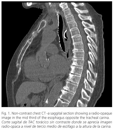
This is an 82 years woman with a history of hypertension, type-2 diabetes mellitus, chronic atrial fibrillation, aortic stenosis, and ischemic heart disease who presented with mild cognitive impairment and needed help with her personal hygiene and dressing. She was brought to the emergency room for sudden chest pain accompanied by dyspnea while eating, which was deemed a probable broncho-aspiration event. ECG, blood tests, and chest radiographs were normal, but the patient was admitted to hospital because of persisting symptoms. The second day after admission, a chest CT showed a radio-opaque foreign body 2.4 x 2.2 cm in diameter, located in the esophagus at the level of the tracheal carina, and that seemed affixed to the back wall of the esophagus. In addition, there was a concentric thickening of the esophageal wall in relation to this foreign body, which made endoscopy needed. Endoscopy showed an impacted foreign body in the mid third of the esophagus that was revealed to be a clam shell encrusted in the esophageal wall with a deep decubitus ulcer (Figs. 1-3).


Discussion
The interest of this case lies in its clinical presentation and its importance in that the patient was an elderly at risk for foreign body ingestion. The presence of a foreign body in the esophagus can lead to serious complications (bleeding, perforation, aspiration, pneumomediastinum, mediastinitis), hence extraction or disimpactation is urgent as the risk increases with delayed extraction. The diagnosis was reached using three-dimensional images obtained from CT.
Recommended references
1. American Society for Gastrointestinal Endoscopy. Guideline for the management of ingested foreign bodies. Gastrointest Endosc 1995; 42: 622-5. [ Links ]
2. Llompart A, Reyes J, Ginard D, Barranco L, Riera J, Gayà J, et al. Abordaje endoscópico de los cuerpos extraños esofágicos. Resultados de una serie retrospectiva de 501 casos. Gastroenterol Hepatol 2002; 25(7): 448-51. [ Links ]

