


Mi SciELO
 Servicios personalizados
Servicios Personalizados
Revista
Articulo
 texto en
  Inglés (pdf)
 Articulo en XML
 Referencias del artículo
 Enviar articulo por email
Indicadores
-
 Citado por SciELO
 -
 Accesos
Links relacionados
-
 Citado por Google
 -
 Similares en
SciELO
Similares en
SciELO -
 Similares en Google
Compartir

 Permalink
Revista Española de Enfermedades Digestivas
versión impresa ISSN 1130-0108
Rev. esp. enferm. dig. vol.102 no.1 Madrid ene. 2010
PICTURES IN DIGESTIVE PATHOLOGY
Rectal polyp as presentation form of colitis cystica profunda
Pólipo rectal como forma de presentación de colitis quística profunda
R. Baltar-Arias1, J. L. Ulla-Rocha1, E. Moreno-López2, E. Fernández-Salgado1, S. Vázquez-Rodríguez1, W. Díaz-Saa1, V. Carrera-González1 and E. Vázquez-Astray1
Departments of 1Gastroenterology, and 2Anesthesiology. Complexo Hospitalario de Pontevedra. Spain
Case report
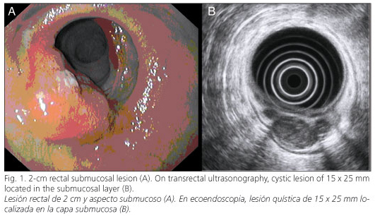
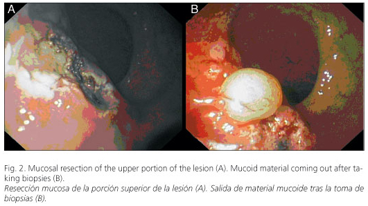
A 63-year-old woman complained of rectal bleeding of hemorroidal type and spontaneous emission of mucus in the feces for 2 months. Laboratory tests, tumoral markers included, were normal. A colonoscopy was performed revealing a 2-cm polypoid lesion (Fig. 1A) with a mucoid and gelatinous material (Fig. 2) coming out of it. Cytology and biopsies were inconclusive. An endoscopic ultrasonogram showed a cystic anechoic 20-mm lesion filled with some non-solid materials located in the submucosal layer of the rectal wall (Fig. 1B), without adjacent adenopathies, and a hypertonic internal anal sphincter. Histology showed mucosal thickening with dilated glands and the presence of inflammatory infiltrates, muscular fibers, and congestive vessels in the muscularis propria, findings that can be seen in rectal prolapse. These results, in addition to endoscopic and ultrasonographic findings, established the diagnosis.
Discussion
Colitis cystica profunda is a rare and benign condition characterized by mucoid cysts in the submucosal layer of the colon (1), mainly in the anterior rectal wall. It is associated with rectal prolapse, isolated rectal ulcer, inflammatory bowel disease, and local trauma (2). Typical symptoms are hematochezia, mucus in the feces, and diarrhea (3). The diagnosis is based on endoscopy, computer tomography, and magnetic resonance imaging. Colonoscopy shows polypoid or sessile lesions covered with normal, edematous or congestive mucosa, or ulcerated areas (4). On transrectal ultrasonography multiple submucosal cysts with hyperechoic bands corresponding to fibrosal areas among them can be seen, without nodal or muscular layer involvement (5). Treatment is conservative (3), except for highly symptomatic cases where surgery is mandatory.
References
1. Kornprat P, Langner C, Pfeifer J, Mischinger HJ. Colitis cystica profunda associated with rectal prolapse: report of a case. Int J Colorectal Dis 2007; 22 (12): 1555-6. [ Links ]
2. De Toro G, Villaseca M, Roa JC. Colitis quística profunda posradioterapia. Caso clínico. Rev Med Chile 2007; 135: 759-63. [ Links ]
3. Higuera Álvarez R, De la Peña García J, San Miguel G, Castro B. Colitis cystica profunda. Rev Esp Enferm Dig 2008; 100: 240-2. [ Links ]
4. Hulsmans FJH, Lok Tio T, Reeders JWAJ, Tytgat GNJ. Transrectal US in the diagnosis of localized colitis cystica profunda. Radiology 1991; 181: 201-3. [ Links ]
5. Valenzuela M, Martín-Ruiz JL, Álvarez-Cienfuegos E, Caballero AM, Gallego F, Carmona I, et al. Colitis cystica profunda: imaging diagnosis and conservative treatment. Report of two cases. Dis Colon Rectum 1996; 39: 587-90. [ Links ]

