


My SciELO
 Custom services
Services on Demand
Journal
Article
 text in
  English (pdf)
 Article in xml format
 Article references
 Send this article by e-mail
Indicators
-
 Cited by SciELO
 -
 Access statistics
Related links
-
 Cited by Google
 -
 Similars in
SciELO
Similars in
SciELO -
 Similars in Google
Share

 Permalink
Revista Española de Enfermedades Digestivas
Print version ISSN 1130-0108
Rev. esp. enferm. dig. vol.103 n.6 Madrid Jun. 2011
https://dx.doi.org/10.4321/S1130-01082011000600007
PICTURES IN DIGESTIVE PATHOLOGY
Endoscopic image of an ileo-colonic intussusception
Imagen endoscópica de una intususcepción íleo-colónica
Antonio Velasco-Guardado1, Juan Ignacio González-Muñoz2, Beatriz Prieto-Bermejo1, Luis Muñoz-Belvis2 and Antonio Rodríguez-Pérez1
Departments of 1Digestive Diseases, and 2General Surgery. Hospital Universitario de Salamanca. Spain
Case report
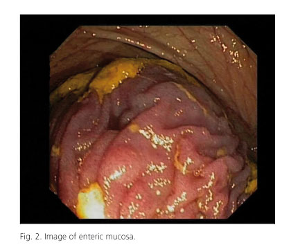
We present a thirty five years old woman with a family history of Peutz-Jeghers syndrome (PJS) (sister, brother and father) and a four-month history of progressive and constant right iliac fossa pain accompanied by weight loss of 5 kg. The patient does not refer nausea, vomiting or fever. The abdomen was soft, painless, without other signs of peritoneal irritation or palpable masses. In the rest of the physical exploration we found hyperpigmented maculae on the lips ("coffee with milk macules"). We performed a colonoscopy that showed a 6 mm adenomatous peduncular polyp in the sigmoid colon and an image compatible with ileo-colonic intussusception over the hepatic flexure (Figs. 1 and 2). An urgent abdominal CT scan was performed that showed ileo-colonic intussusception without obstruction signs or free fluid. We contacted with the Surgery Service and the patient was operated six hours after the colonoscopy. After a lower middle laparotomy we found an ileo-ileal intussusception that was reduced by hand, and we performed an enteroscopy through enterotomy made 30 cm from ileocecal valvula. Hundreds of milimetric polyps were observed through the entire explored bowel. Seven polyps were removed using a polypectomy snare. The polyp's size varied between 0.5 and 4 cm and the anatomopathologic study showed hamartomatous polyps.

Discussion
Finding an ileo-colonic intussusception during the colonoscopy exploration is unusual because most of the time the intussusception causes obstructions symptoms that require an image study like a CT scan. Anemia, rectal bleeding, abdominal pain, obstruction and/or intussusception are common complications in patient with PJS (1-3).
References
1. Gammon A, Jasperson K, Kohlmann W, Burt RW. Hamartomatous polyposis syndromes. Best Pract Res Clin Gastroenterol 2009;23(2):219-31. [ Links ]
2. Lynch HT, Lynch JF, Lynch PM, Attard T. Hereditary colorectal cancer syndromes: molecular genetics, genetic counseling, diagnosis and management. Fam Cancer 2008;7(1):27-39. [ Links ]
3. González Muñoz JL, Angoso Clavijo M, Esteban Velasco C, Rodríguez Pérez A, Muñoz Bellvis L, Gómez Alonso A. Diagnóstico de síndrome de Peutz-Jeghers. Rev Esp Enferm Dig 2007;99(3):167. [ Links ]

