


My SciELO
 Custom services
Services on Demand
Journal
Article
 text in
  English (pdf)
 Article in xml format
 Article references
 Send this article by e-mail
Indicators
-
 Cited by SciELO
 -
 Access statistics
Related links
-
 Cited by Google
 -
 Similars in
SciELO
Similars in
SciELO -
 Similars in Google
Share

 Permalink
Revista Española de Enfermedades Digestivas
Print version ISSN 1130-0108
Rev. esp. enferm. dig. vol.103 n.6 Madrid Jun. 2011
https://dx.doi.org/10.4321/S1130-01082011000600008
PICTURES IN DIGESTIVE PATHOLOGY
Telangiectasias bleeding in patient with multiple sclerosis
Hemorragia por telangiectasias en paciente con esclerosis sistémica
Alejandro Martínez-Caselles1, Cristina Martínez-Pascual1, María José Moreno-Martínez2, Antonio Sánchez-Torres1 and Luis Fernando Carballo-Álvarez1
Departments of 1Digestive Diseases, and 2Rheumatology. Hospital Universitario Virgen de la Arrixaca. Murcia, Spain
Scleroderma is a disease characterized by multiorgan fibrosis due to connective tissue proliferation and vasculitis of small vessels. Sclerodermia can be localized or systemic, and the latter can compromise skin in a limited or diffuse type. Digestive tract involvement is common in the systemic form (82%) and is often characterized by dysphagia and gastroesophageal reflux disease.
Case report
A 50 year old male, with no relevant family history, who was diagnosed of subclinical hypothyroidism and early scleroderma has been follow-up by Rheumatology and Dermatology. Due to the finding of necrosis in his finger pads since the previous year examinations were initiated. Capillaroscopy was consistent, ANA were positive (1/640) and anticentromere antibody, RNP and Scl-70 were negative. Therefore, treatment was started with bosentan and aspirin.
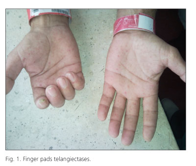
The patient was referred to the Emergency ward of our hospital because of melena and epistaxis. At admission, hemoglobin level was 8.5 mg/dL, with normal platelet count, normal coagulation and an ESR of 48 mm/h. Examination revealed multiple telangiectasias on his fingertips (Fig. 1), in oral mucosa and perioral region. Gastroscopy was performed which showed multiple small vascular appearance lesions (less than 5 mm) in esophageal, gastric and duodenal mucosa, with no signs of recent bleeding or clots (Fig. 2). In colonoscopy, multiple telangiectases were visualized from the anus to the cecum (Fig. 3). Neither melena nor decrease in hematocrit was presented during admission and he was discharged with supplemental oral iron and folic acid.


Discussion
Differential diagnosis of Rendu-Osler-Weber -hereditary hemorrhagic telangiectasia (HHT)- was performed. This autosomal dominant inheritance disease is characterized by a deficiency of vascular wall causing arteriovenous malformations. Multiple telangiectasias located mainly in the nasal and oral mucosa and nail beds are common. Diagnosis of this disease is based on the observation of mucocutaneous telangiectasia, visceral lesions, epistaxis and first-grade family history. Three or four of these criteria should be present to consider the disease.
Our patient had no first-degree relative HHT diagnosed. On the other hand, he had mucocutaneous and digestive tract telangiectasia, and also recently reported episodes of epistaxis. However, he had a vasculitis compatible capillaroscopy and high acute phase reactants which are not characteristic of Rendu-Osler-Weber. Raynaud with pathological capillaroscopy allow early diagnosis of systemic sclerosis.
Intestinal bleeding is a known complication although eso-phageal manifestations are more common and motility disorders are most common in stomach in scleroderma. In fact, there have been reports of bleeding, sometimes severe, from gastric mucosa telangiectases. Since melena is not always possible to detect in these patients, test for fecal occult blood would be advisable and, if positive, colonoscopy should be performed, specially when anemia is detected associated with this disease.
Recommended References
1. Sánchez-Salas MP, et al. Telangiectasias de la piel y la mucosa oral. Piel 2008;23(2):90-2. [ Links ]
2. Begbie ME, Wallace GMF, Showlin CL. Hereditary haemorrhagic telangiectasia (Osler-Weber-Rendu syndrome): a view from the 21st century. Postgrad Med J 2003;73:18-24. [ Links ]
3. Vautier G, McDermott E, Carty JE, et al. Small bowel telangiectasia in scleroderma. Ann Reum Dis 1995;54(1):78. [ Links ]

