


Mi SciELO
 Servicios personalizados
Servicios Personalizados
Revista
Articulo
 texto en
  Inglés (pdf)
 Articulo en XML
 Referencias del artículo
 Enviar articulo por email
Indicadores
-
 Citado por SciELO
 -
 Accesos
Links relacionados
-
 Citado por Google
 -
 Similares en
SciELO
Similares en
SciELO -
 Similares en Google
Compartir

 Permalink
Revista Española de Enfermedades Digestivas
versión impresa ISSN 1130-0108
Rev. esp. enferm. dig. vol.103 no.6 Madrid jun. 2011
https://dx.doi.org/10.4321/S1130-01082011000600011
LETTERS TO THE EDITOR
Endoscopic extraction of a pig-tail catheter after urological surgery with Mainz II pouch urinary diversion
Extracción endoscópica de catéter pig-tail tras cirugía urológica con derivación urinaria tipo Mainz II
Key words: Endoscopic extraction. Colorectal foreign body. Ureterosigmoidostomy.
Palabras clave: Extracción endoscópica. Cuerpo extraño colorrectal. Ureterosigmoidostomía.
Dear Editor,
Endoscopic extraction of foreign bodies in the gastrointestinal tract is the second cause of endoscopic emergencies, and it is compulsory if the objects may induce injuries or severe complications (1).
We report the case of a patient who presented with intraperitoneal migration of a pig-tail catheter placed during urological surgery. Endoscopic removal was successful resolving the associated peritoneal irritation.
Case report
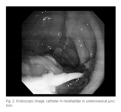
A 50 year-old woman was diagnosed of solid, poorly differentiated, high grade urothelial carcinoma, which infiltrates lamina propria and muscularis mucosa. She underwent radical cystectomy and ureterosigmoidostomy (Mainz II), being both ureters reimplanted on the left side of a sigma-rectum pouch, and two catheters on the other side to the urostomy pouch. Ten days after the procedure, the patient presents with abdominal pain on the right side, signs of peritoneal irritation and leukocytosis. It is observed that the left catheter has migrated underneath the skin. An urgent abdominal CT is performed showing the pig-tail catheter in left renal pelvis. It follows a normal pathway from the anterior to the posterior side of the neobladder, but then it goes up, first anterior to cecum and afterwards to the medium line, over the bowels and in the end, turning to the right flank. The end is located just posterior to the right rectus abdominis muscle (Fig. 1). Free air suggesting perforation is not observed. An urgent colonoscopy is performed. The co-lonoscope is introduced to 20 cm from anus, showing the neobladder with a catheter in the ureterovesical junction going out the posterior side, being the distal end not identified (Fig. 2). The catheter is withdrawn with forceps without immediate complications. Forty-eight hours after the procedure the patient remains asymptomatic, without abdominal pain and she is discharged from hospital.

Discussion
Colorectal foreign bodies usually reach the colon through the anus (2), being less frequent to find an ingested foreign body in the distal segments of the colon. They may cause many complications, such us rectal or colonic perforation, pararectal infections, peritonitis and intraabdominal abcesses, anal sphincter laceration, perirectal tissues injuries, adjacent organs lesions, intestinal obstruction, lower gastrointestinal bleeding and different kinds of fistulae: rectovesical, rectovaginal, enterocutaneous and perianal, even septic shock and death (3). Therefore, if the case doesn't resolve spontaneously, objects causing lesions or complications should be immediately removed. In the absence of perforation signs or peritonitis, manual extraction should be attempted. Most foreign bodies can be manually or endoscopically removed (4) and the transanal extraction can be performed under sedation; local, spinal or gene-ral anesthesia, being anal dilation frequently enough, with very few short and long-term complications.
A flexible sigmoidoscopy occasionally allows object extraction, describes the characteristics of the object and helps its distal advance for a manual removal and identifies rectosigmoid lesions (5).
Surgery is the treatment of choice in the presence of complications or endoscopic or manual extraction failure (6).
We consider this case report is exceptional, because a pig-tail catheter was placed in each ureter after radical bladder resection with sigmoid anastomosis. One of these catheters migrated underneath the skin and into the peritoneal cavity, being responsible for the peritoneal irritation signs.
Successful endoscopic extraction was possible due to the location of an end of the catheter in the neobladder.
Mar Lozano-Maya, Rocío Campos-Cantero, Rocío Plaza-Santos, José María Alberdi-Alonso,
Yolanda Real-Martínez and Mercedes Aldeguer-Martínez
Department of Digestive Diseases. Hospital Infanta Leonor. Madrid, Spain
References
1. Rodríguez Hermosa JI, Codina Cazador A, Ruiz B, Sirvent JM, Roig J, Farrés R. Management of foreign bodies in the rectum. Colorectal Dis 2007;9(6):543-8. [ Links ]
2. Gea F, Rábago L, Soler F, Mora P. Cuerpo extraño en rectosigma. Rev Esp Enferm Dig 1991;79:445-6. [ Links ]
3. Cohen JS, Sackier JM. Management of colorectal foreign bodies. J R Coll Surg Edinb 1996;41:312-5. [ Links ]
4. Ayantunde AA, Oke T. A review of gastrointestinal foreign bodies. Int J Clin Pract 2006;60(6):735-9. [ Links ]
5. Golberg JE, Steele SR. Rectal foreign bodies. Surg Clin North Am 2010;90(1):173-84. [ Links ]
6. Rodríguez Hermosa JI, Codina Cazador A, Sirvent JM, Martín A, Gironés J, Garsot E. Surgically treated perforations of the gastrointestinal tract caused by ingested foreign bodies. Colorectal Dis 2008;10 (7):701-7. [ Links ]

