Mi SciELO
Servicios Personalizados
Revista
Articulo
Indicadores
-
 Citado por SciELO
 -
 Accesos
Links relacionados
-
 Citado por Google
 -
 Similares en
SciELO
 -
 Similares en Google
Compartir
Revista Española de Enfermedades Digestivas
versión impresa ISSN 1130-0108
Rev. esp. enferm. dig. vol.104 no.5 Madrid may. 2012
https://dx.doi.org/10.4321/S1130-01082012000500015
LETTERS TO THE EDITOR
Primary biliary sarcoidosis mimicking Klatskin tumor: case report and review of literature
Sarcoidosis biliar primaria simulando un tumor de Klatskin: aportación de un caso y revisión de la literatura
Key words: Sarcoidosis. Klatskin tumor. Biliary obstruction.
Dear Editor,
Sarcoidosis is a multi-systemic granulomatous disease of unknown aetiology. It is more common in young adults and typically presents as a pulmonary granulomatous disease with its associated systemic clinical manifestations (1). Gastrointestinal sarcoidosis most commonly affects the liver, although this is rare without primary pulmonary involvement (2). Extra-hepatic biliary tract involvement is usually due to extrinsic compression caused by infiltrative sarcoid granulomas, however, in isolation it can be a major diagnostic challenge.
Case report
A 36 year old gentleman of Indo-Asian origin presented with a short history of weight loss of 12 kg in 1 year and mild jaundice associated with darkened colored urine. He was born in Pakistan and had resided in the UK for the last 6 years. He was a non-drinker and had never smoked. There was no family history of malignancy, and no known exposure to hepatitis B or C. There were no other medical co-morbidities.
On clinical examination, apart from being mildly jaundiced there were no other abnormal clinical findings.
Initial serum biochemistry showed deranged liver function tests (elevated total bilirubin 58 mg/dL (normal < 21), raised alanine transaminase (ALT 217 IU/L), raised alkaline phosphatase (ALP 790 IU/L). Both Cancer Antigen 19.9 and carcinoembryonic antigen (CEA) were within normal limits as was his serum a-fetoprotein (AFP). Hepatitis serology (A to E) was negative.
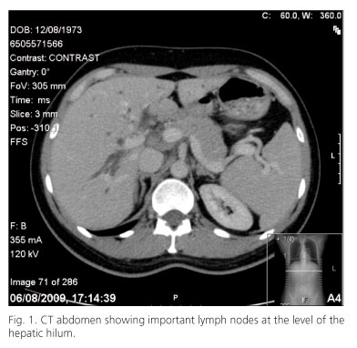
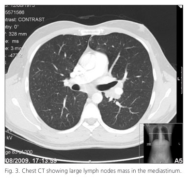
An ultrasound scan of his biliary tract demonstrated intra-hepatic ductal dilatation more marked on the left lobe of the liver, associated with a possible hilar mass. The extra-hepatic common bile duct was normal as was the liver echo-texture. Subsequent contrast enhanced multi-slice CT of the chest and abdomen revealed prominent intrahepatic biliary dilatation and all the segmental ducts tapering in diameter towards the liver hilum. The extrahepatic biliary tree was not dilated. The most proximal common duct appeared to have luminal soft tissue although there was no measurable mass lesion. Significantly enlarged lymph nodes were demonstrated in the liver hilum (Fig. 1), left gastric territory, and in the chest (left lung hilum, pre-tracheal, subcarinal, and para-tracheal nodes) (Fig. 2). There was no evidence of vascular involvement or encasement. The appearance was suggestive of hilar cholangiocarcinoma with disconnection of segmental ducts, together with upper abdominal, hilar and mediastinal lymphadenopathy.
In the absence of an obvious hilar mass the differential at this stage included autoimmune biliary disease, hilar cholangiocarcinoma, and lymphoma in view of the lymphadenopathy. Two weeks following the initial consult his serum total bilirubin remained static at 50 mg/dL. Although his serum IgG was moderately elevated at 18 (normal range), serum IgG4 levels were normal.
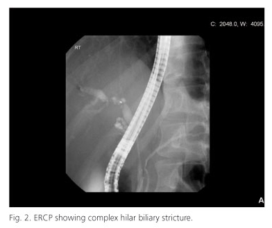
A magnetic resonance imaging/magnetic resonance cholangiopancreatography performed to define the biliary anatomy more clearly prior to biliary drainage revealed a complex hilar stricture with dissociation of ducts suggestive of either a hilar cholangiocarcinoma or auto-immune etiology. The vascular structures were unaffected. Subsequent endoscopic retrograde cholangiopancreatography (ERCP) confirmed a hilar stricture (Fig. 3). Both the brush cytology from the stricture and exfoliative bile sample revealed scattered groups of benign duct epithelial cells with no malignant cells. The cells also failed to stain for IgG4.
"Spy-glass" choledochoscopy was of limited help as it failed to traverse through a "hard" occlusion at the level of the hilum, although the epithelium itself appeared normal. Normal IgG4 levels suggested that the mass was likely to be a hilar cholangiocarcinoma which was amenable to surgical resection (extended left hepatectomy, complete excision of the extra-hepatic biliary tree, and radical lymphadenectomy). In view of his age, an endoscopic ultrasound examination (EUS) was suggested to obtain tissue from his upper abdominal and mediastinal lymph nodes for cytological confirmation prior to embarking on resection, as positive cytology in the latter would preclude resection.
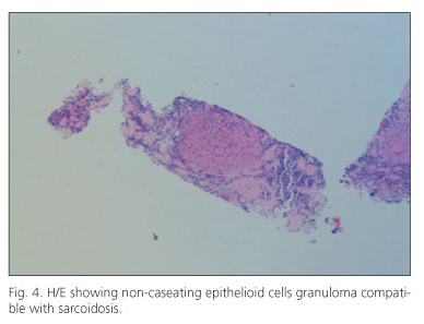
EUS guided biopsy of the mediastinal nodes reported non-caseating granulomas, negative for acid-fast bacilli in the Ziehl-Neelsen stain, suggestive of sarcoidosis (Figs. 4 and 5), although the celiac node biopsy demonstrated reactive changes only.
The patient was commenced on a 6 week course of high dose steroids after which the cholestasis disappeared. A follow up CT scan showed a marked reduction in the size of the hepatic hilar and mediastinal lymph nodes.
Discussion
Biliary obstruction is a common entity caused by benign and malignant lesions. The level of obstruction may be intrahepatic or extrahepatic. Common conditions are: pancreatic head carcinoma, cholangiocarcinoma, periampullary tumors, metastases, common bile duct stones, inflammatory and iatrogenic strictures. Rare causes include tuberculosis, sarcoidosis, celiac disease and Crohn's disease.
Differential diagnosis: in the presence of a biliary stricture, cholangiocarcinoma can not be excluded by the negativity of the cytology. Autoimmune biliary disease was excluded by the presence of a normal IgG4, while the presence of intra-abdominal plus mediastinal lymph nodes raised the possibility of lymphoma versus sarcoidosis. The differentiation between these two entities can be achieved only through histopathology as it was the case in our patient in which an EUS guided biopsy of the mediastinal nodes demonstrated the presence of non-caseating granulomas compatible with sarcoidosis.
Sarcoidosis is a member of a group of disorders that lead to non neoplastic biliary obstruction (2); other such conditions include Crohn's disease of the pancreas, tuberculosis of the biliary lymph nodes, primary sclerosing cholangitis, AIDS cholangitis, Mirizzi syndrome, metastases, melanoma, lymphoma, leukemia, autoimmune pancreatitis and carcinoid tumors. Sarcoidosis is a chronic multisystemic granulomatous condition of unknown etiology, with an extremely rare gastrointestinal and hepatobiliary involvement. It is most frequent in white and black young people of North Europe. Although all organs and systems can be affected, the lungs and intrathoracic lymph nodes are the most common sites of involvement. Diagnosis is established by the presence of a compatible clinical illness (chronic respiratory symptoms, and few constitutional symptoms) and by histological demonstration of non-caseating epithelioid cell granulomas in the affected organs. At least two organs need to be involved in order to achieve a diagnosis.
The liver is the most common gastrointestinal site of involvement, as seen in series of autopsies -nearly 70% of livers were found with hepatic granulomas, but most of them were without clinical gastrointestinal symptoms (3). Liver sarcoidosis in isolation has been documented in only about 13% of patients with systemic sarcoidosis (4).
In the liver, the disease can cause presinusoidal portal hypertension, advancing quickly to fibrosis and micronodular cirrhosis with progressive destruction of the interlobular bile ducts, similar to primary biliary cirrhosis, but without it's non suppurative destructive cholangitis. Non-caseating granulomas tend to be portal or peri-portal and involvement of the extrahepatic biliary tree via nodal infiltration with extrinsic compression or direct invasion is rare (5). Our case definitely is not a liver sarcoidosis but an involvement of the extrahepatic biliary duct, mimicking a hilar cholangiocarcinoma.
Intra-abdominal lymphadenopathy is common in sarcoidosis and can exist in the perihilar lymph nodes and in those around the extrahepatic biliary tract, but obstruction of the common bile duct is rare (6). In most cases, enlarged lymph nodes do not cause obstruction of the biliary ducts, and thus pruritus is typically attributed to direct hepatic involvement (7). As is often the case, despite state-of-the-art imaging and diagnostic modalities it is often extremely difficult to differentiate hilar obstruction due to benign disease from cholangiocarcinoma. Final histological diagnosis in this particular case was achieved by an EUS guided biopsy of a mediastinal lymph node.
The presence of enlarged lymph nodes may lead to confusion, as they can be interpreted as metastatic disease, but multiple enlarged lymph nodes, with moderately elevated total bilirubin levels, in the presence of dominant systemic symptoms should lead to the differential diagnosis of a benign inflammatory disease. However, this case highlights the fact that biliary sarcoidosis may exist in the absence of constitutional systemic symptoms, or indeed hepatic or pulmonary sarcoidosis (8). The treatment for biliary sarcoidosis is the same as for systemic sarcoidosis. Sarcoidosis is very sensitive to corticosteroid therapy. Once the diagnosis is made in the biliary tree, decompression is important, and most patients should enter a long-lasting remission. Steroids can improve liver function tests but they do not alter the granulomas. Most corticosteroid regimens for the treatment of gastrointestinal sarcoidosis involve 40-60 mg daily for 8-12 weeks and then a taper down to 10-20 mg for up to 6 months. Methotrexate, ursodeoxycholic acid and cyclosporine have also been utilized, with varying degrees of clinical and biochemical improvement. Serum angiotensin-converting enzyme is an insensitive and non-specific diagnostic test and a poor therapeutic guide in sarcoidosis, particularly in the liver or biliary tree. Biliary obstruction from sarcoidosis resembling Klatskin cholangiocarcinoma will respond to balloon dilation, placement of a temporary biliary endoprosthesis, and corticosteroid therapy (9). In our case the corticosteroid therapy was enough to achieve a dramatic improvement in biliary obstruction.
Hector Daniel González, Samir John Sahay and Sakhawat Hussain Rahman
Centre for HPB Surgery & Liver Transplantation. London
References
1. Newman LS, Rose CS, Maier LA. Sarcoidosis. N Engl J Med 1997; 336:1224-34. [ Links ]
2. Ganne-Carrie N, Guettier C, Ziol M, Beaugrand M, Trinchet JC. Sarcoidose et foie. Ann Med Interne (Paris). 2001;152:103-7. [ Links ]
3. Rudzki C, Ishak KG, Zimmerman HJ. Chronic intrahepatic cholestasis of sarcoidosis. Am J Med 1975; 59: 373-87. [ Links ]
4. Mueller S, Boehme MW, Hofmann WJ, Stremmel W. Extrapulmonary sarcoidosis primarily diagnosed in the liver. Scand J Gastroenterol 2000;35:1003-8. [ Links ]
5. Ishak KG. Sarcoidosis of the liver and bile ducts. Mayo Clin Proc 1998; 73:467-72. [ Links ]
6. Valla D, Pessegueiro Miranda H, Degott C, Lebrec D, Rueff B, Benhamou JP. Hepatic sarcoidosis with portal hypertension. A report of seven cases with a review of the literature. Q J Med 1987;63:531-44. [ Links ]
7. Papo T, Piette JC, Valla D. Sarcoidosis, liver transplantation, and cyclosporine. Ann Intern Med 1993;119:1148; author reply 1148-9 . [ Links ]
8. Harder H, Büchler MW, Fröhlich B, Ströbel P, Bergmann F, Neff W, Singer MV. Extrapulmonary sarcoidosis of liver and pancreas: A case report and review of literature. World J Gastroenterol 2007;13:2504-9. [ Links ]
9. Petersen JM. Klatskin-like biliary sarcoidosis: a cholangiocscopic diagnosis. Gastroenterol and Hepatol 2009;5:137-41. [ Links ]


