


Mi SciELO
 Servicios personalizados
Servicios Personalizados
Revista
Articulo
 texto en
  Inglés (pdf)
 Articulo en XML
 Referencias del artículo
 Enviar articulo por email
Indicadores
-
 Citado por SciELO
 -
 Accesos
Links relacionados
-
 Citado por Google
 -
 Similares en
SciELO
Similares en
SciELO -
 Similares en Google
Compartir

 Permalink
Revista Española de Enfermedades Digestivas
versión impresa ISSN 1130-0108
Rev. esp. enferm. dig. vol.105 no.8 Madrid sep. 2013
https://dx.doi.org/10.4321/S1130-01082013000800007
PICTURES IN DIGESTIVE PATHOLOGY
Pneumatosis cystoides, CT colonoscopy and endoscopic correlation
Neumatosis quística en colonografía TC y correlación endoscópica
Daniel Rodríguez-Sánchez, María Elena Sáez-Martínez, Regina María Sánchez-Jiménez, Juan de-Dios-Berná-Mestre and Florentina Guzmán-Aroca
Department of Radiology. Hospital Clínico Universitario Virgen de la Arrixaca. Murcia, Spain
Case report
A 52-year-old male participating in a colorectal cancer screening programme and testing positive for faecal occult blood underwent a colonoscopy to the caecum, which detected multiple polypoid nodulations in the right colon wall, most of them confluent. The mucosa was conserved. A single biopsy was performed, which revealed injury collapse. Suspected with microperforation, the patient had a simple abdominal X-ray and a CT colonography to complete the study.
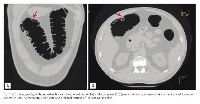
The CT colonography (Figs. 1A: coronal plane, and 1B: axial plane; arrows) showed numerous air-containing cyst formations dependent on the ascending colon wall and proximal portion of the transverse colon. The 3D-type reconstruction of endoscopic images (Figs. 2A and 2B) showed injuries superimposable to those visible on optical colonoscopy (Figs. 2C and 2D). The findings were compatible with cystic pneumatosis of the colon.

Discussion
Intestinal cystic pneumatosis is a rare condition characterised by gas-filled multilocular cysts located in the submucosa and subserosa of the gastrointestinal tract. It is a clinical or radiological entity, not a real pathology, and there may be multiple causes. Depending on the factors involved in its aetiology it is classed as idiopathic (15 %) or secondary (85 %). Although it may appear in any location from the oesophagus to the rectum it most commonly affects the intestine (1,2). When located in the colon, optical colonoscopy shows multiple polypoid lesions covered with normal-looking mucosa. These findings do not help distinguish it from other entities such as lymphoid hyperplasia, hyperplastic polyposis or profound cystic colitis (3). However, CT reveals the presence of air cysts in the bowel wall. Furthermore, CT colonography allows images on different spatial planes and volumetric reconstructions in the form of virtual colonoscopy for proper assessment of its extent and location (4,5).
References
1. Arikanoglu Z, Aygen E, Camci C, Akbulut S, Basbug M, Dogru O, et al. Pneumatosis cystoides intestinalis: A single center experience. World J Gastroenterol 2012;18:453-7. [ Links ]
2. Kim KM, Lee CH, Kim KA, Park CM. CT Colonography of pneumatosis cystoides intestinalis. Abdom Imaging 2007;32:602-5. [ Links ]
3. Ivanovic A, Kovac J, Masulovic D, Stefanovic A, Jaksic E, Saranovic D. Education and imaging. Gastrointestinal: The role of multidetector computer tomography in diagnosis of pneumatosis cystoides intestinalis. J Gastroenterol Hepatol 2012;27:182. [ Links ]
4. Kim YN, Lee SJ, Kim MJ, Kim YH, Jang DK. Computed tomography colonographic findings of pneumatosis cystoides coli. J Comput Assist Tomogr 2008;32:65-8. [ Links ]
5. Kim BN, Jeong JY, Sohn DK, Han KS, Hong CW, Chang HJ, et al. Pneumatosis cystoides coli of the ascending colon: Colonoscopic and CT colonographic features. Endoscopy 2007;39(Supl. 1):73-4. [ Links ]

