


My SciELO
 Custom services
Services on Demand
Journal
Article
 text in
  English (pdf)
 Article in xml format
 Article references
 Send this article by e-mail
Indicators
-
 Cited by SciELO
 -
 Access statistics
Related links
-
 Cited by Google
 -
 Similars in
SciELO
Similars in
SciELO -
 Similars in Google
Share

 Permalink
Revista Española de Enfermedades Digestivas
Print version ISSN 1130-0108
Rev. esp. enferm. dig. vol.106 n.3 Madrid Mar. 2014
PICTURES IN DIGESTIVE PATHOLOGY
Deep infiltrating rectosigmoid endometriosis. Diagnostic keys
Endometriosis rectosigmoidea infiltrante profunda. Claves para su diagnóstico
Ma Alcázar Iribarren-Marín1, Herminia Pérez-Vega1, Manuel Martínez-Moya1, Lourdes Gómez-Izquierdo2 and Cristina Martínez-Polanco1
1Diagnostic Imaging Unit. Radiology Department. 2Pathological Anatomy Service. Hospital Universitario Virgen del Rocío. Seville, Spain
Case report
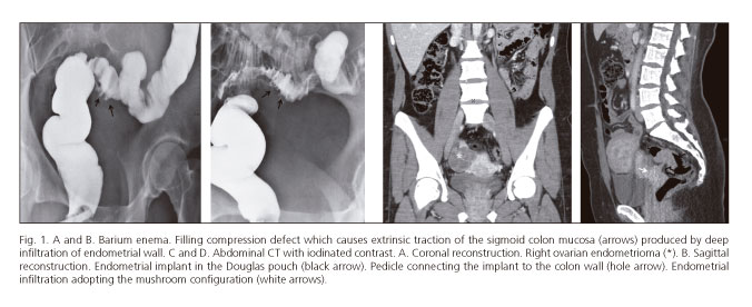
A 36 year-old woman was admitted with dysmenorrhea associated with rectal bleeding. The colonoscopy showed a stenosis at the rectosigmoid (RS) junction which did not exceed the endoscope. A barium enema (Figs. 1 A and B) showed traction of the sigmoid mucosa at the lower margin of the RS junction. CT examination (Figs. 1 C and D) showed an endometrial implant in the Douglas pouch, the pedicle connected to the colon wall and the "mushroom cap" configuration of the endometrial implant in the RS wall, all with a density similar to that of muscle tissue. A sigmoidectomy was performed (Fig. 2) with histological (Fig. 3) and immunohistochemical analysis, which demonstrated deep infiltrating rectosigmoid endometriosis (DIRSE) as the resulting diagnosis.


Discussion
Endometriosis is defined as the presence of functioning ectopic endometrial tissue (endometrial glands and stroma) outside the uterus and at a distance from and unconnected with uterine endometrial tissue. The ectopic endometrium is sensitive to hormonal variation during the menstrual cycle. These resultsin chronic inflammation that is the cause of cyclic pelvic pain found in these patients. In the DIRSE, the ectopic tissue would respond to the ovarian hormonal cycles causing inflammation, bleeding, fibrosis and metaplasia or hyperplasia of the smooth intestinal muscle possibly affecting the serosa, submucosa and rarely the mucosa, which gives rise to a thickening of the intestinal wall and in some cases may evolve into an obstruction (1-3). The "mushroom cap" sign has been described in the presumed diagnosis because the tumor grows towards the RS colonic lumen (4). The differential diagnosis is reached from the neoplasic processes seen in this area.
References
1. Chamiè LP, Blasbalg R, Pereira RM, Warmbrand G, Serafini, PC. Findings of pelvic endometriosis at transvaginal US, MR imaging and laparoscopy. RadioGraphics 2011;31:E77-100. [ Links ]
2. Nasim H, Sikafi D, Nasr A. Sigmoid endometriosis and a diagnostic dilema-A case report and literature review. Int J Surg Case Rep 2011;2:181-4. [ Links ]
3. Fernández-Rey CL, Álvarez-González SA, Díaz-Solís P, Blanco-González A, Costilla-García S. Endometriosis ileal como causa de obstrucción de intestino delgado: diagnóstico por tomografía computarizada multicorte. Rev Esp Enferm Dig 2009;101:872-4. [ Links ]
4. Yoon JH, Choi D, Jang KT, Kim CK, Kim H, Lee SJ, et al. Depp rectosigmoid endometriosis: "mushroom cup" sing on T2-weighted MR imaging. Abdom Imaging 2010;35:726-31. [ Links ]

