


My SciELO
 Custom services
Services on Demand
Journal
Article
 English (pdf)
 Article in xml format
 Article references
 Send this article by e-mail
Indicators
-
 Cited by SciELO
 -
 Access statistics
Related links
-
 Cited by Google
 -
 Similars in
SciELO
Similars in
SciELO -
 Similars in Google
Share

 Permalink
Revista Española de Enfermedades Digestivas
Print version ISSN 1130-0108
Rev. esp. enferm. dig. vol.106 n.3 Madrid Mar. 2014
PICTURES IN DIGESTIVE PATHOLOGY
Cavernous hemangioma of small bowel: A rare cause of digestive hemorrhage
Hemangioma cavernoso del intestino delgado: una causa rara de hemorragia digestiva
Dália Fernandes1,3, Isabel Dionísio2, Sofia Neves2 and Patrícia Duarte1,3
Departments of 1Gastroenterology and 2Surgery. Cova da Beira Hospital Center. Covilhã, Portugal. 3Health Sciences Faculty. University of Beira Interior. Covilhã, Portugal
Introduction
Gastrointestinal hemangiomas are rare benign tumors, representing 0.05 % of all gastrointestinal tumors, that may appear isolated or as part of systemic vascular disorders (1). They are more frequent in the jejunum. Their main clinical manifestation is gastrointestinal bleeding, usually insidious in capillary hemangiomas, although it can be acute and severe in the cavernous type (2). Other forms of presentation can be obstruction, intussusception, intramural hematoma, perforation and platelet sequestration (3). Endoscopy is the method of choice in the diagnosis of such lesions, and the capsule endoscopy the gold standard for small bowel lesions (4). Surgical resection is the ideal treatment.
Case report
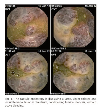
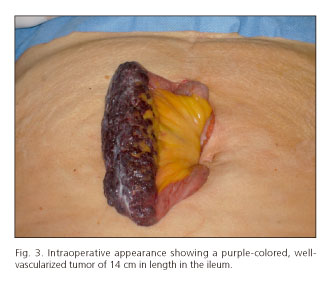
We presented a 56-year-old woman who was admitted to the emergency room with hematochezia and syncope, of 24 hours of duration. She denied abdominal pain and referred recent use of ibuprofen. Physical examination on admission only revealed cutaneous-mucous pallor. Laboratory analyses revealed microcytic and hypochromic anemia (HGB 9.4 g/dL, MCV 78.1 fL and MCHC 33.7 g/dL). Upper gastrointestinal endoscopy showed none bleeding lesion. Fibrosigmoidoscopy showed abundant blood and clots on the lumen, but colonoscopy with ileoscopy on the following day detected none trace of blood or bleeding lesion. One week later a capsule endoscopy was perfomed and identified a large, violet-colored and circumferential lesion in the ileum, conditioning luminal stenosis, without active bleeding (Fig. 1). Computed tomography enterography demonstrated a 14 centimeter-long irregular thickening of the ileum, with focal calcifications (Fig. 2). A laparotomy was performed and the vascular tumor was confirmed and resected (Fig. 3). Histological examination revealed a cavernous hemangioma (Fig. 4). The surgery and postoperative period was uneventful.


Discussion
Although there are some case reports of gastrointestinal bleeding due to hemangiomas in the literature, this case stands out because of large size of hemangioma and its presentation as overt gastrointestinal bleeding at an advanced age.
References
1. Boyle L, Lack EE. Solitary cavernous hemangioma of small intestine. Case report and literature review. Arch Pathol Lab Med 1993;117:939-41. [ Links ]
2. Levy AD, Abbott RM, Rohrmann CA Jr, Frazier AA, Kende A. Gastrointestinal hemangiomas: Imaging findings with pathologic correlation in pediatric and adult patients. AJR Am J Roentgenol 2001;177:1073-81. [ Links ]
3. Pera M, Márquez L, Dedeu JM, Sánchez J, Garcia M, Ramón JM, et al. Solitary cavernous hemangioma of the small intestine as the cause of long-standing iron deficiency anemia. World J Gastrointest Surg 2012;16:2288-90. [ Links ]
4. De Mascarenhas-Saraiva MN, da Silva Araújo Lopes LM. Small-bowel tumors diagnosed by wireless capsule endoscopy: report of five cases. Endoscopy 2003;35:865-8. [ Links ]

