


Mi SciELO
 Servicios personalizados
Servicios Personalizados
Revista
Articulo
 texto en
  Inglés (pdf)
 Articulo en XML
 Referencias del artículo
 Enviar articulo por email
Indicadores
-
 Citado por SciELO
 -
 Accesos
Links relacionados
-
 Citado por Google
 -
 Similares en
SciELO
Similares en
SciELO -
 Similares en Google
Compartir

 Permalink
Revista Española de Enfermedades Digestivas
versión impresa ISSN 1130-0108
Rev. esp. enferm. dig. vol.106 no.8 Madrid dic. 2014
Peritoneal tuberculosis. radiographic diagnosis
Tuberculosis peritoneal. diagnóstico radiológico
Carolina Ospina-Moreno, Jorge González-Gambau, Inmaculada Montejo-Gañán, Alba Castán-Senar, Luis Sarría-Octavio-de-Toledo and Elena Martínez-Mombila
Department of Radiodiagnosis. Hospital Universitario Miguel Servet. Zaragoza, Spain
ABSTRACT
Peritoneal tuberculosis (TB) is an extrapulmonary form of presentation of tuberculosis. HIV infection is a primary risk factor for this condition. Diagnosis requires microbiological or histopathological confirmation in addition to supporting radiological imaging studies. Abdominal ultrasonography and CT are useful to obtain a radiographic diagnosis, with typical findings including diffuse peritoneal thickening, presence of ascites in varying volumes, adenopathies, and caseating nodes. We report 2 cases of patients with ascites and nodular peritoneal thickening on diagnostic images, as well as high CA-125 levels in laboratory tests. In both patients, a diagnosis of peritoneal tuberculosis was reached following a US-guided peritoneal biopsy.
Key words: Peritoneal tuberculosis. Cancer antigen (CA) 125. Ascites. Peritoneal carcinomatosis.
RESUMEN
La tuberculosis (TBC) peritoneal es una de las formas de presentación extrapulmonar de la tuberculosis. La infección por VIH es uno de los principales factores de riesgo para esta enfermedad. El diagnóstico requiere una confirmación microbiológica o histopatológica, además de pruebas radiológicas que lo apoyen. En el diagnóstico radiológico son útiles la ecografía y la TC abdominal, los hallazgos característicos son el engrosamiento peritoneal difuso, la presencia de ascitis en cantidades variables, adenopatías y nódulos caseificantes. Presentamos 2 casos de pacientes con ascitis y engrosamiento nodular del peritoneo en las pruebas de imagen y un CA 125 elevado en las pruebas de laboratorio. En ambos casos se llegó al diagnóstico de tuberculosis peritoneal después de realizar biopsia peritoneal guiada por ecografía.
Palabras clave: Tuberculosis peritoneal. Antígeno CA 125. Ascitis. Carcinomatosis peritoneal.
Case report 1
A 40-year-old male patient with a history of HCV-related cirrhosis and HIV without treatment. He arrives at the ER complaining of increased abdominal circumference, jaundice, dyspnea on exertion, abdominal pain and fever for 1 month. The physical examination reveals non-tense ascites, non-tender hepatomegaly, collateral circulation, telangiectasias, and edema in the lower limbs. In laboratory tests, tumor markers are notably high: CA 125 519 (n 0-35), CA 19.9 278 (n 0-37). Abdominal ultrasonography shows moderate ascites with thickened, nodular-looking parietal peritoneum fascia. In view of sonographic findings, a thoracoabdominal CT scan is performed, which reveals hypodense mediastinal adenopathies suggestive of caseating necrosis; hepatomegaly; abundant ascites with irregular, nodular thickening of the peritoneal fascia; and infiltration areas in peritoneal fat (Fig. 1). A US-guided peritoneal biopsy was carried out (Fig. 2), which found granulomatous peritonitis with focal caseating necrosis, consistent with TB. Ascitic fluid samples were obtained for culture, which resulted in Mycobacterium tuberculosis growth.


Case report 2
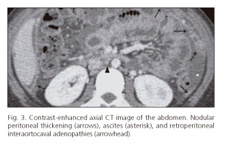
A 19-year-old female patient, without a relevant medical history, is seen in the ER because of intermittent fever, asthenia, and progressive abdominal distension for 6 months. The physical exam reveals hepatomegaly and positive ascitic wave. Laboratory test highlights include: Mantoux +, CA 125 797.7, white blood cells 4,000, Hb 9.7. A thoracoabdominal CT scan shows: Ascites and serosal thickening with multiple nodular lesions suggestive of peritoneal implants, primarily in the omentum. Mild splenomegaly. Multiple mesenteric, aortoiliac, retrocrural, and periceliac adenopathies (Fig. 3). A US-guided peritoneal biopsy is taken from the thickest peritoneal area, and samples are obtained for culture, which showed Mycobacterium tuberculosis growth. Tuberculostatics are initiated, which results in clinical improvement and in ascites and peritoneal thickening clearance (Fig. 4).

Discussion
TB remains a primary public health challenge worldwide, the HIV epidemics having facilitated its recurrence and spread (1). On the other hand, because of the coexistence of HIV infection, extrapulmonary forms of tuberculosis, including peritoneal tuberculosis, which represents nearly 3.5 % of presentations, are now increasingly common. Risk factors for peritoneal TB include, besides HIV, cirrhosis, diabetes, neoplasms, and peritoneal dialysis (2). Its diagnosis requires a histological demonstration of TB with caseating necrosis, presence of acid-alcohol-fast bacilli (AAFB), or an M. tuberculosis-positive culture from ascitic fluid, peritoneal biopsy, or mesenteric lymph nodes. Imaging techniques allow a diagnostic approximation. Ultrasonography reveals ascites (which may contain fine echogenic filaments), and diffuse or irregular peritoneal thickening with nodules with a hypoechoic center suggestive of caseation (3). CT is the most sensitive technique for the assessment of peritoneal disease. This technique unveils a usually consistent peritoneal thickening with parietal peritoneum enhancement following the administration of a contrast. It is associated with mesenteric and peripancreatic adenopathies in 90 % of patients (4). When there is a nodular aspect to the peritoneal thickening, a differential diagnosis with peritoneal carcinomatosis is due (3). Tree clinical forms have been described:
-Wet form: Characterized by abundant ascites with a high attenuation value (20 to 40 HU), as it is an exudate with high protein contents.
-Dry form: Characterized by loculated ascites with predominant adhesions, fibrosis, peritoneal thickening, and caseated nodes.
-Fibrotic form: Associated with low-volume ascites and intestinal adhesions to the mesentery, thus making up a fibrotic mass (1,5).
However, peritoneal TB may present as a combination of all the 3 types above.
Our two patients had the wet form of peritoneal TB in view of both radiological and microbiological findings. The first report was of a patient with 2 risk factors for peritoneal TB, such as HCV-related liver cirrhosis and infection with HIV. The history of untreated HCV-related cirrhosis led to consider decompensation as the potential cause for ascites (6), but sonographic findings such as a thickened peritoneum prompted us to rule out a different underlying origin. Various differential diagnoses are then considered, including peritoneal carcinomatosis, malignant mesothelioma, primary peritoneal lymphoma, and peritoneal tuberculosis. These 4 conditions are characterized by ascites and an irregularly thickened peritoneum (4). Malignant mesothelioma is a rare entitiy associated with asbestos exposure (7). Primary peritoneal lymphoma, while uncommon, may be considered in this case as it almost exclusively involves HIV-infected patients (4).
Increased tumor markers, including CA-125 and CA-19.9, raise suspicion of a causing malignancy, but elevated levels may also be seen in selected benign conditions. Thus, increased CA-19.9 levels suggest a pancreatic or colonic neoplasm, but other possible origins include liver cirrhosis and biliary disorders in association with cholestasis and liver cytolysis (8). CA-125 is commonly used as a marker of ovarian cancer progression and may also be elevated by other malignancies such as breast, lung, and gut adenocarcinoma, as well as lymphoma and leiomyosarcoma. It may be also increased in benign conditions with serosal compromise. One such instance is peritoneal TB, as seen in both case reports, this being a useful tool for diagnosis and follow-up. It may also increase in TB with pleural and pericardial involvement (9).
In contrast with the first report, it was in the second case that we found adenopathies as the most striking finding, and both patients presented with peritoneal thickening and ascites. Adenopathies prompt to include lymphoproliferative disorders in the differential diagnosis; peritoneal infiltration may be a manifestation of non-Hodgkin lymphoma in association with peritoneal thickening and ascites (10).
In both our cases peritoneal carcinomatosis was considered in the differential diagnosis as a result of radiographic findings and high CA-125 levels, hence peritoneal biopsy was decided upon to obtain samples for histological and microbiological studies, including mycobacteria.
In conclusion, a diagnosis of peritoneal tuberculosis must be considered in patients with ascites, peritoneal thickening of unknown origin, and increased CA-125 levels. Confirmation is obtained by combining radiological and microbiological findings with the study of peritoneal fluid or of a sample collected by means of percutaneous biopsy.
References
1. Lee WK, Van Tonder F, Tartaglia CJ, Dagia C, Cazzato RL, Duddalwar VA, et al. CT appearances of abdominal tuberculosis. Clinical Radiology 2012;67:596-604. [ Links ]
2. Sanai FM, Bzeizi KI. Systematic review: Tuberculous peritonitis - Presenting features, diagnostic strategies and treatment. Aliment Pharmacol Ther 2005;22:685-700. [ Links ]
3. Jadvar H, Mindelzun RE, Olcott EW, Levitt DB. Still de great mimicker: Abdominal tuberculosis. AJR 1997;168:1455-60. [ Links ]
4. Levy A, Shaw JC, Sobin LH. Secondary Tumors and tumorlike lesions of the peritoneal cavity: Imaging features with pathologic correlation. Radiographics 2009; 29:347-73. [ Links ]
5. Pickhardt PJ, Bhalla S. Unusual nonneoplastic peritoneal and subperitoneal conditions: CT findings. Radiographics 2005;25:719-30. [ Links ]
6. Benvegnú L, Gios M, Boccato S, Alberti A. Natural history of compensated viral cirrhosis: A prospective study on the incidence and hierarchy of mayor complications. Gut 2004;53:744-9. [ Links ]
7. P. Boffetta. Epidemiology of peritoneal mesotelioma a review. Ann Oncol 2007;18: 985-90. [ Links ]
8. González E. Marcador tumoral CA 19.9 aumentado sin evidencia de malignidad. MEDICINA (Buenos Aires) 2007;67:285-6. [ Links ]
9. Huang, WC. Tseng CW, Chang KM, Hsu JY, Chen JH, Shen GH. Usefulness of tumor marker CA-125 serum levels for the follow-up of therapeutic responses in tuberculosis patients with and without serositis. Jpn J Infect Dis 2011;64:367-72. [ Links ]
10. Perry P, Bhalla S. Primary neoplasms of peritoneal and subperitoneal origin: CT findings. Radiographics 2005;25:983-95. [ Links ]
![]() Correspondence:
Correspondence:
Carolina Ospina-Moreno
Department of Radiodiagnosis
Hospital Universitario Miguel Servet
Avda. Isabel La Católica, 3
50009, Zaragoza, Spain
e-mail: carolinao29@yahoo.com
Received: 31-03-2014
Accepted: 07-04-2014

