


Mi SciELO
 Servicios personalizados
Servicios Personalizados
Revista
Articulo
 texto en
  Inglés (pdf)
 Articulo en XML
 Referencias del artículo
 Enviar articulo por email
Indicadores
-
 Citado por SciELO
 -
 Accesos
Links relacionados
-
 Citado por Google
 -
 Similares en
SciELO
Similares en
SciELO -
 Similares en Google
Compartir

 Permalink
Revista Española de Enfermedades Digestivas
versión impresa ISSN 1130-0108
Rev. esp. enferm. dig. vol.107 no.5 Madrid may. 2015
LETTERS TO THE EDITOR
Sporadic neurofibroma on the esophagogastric junction. A case report
Neurofibroma esporádico sobre unión esofagogástrica. A propósito de un caso
Key words: Neurofibroma. Neurofibromatosis. von Recklinghausen. GIST.
Palabras clave: Neurofibroma. Neurofibromatosis. von Recklinghausen. GIST.
Dear Editor,
We present a case of a tumour on the esophagogastric junction, which underwent surgical resection with an anatomopathological study compatible with a solitary neurofibroma, in a patient with no neurofibromatosis criteria.
Case report
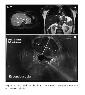
A 51-years-old male with no medical history of interest comes to our outpatient clinic to evaluate a groin hernia with surgical criteria. Incidentally, on the chest X-ray for the preoperatory study, a pulmonary nodule is found consequently giving a chest CT scan with the finding of some unspecific pulmonary nodes and a 3 cm infradiafragmatic nodular tumour on the esophagogastric junction (EGJ) of uncertain character. The analysis goes further with an abdominal MRI with the finding of the named lesion as an exophytic growth, compatible with a stromal tumour at the EGJ. An upper echoendoscopy is done where a 40x32 mm, submucous lesion dependent of the fourth layer is found with regular edges, lightly heterogeneous and with cystic images inside, compatible with a gastrointestinal stromal tumour (GIST) (Fig. 1). An intervention is done identifying the previously referred tumour at Hiss angle. A local gastric resection with a suitable margin and anti-reflux (Collis-Nissen) operation is done. The patient is discharged at the fifth postoperative day and without incidents. After 6 months is without symptoms and disease free.
The anatomopathological study shows a delimited growth, with spindle cells and an expanded nucleus without pleomorphism, or an elevated mitotic index and in fibrous stroma. The immunohistochemistry shows a nucleus-cytoplasmic positivity for S100, CD56 focal positive and negative for desmin, actin, CD34 and C-Kit. Absence of mutation in PDGFRA.
Discussion
Neurofibromas are rare benign tumours that are usually associated to neurofibromatosis (NF), although its occurrence is described as sporadic growing in the oral cavity, parotid gland (1), cheek (2), omentum (3) and penis (4). Despite its benignity a malignancy potential between 4 and 10% has been described in patients with a NF type 1 (5), without truly knowing the malignancy transformation rate when presented as a sporadic lesion.
In the absence of NF, the differential diagnosis of an image with the described characteristics should go towards the GIST as first possibility. At the initial approach the CT chest scan plays an essential role, being indispensable to extent the study with an upper echoendoscopy or a MRI in order to establish the affectation degree of the gastric wall.
In the anatomopathological study of our case the GIST suspicion was discarded due to the negativity of CD34, C-Kit and the PDGFRA mutation absence. The positivity for S100 supports the neurofibromas diagnose.
We consider that the surgical resection is the best treatment option since it eliminates the risk of malign transformation and on the other side it allows to distinguish an unexpected lesion, leading to a definitive diagnosis with the histological study.
Carlos García-Vasquez1, Camilo Castellón-Pavon1, Marta de-Mingo1,
Santos Jiménez-de-los-Galanes1, Pedro Pacheco1, José de-Jaime1,
María Domingo-Ajenjo1 and Carlos Prada-Puente2
1Department of General and Digestive Surgery.
2Department of Pathological Anatomy. Hospital Universitario Infanta Elena.
Valdemoro, Madrid. Spain
References
1. Souaid JP, Nguyen VH, Zeitouni AG, et al. Intraparotid facial nerve solitary plexiform neurofibroma: A first paediatric case report. Int J Pediatr Otorhinolaryngol 2003;67:1113-5. [ Links ]
2. Gómez-Oliveira G, Fernández-Alba Luengo J, Martín-Sastre R, et al. Plexiform neurofibroma of the cheek mucosa. A case report. Med Oral 2004;9:263-7. [ Links ]
3. Fu CY, Lin CH, Peng YJ, et al. Acute abdominal pain caused by spontaneous hemorrhagic infarction of a solitary plexiform neurofibroma of lesser omentum. Z Gastroenterol 2008;46:344-7. [ Links ]
4. Garaffa G, Bettocchi C, Christopher N, et al. Plexiform neurofibroma of the penis associated with erectile dysfunction due to arterial steeling. J Sex Med 2008;5:234-6. [ Links ]
5. Merchán Rodríguez R, Cacabelos Pérez P, Delgado C, et al. Neurofibrosarcoma con metástasis pulmonares en paciente con neurofibromatosis tipo I. An Med Interna (Madrid) 2008;25:152-3. [ Links ]

