


My SciELO
 Custom services
Services on Demand
Journal
Article
 English (pdf)
 Article in xml format
 Article references
 Send this article by e-mail
Indicators
-
 Cited by SciELO
 -
 Access statistics
Related links
-
 Cited by Google
 -
 Similars in
SciELO
Similars in
SciELO -
 Similars in Google
Share

 Permalink
Revista Española de Enfermedades Digestivas
Print version ISSN 1130-0108
Rev. esp. enferm. dig. vol.107 n.6 Madrid Jun. 2015
ORIGINAL PAPERS
Obscure gastrointestinal bleeding: Which factors are associated with positive capsule endoscopy findings?
Iolanda Ribeiro1, Rolando Pinho1, Adélia Rodrigues1, Joana Marques2, Carlos Fernandes1 and João Carvalho1
Departments of 1Gastroenterology and 2Immunohemotherapy. Centro Hospitalar Vila Nova Gaia. Portugal
ABSTRACT
Background: Capsule endoscopy is a first line examination to evaluate obscure gastrointestinal bleeding. The identification of factors associated with the detection of lesions by capsule endoscopy could improve resource utilization and patient selection.
Objectives: To identify factors associated with positive capsule endoscopy findings in patients with obscure gastrointestinal bleeding.
Methods: Retrospective, single-center study, including 203 patients (214 capsule endoscopy procedures) submitted to capsule endoscopy in the setting of obscure gastrointestinal bleeding. Type of obscure gastrointestinal bleeding, number of units of packed red blood cells transfused, type of positive finding, number of endoscopy studies performed prior to capsule endoscopy, comorbidities, medication and Charlson index were evaluated. Overt bleeding was subdivided into ongoing and previous gastrointestinal bleeding. Only lesions with high hemorrhagic potential (P2) were classified as positive findings.
Results: The mean age was 62.2 years and 59.7% of patients were female. Most patients were referred for occult gastrointestinal bleeding (64.5%), while 35.5% were referred for overt gastrointestinal bleeding (63.2% previous-overt gastrointestinal bleeding). The most frequent positive findings included ulcers/erosions (34%) and angioectasias (32%). In univariate analysis, the identification of positive findings was significantly higher in those with ongoing-overt bleeding (p < 0.001), advanced age (p = 0.003), increasing number of pre-capsule endoscopies (p < 0.001), increasing transfusion requirements (p < 0.001), moderate/severe renal disease (p = 0.009) and antiplatelet drugs (p = 0.021) and NSAID intake (p = 0.005). In multivariate analysis, positive findings were significantly higher only in those with ongoing-overt bleeding (odds ratio [OR] 18.68, 95% confidence interval [CI] 3.98-85.6, p < 0.001), higher transfusion requirements (OR 1.23, 95% CI 1.1-1.4, p < 0.001) and NSAID intake (OR 5.9, 95% CI 2.1-17.3, p = 0.001).
Conclusions: Capsule endoscopy should be used early in obscure gastrointestinal bleeding. Ongoing-overt bleeding, higher transfusion requirements and NSAIDs intake were associated with positive findings on capsule endoscopy.
Key words: Obscure gastrointestinal bleeding. Capsule endoscopy. Positive findings.
Introduction
Obscure gastrointestinal bleeding (OGIB) is a common diagnostic challenge faced by gastroenterologists. It represents approximately 5% of all gastrointestinal bleedings and it is defined as bleeding of unknown origin that persists or recurs after an initial negative endoscopic evaluation, including upper and lower endoscopy (1).
OGIB can be classified as obscure-overt and obscure-occult gastrointestinal bleeding (GIB). Obscure-overt GIB refers to recurrent or persistent visible bleeding (hematoquezia, melena or hematemesis) and obscure-occult GIB is defined as recurrent or persistent iron-deficiency and/or positive fecal occult blood (1).
Capsule endoscopy (CE) has emerged as a key diagnostic tool for OGIB. It enables visualization of the entire small bowel and it is safe and non-invasive. CE has been shown to be superior to push enteroscopy (2), small bowel follow-through (3) and computed tomography (4) scan in detecting small bowel bleeding lesions. However, limitations include inability to provide therapy, false negative results and the potential for erratic passage resulting in missed lesions (1).
Although CE is the first line examination to evaluate OGIB, there are few published data on which clinical factors predict the ability of CE to detect pathology in routine clinical practice (5-8). Increasing age, a higher number of pre-capsule upper endoscopies and transfusions, connective tissue diseases, ongoing-overt OGIB and anti-coagulation have been associated with a higher diagnostic yield. Such predictive features could improve resource utilization and patient selection, avoiding unnecessary investigations in high risk groups (5).
The aim of this study was to determine whether any clinical or non-clinical feature predicts an increased likelihood of a diagnostic CE in patients with OGIB.
Material and methods
Patient selection and data collection
A retrospective study was performed on 232 CE made between January 2005 and February 2013, in patients with OGIB. All patients had undergone at least one upper endoscopy (EGD) and colonoscopy prior to CE, which had not detected the source of bleeding.
Eighteen CE were excluded because: The small bowel transit time was < 1 hour, poor small bowel visualization and CE technical problems.
Data collected included demographic variables, type of OGIB (occult versus overt), type of positive findings, number of units of packed red blood cells transfused (PRBC), EGD and colonoscopies performed prior to CE, comorbidities (cardiovascular, respiratory, renal, haematological, liver and rheumatologic diseases), and medications (anticoagulants, antiplatelets and non-steroidal anti-inflammatory drugs [NSAID]). Comorbidities included in the Charlson index were collected as per definitions outlined by the Charlson index authors (9). The Charlson comorbidity index was calculated. Charlson comorbiditiy index is a simple, readily available method of estimating risk of death from comorbid disease. It includes a total of 22 conditions, such as heart disease, AIDS or cancer. Each condition is assigned a score of 1, 2, 3, or 6, depending on the risk of dying associated with each one. Scores are summed to provide a total score to predict mortality (9).
Overt OGIB was subdivided into ongoing-overt OGIB (melenas or hematoquezias during the procedure) and previous-overt OGIB (melenas or hematoquezias in the past, but not during the procedure).
Lesions detected on CE, were classified according to three categories, according to their bleeding potential, as previously reported (2): P0 lesions (with no potential for bleeding: Submucosal veins, diverticula without presence of blood or nodules without mucosal breaks), P1 lesions (with uncertain hemorrhagic potential: Red spots, small or isolated angiomata and erosions without bleeding), and P2 lesions (with high potential for bleeding: Typical angiomata, multiple erosions, ulcers, visible blood, tumors and varices). Only P2 lesions were considered positive findings, because the bleeding risk and the therapeutic impact are significantly higher than P0 or P1 lesions (2). In case of multiple positive findings, a single primary finding believed to be the most probable cause of OGIB was considered. CE with no findings or with lesions classified as P0 and P1, were considered negative in this study.
The classification of small bowel lesions in P0, P1 and P2 is not a validated classification but a result of a consensus of experts (2). However, this classification was used as it is a simple classification used in several series (2,10), allowing the capsule reader to estimate the relevance of each lesion detected at CE and avoiding the over-interpretation of the clinical relevance of some small-bowel lesions.
CE procedure
The Given® Video Capsule system and MiroCam® Video Capsule systems were used in the study. Patients were asked to suspend iron supplements 8 days before the procedure and were told to not eat fibers the 3 days before. They were allowed a liquid diet for lunch in the previous day and were fasted overnight for 12 hours. Written informed consent was obtained from all patients. After capsule ingestion, patients were allowed to eat a light snack 4 hours later.
Each CE recorded at least 8 hours during daytime. The recorded information was downloaded into the respective workstation and images were then reviewed and a report was written.
Statistical analysis
The data was analyzed using Statistical Software Package version 19.0. Descriptive statistics were used to describe the patient's demographic features, clinical characteristics and endoscopic findings. Categorical variables were presented as percentages and numeric variables as means. Results are expressed as percentages or means ± standard deviation (SD) for continuous variables. Chi-square test and t-student test, were used to compare non-continuous and continuous data, respectively. Univariate analysis was performed, in order to identify factors that predicted a higher diagnostic yield. Significant factors in the univariate analysis were evaluated in a multivariate analysis. p < 0.05 was considered to be statistically significant.
Results
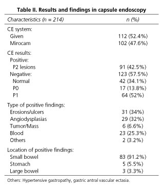
A total of 232 CE examinations were reviewed (Table I). After exclusion of eighteen CE, 214 CE (203 patients) were included for analysis. Eleven patients repeated CE for recurrent OGIB after a first negative CE. The mean age was 62.2 years (standard deviation [SD], 16.9 years), and 58.9% of patients were female (n = 126). The Given® capsule was used in 52.4% of the examinations (n = 112) and the cecum was reached in 87% (n = 186) of all procedures. There was only one complication, a capsule retention in a patient with incidental Crohn's disease that required surgery for removal.

Most patients were referred for occult OGIB [64.5% (n = 138)], while 35.5% (n = 76) were referred for overt OGIB. In the group of overt OGIB, 63.2% (n = 48) of patients had previous-overt OGIB and 36.8% (n = 28) had ongoing-overt OGIB.
The mean number of EGD and colonoscopies performed before CE was 1.3 (SD 0.62) and 1.2 (SD 0.43), respectively. Most patients (55.6%) received transfusion of at least 1 unit of packed red blood cells (PRBC) and 12.1% received ten or more PRBC (Table I). Concerning medications, 28% of patients used anti-platelet drugs, 13.6% used anticoagulants and 10.7% used NSAIDs. The most common comorbid diseases were congestive heart failure (25.7%), osteoarticular disease (13.6%), diabetes with end organ damage (12.1%) and moderate-severe renal disease (9.3%). The mean Charlson index score was 1.35 (SD 1.72).
Positive results (P2 lesions) were seen in 42.5% of cases (n = 91) (Table II). Most of these positive findings included ulcers/erosions (34%; n = 31), angioectasias (32%; n = 29), blood (25.3%; n = 23) and tumors (6.6%; n = 6) (Table II). Most positive findings were located in the small bowel (91.2%), and the remaining in the stomach (5.5%) and colon (3.3%).
In patients with ongoing-overt OGIB, positive findings were seen in 93%, in patients with previous-overt OGIB in 46.8% and in occult OGIB only in 31.2% (Fig. 1). The ability to identify a definite bleeding source was significantly higher in ongoing-overt GIB compared with the other types of bleedings (p < 0.001).

Ongoing overt OGIB (p < 0.001), advanced age (p = 0.003), greater number of pre-capsule EGD (p < 0.001) and colonoscopies (p < 0.001), more transfusion requirements (p < 0.001), moderate/severe renal disease (p = 0.009) and antiplatelet drug (p = 0.021) and NSAID intake (p = 0.005) were all significantly associated with the identification of positive findings in CE in the univariate analysis (Table III). The type of CE did not influence the likelihood of positive findings. In the multivariate analysis only ongoing-overt bleeding (OR 18.68, 95% CI) 3.98-85.6, p < 0.001), higher transfusion of PRBC (OR 1.23, 95% CI 1.1-1.4, p < 0.001) and NSAID intake (OR 5.9, 95% CI 2.1-17.3, p = 0.001) were associated with positive findings (Table IV).


Of the 85 patients with positive CE findings, further investigation and/or therapeutic intervention were undertaken in 51% (n = 43), which included in most cases a device-assisted enteroscopy (82%, n = 38), EGD (10.8%; n = 5), surgery (6.5%; n = 3) and colonoscopy (6.5%; n = 3).
Discussion
Capsule endoscopy (CE) was introduced for clinical use in 2001 as a promising tool to examine the entire small bowel, thereby closing the gap between a conventional EGD and a colonoscopy (11). Nowadays, OGIB is the main indication for CE, with a diagnostic yield ranging from 30 to 92%, depending on the definition of positive findings and the type of bleeding investigated (12). In our series, the overall diagnostic yield of CE was 42%, similar to the percentage reported by a recent study by Shahidi et al. (5), which also considered CE to be positive only when lesions with high bleeding potential were detected.
The definition of what represents a positive finding on CE is an important aspect and frequently the subject of debate, varying between studies and influenced by subjective interpretation of positive findings (5-8,12,13). The construction of a universal nomenclature for CE findings will be an important advance in the use of this technology (13). In our study, only typical angioectasias, ulcers and multiple erosions, tumors and varices were considered positive findings, because they have high bleeding potential. Visible blood was also considered a positive finding, because it alters management and frequently leads to diagnosis. The other types of lesions were classified as negative findings because of their limited or low bleeding potential.
Some studies also indicate that the diagnostic yield is dependent on the type of bleeding, being highest when CE is done in the first 48 hours of hemorrhage (6,12-14). In a recent series, the diagnostic yield was 78.2% in ongoing-overt bleeding, 48.5% in previous-overt bleeding and 36.4% in occult bleeding (12). In our study, the ability of CE to identify a positive bleeding source was significantly higher in patients with ongoing-overt OGIB (93%, p < 0.001) than in patients with previous-overt OGIB (46.8%) and patients with occult GIB (31%). The high rate of positive findings in the setting of ongoing-overt OGIB is explained by the fact that, in this setting, the presence of fresh blood is considered a positive finding as it delineates subsequent management, usually urgent DAE which also has a higher diagnostic yield close to bleeding episode (14). This highlights the importance of using CE early in the diagnosis of OGIB. Other studies (5,7) didn't find this association, because they didn't stratify overt bleeding in ongoing and previous bleeding, which emphasis the importance of differentiation of this type of hemorrhage.
In contrast with other studies, occult OGIB was associated with less positive findings, which can be attributed to the classification of what represents a positive diagnosis. Including P1 lesions in the positive lesion group, we found a higher positive diagnostic yield in patients with occult OGIB (61%).
Approximately 9% of positive CE lesions were located within the reach of conventional upper and lower endoscopies, which emphasis the value of repeating EGD and colonoscopies before more invasive or expensive procedures like device-assisted enteroscopy or CE. This is in accordance with previous studies that showed that up to 24% of pathologic lesions were found within the stomach and colon (15).
Another factor associated with a higher diagnostic yield was an increasing number of PRBC. We found that more than 50% of patients with positive CE findings received more than 3 PRBC before the procedure, a number much higher than those with a negative CE bleeding source (21.2%). Two other studies found an association between higher transfusion requirements and positive findings (5,16). Presumably the transfusion requirement may be a marker of ongoing or severe pathology within the gastrointestinal tract.
Finally, we also found a positive association with NSAID ingestion. This type of medication is associated with erosions and ulcers of the GI tract, the most frequent positive finding in our study. Some series concluded that anticoagulation and anti-platelet drugs are significant predictors of a positive diagnostic finding, an association not found in our study. This may be related to a limitation of patient-reported medication use.
A higher number of endoscopic procedures has been associated with higher diagnostic yield in other studies (5), but it was only associated with a positive diagnosis on univariate analysis in our series. As CE becomes more widely available, it is performed earlier in the course of investigation, which might decrease the number of endoscopies preformed before CE (7).
Advanced age, moderate/severe renal disease and anti-platelet drugs are predictive factors of positive diagnosis in some series (16,17). However we have only found an association with these factors in the univariate analysis. These findings can be explained by the more frequent occurrence of angioectasia in elderly patients and patients with renal failure, which is one of the main bleeding lesions found in our series.
Our series has some limitations. It is a retrospective study, with a small cohort and with bias in identification of comorbidities in medical records. In more than 50% of patients further endoscopic assessment was made, which demonstrates the utility of CE in OGIB. However, the outcome of these patients was not evaluated.
In summary, high diagnostic yield and safety have established CE as an important diagnostic tool for OGIB. In this study we found that erosions/ulcers and angioectasias are the most important lesions found in CE. Ongoing-overt bleeding, increasing transfusion requirements and NSAIDs intake were associated with more positive findings on CE. However, it is important to create a consensus definition of positive finding on CE. Defining the relevance of each lesion detected at capsule endoscopy may help CE readers to improve the quality of their report and consequent decision making, may allow a more correct interpretation of results in clinical studies and improve patient selection for CE.
References
1. ASGE Standards of Practice Committee, Fisher L, Lee Krinsky M, et al. The role of endoscopy in the management of obscure gastrointestinal bleeding. Gastrointest Endosc 2010;3:471-9. [ Links ]
2. Saurin JC, Delvaux M, Vahedi K, et al. Clinical impact of capsule endoscopy compared to push enteroscopy: 1-year follow-up study. Endoscopy 2005;37:318-23. [ Links ]
3. Costamagna G, Shah SK, Riccioni ME, et al. A prospective trial comparing small bowel radiographs and video capsule endoscopy for suspected small bowel disease. Gastroenterology 2002;123:999-1005. [ Links ]
4. Hara AK, Leighton JA, Sharma VK, et al. Small bowel: Preliminary comparison of capsule endoscopy with barium study and CT. Radiology 2004;230:260-5. [ Links ]
5. Shahidi NC, Ou G, Svarta S, et al. Factors associated with positive findings from capsule endoscopy in patients with obscure gastrointestinal bleeding. Clin Gastroenterol Hepatol 2012;10:1381-5. [ Links ]
6. Pennazio M, Santucci R, Rondonotti E, et al. Outcome of patients with obscure gastrointestinal bleeding after capsule endoscopy: Report of 100 consecutive cases. Gastroenterology 2004;126:643-53. [ Links ]
7. Selby W. Can clinical features predict the likelihood of finding abnormalities when using capsule endoscopy in patients with GIB of obscure origin? Gastrointest Endosc 2004;59:782-7. [ Links ]
8. Lepileur L, Dray X, Antonietti M, et al. Factors associated with diagnosis of obscure gastrointestinal bleeding by video capsule enteroscopy. Clin Gastroenterol Hepatol 2012;10:1376-80. [ Links ]
9. Charlson M, Peter P, Kathy LA, et al. A new method of classifying prognostic comorbidity in longitudinal studies: Development and validation. J Chronic Dis 1987;40:373-83. [ Links ]
10. Kawamura H, Sakai E, Endo H, et al. Characteristics of the small bowel lesions detected by capsule endoscopy in patients with chronic kidney disease. Gastroenterol Res Pract 2013;2013:1-8. [ Links ]
11. Hindryckx P, Botelberge T, De Vos M, et al. Clinical impact of capsule endoscopy on further strategy and long-term clinical outcome in patients with obscure bleeding. Gastrointest Endosc 2008;68:98-104. [ Links ]
12. Goenka MK, Majumder S, Kumar S, et al. Single center experience of capsule endoscopy in patients with obscure gastrointestinal bleeding. World J Gastroenterol 2011;17:774-8. [ Links ]
13. Carey EJ, Leighton JA, Heigh RI, et al. A single-center experience of 260 consecutive patients undergoing capsule endoscopy for obscure gastrointestinal bleeding. Am J Gastroenterol 2007;102:89-5. [ Links ]
14. Pinto-Pais T, Pinho R, Rodrigues A, et al. Emergency single-balloon enteroscopy in overt obscure gastrointestinal bleeding: Efficacy and safety. United European Gastroenterol J 2014;2:490-6. [ Links ]
15. Fry LC, Bellutti M, Neumann H, et al. Incidence of bleeding lesions within reach of conventional upper and lower endoscopes in patients undergoing double-balloon enteroscopy for obscure gastrointestinal bleeding. Aliment Pharmacol Ther 2009;29:342-9. [ Links ]
16. Sidhu R, Sanders DS, Kapur K, et al. Factors predicting the diagnostic yield and intervention in obscure gastrointestinal bleeding investigated using capsule endoscopy. J Gastrointest Liver Dis 2009; 18:273-8. [ Links ]
17. Shyung L-R, Lin S-C, Chang W-H. Capsule endoscopy in elderly patients with obscure gastrointestinal bleeding: Retrospective analysis of 152 cases. Int J Gerontol 2010;4:23-7. [ Links ]
![]() Correspondence:
Correspondence:
Iolanda Ribeiro.
Department of Gastroenterology.
Centro Hospitalar Vila Nova Gaia.
Portugal
e-mail:
iolandatribeiro@hotmail.com
Received: 25-10-2014
Accepted: 04-03-2015

