


My SciELO
 Custom services
Services on Demand
Journal
Article
 text in
  English (pdf)
 Article in xml format
 Article references
 Send this article by e-mail
Indicators
-
 Cited by SciELO
 -
 Access statistics
Related links
-
 Cited by Google
 -
 Similars in
SciELO
Similars in
SciELO -
 Similars in Google
Share

 Permalink
Revista Española de Enfermedades Digestivas
Print version ISSN 1130-0108
Rev. esp. enferm. dig. vol.107 n.8 Madrid Aug. 2015
PICTURES IN DIGESTIVE PATHOLOGY
Overt obscure gastrointestinal bleeding secondary to small bowel gastrointestinal stromal tumor diagnosed by videocapsule endoscopy
Tumor de GIST de intestino delgado como causa de hemorragia digestiva de origen oculto manifiesta diagnosticado por cápsula endoscópica
Gabriel Carrilero-Zaragoza1, Guillermo Carbonell-López-del-Castillo2, Gema Ruiz-García3, Juan Egea-Valenzuela1, Paula Tomas-Pujante1, Elena Iglesias-Jorquera1, Josefa Parra-García3, Antonio Sánchez-Torres1, Elena Navarro-Noguera1, Esther Estrella-Díaz1 and Luis Fernando Carballo-Álvarez1
Services of 1Digestive Diseases, 2Radiodiagnosis, and 3Pathology. Hospital Universitario Virgen de la Arrixaca. Murcia, Spain
An 82 year-old male was admitted to our hospital presenting rectal bleeding with hemodynamic and analytic compromise (hemoglobin: 7.3 g/dl). He had personal history of percutaneous closure of the left atrial appendage, severe pulmonary hypertension and was under double-antiaggregation therapy (triflusal and aspirin).
The patient required red blood cells transfusion and upper and lower emergency endoscopies were performed, not being able to find the bleeding lesion. As the origin of the hemorrhage was suspected in the small bowel, videocapsule endoscopy was performed in the following hours, observing a huge jejunal tumor with a big ulcer on its surface (Fig. 1).

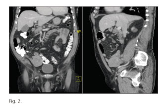
Diagnostic study was completed with an abdominal computed tomography in which a 10x5.3 cm tumor was observed in the jejunoileal area, suggestive of lymphoma vs. gastrointestinal stromal tumor (GIST) (Fig. 2).
Once the patient was stable and with no more episodes of bleeding, surgical intervention was carried out to remove the tumor and the affected segment of the small bowel. Histologic examination of the specimen reported a definitive diagnosis of GIST (Fig. 3).

Discussion
Submucosal tumors have been described as a cause for gastrointestinal bleeding located in the small bowel (1), but it is a rare condition especially in cases of acute bleeding (2).
However, chronic anemia secondary to gastrointestinal occult bleeding is the most frequent form of presentation of GISTs of jejunum and ileum (3,4), as in this case due to ulceration of the mucosa.
References
1. Tran T, Davila JA, El-Serag HB. The epidemiology of malignant gastrointestinal stromal tumors: An analysis of 1,458 cases from 1992 to 2000. Am J Gastroenterol 2005;100:162. [ Links ]
2. Aberle J, Kilie E, Guth S, et al. An unusual cause of acute lower gastrointestinal bleeding. Acta Gastroenterol Belg 2006;69:221-3. [ Links ]
3. Oyanedel R, O'Brien A, Pizarro A, et al. Tumor estromal gastrointestinal (GIST): formas de presentación. Rev Chil Radiol 2005; 11:13-8. [ Links ]
4. Bucher P, Egger JF, Gervaz P, et al. An audit of surgical management of gastrointestinal stromal tumours (GIST). Eur J Surg Oncol 2006;32:310-4. [ Links ]

