


Mi SciELO
 Servicios personalizados
Servicios Personalizados
Revista
Articulo
 texto en
  Inglés (pdf)
 Articulo en XML
 Referencias del artículo
 Enviar articulo por email
Indicadores
-
 Citado por SciELO
 -
 Accesos
Links relacionados
-
 Citado por Google
 -
 Similares en
SciELO
Similares en
SciELO -
 Similares en Google
Compartir

 Permalink
Revista Española de Enfermedades Digestivas
versión impresa ISSN 1130-0108
Rev. esp. enferm. dig. vol.107 no.10 Madrid oct. 2015
LETTERS TO THE EDITOR
Leiomyoma of the round ligament of the liver: Report of one case
Leiomioma del ligamento redondo hepático: descripción de un caso
Key words: Tumour. Neoplasms. Leiomyoma. Round ligament of the liver.
Palabras clave: Tumor. Neoplasias. Leiomioma. Ligamento redondo hepático.
Dear Editor,
Tumoral conditions in the round ligament of the liver are very uncommon and exhibit nonspecific manifestations, hence a high level of suspicion is necessary for their diagnosis.
Case report
We report the case of a 47-year-old woman with no pertinent history presented with months standing abdominal pain in the left iliac fossa in the absence of other manifestations. Abdominal ultrasounds identified a liver of normal size with multiple round images in both lobes suggestive of non-complicated angiomas, although one on the edge of the left lobe is outstanding for being pedunculated, and one in segments VI-VII, approximately 10 centimeters in size, is heterogeneous in echotexture, both being consistent with atypical angiomas.
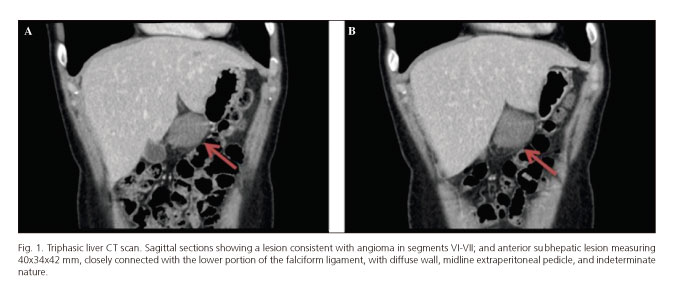
In the presence of these findings, the patient is referred for assessment to our Hepatobiliopancreatic Surgery and Liver Transplant clinic, where the decision is made to complete the previous workup with triphasic liver CT (Fig. 1), which identifies multiple lesions compatible with angiomas and a lesion of indeterminate nature closely connected with the lower portion of the falciform ligament. An abdominal MRI scan is performed to characterize this lesion (Fig. 2), which may consist of a desmoid tumor or a primary or secondary peritoneal growth.

With this radiographic diagnosis surgery is decided upon, and the procedure reveals a benign-looking mass on the round ligament, and an angioma about 10 centimeters in diameter in segments VI-VII, in addition to numerous smaller angiomas in both lobes. The round ligament is excised, with an intraoperative biopsy confirming the lesion's benignity, and a VI-VII segmentectomy is carried out with electrocoagulation.
The patient has a favorable postoperative course and is uneventfully discharged on the fourth day after surgery. A definitive pathology report confirms the presence of a cavernous hemangioma of the liver and a leiomyoma of the round ligament.
Discussion
The most common pathology found at the round ligament of the liver is the dilation of one or several paraumbilical veins in cirrhotic patients with portal hypertension, resulting in portosystemic shunt. More rarely, the round ligament may atypically harbor abdominal conditions of tumoral, infectious or inflammatory origin. Primary tumors are exceptional -ipomas, sarcomas, stromal tumors, PEComas, mesenchymal tumors derived from perivascular epithelioid cells (angiomyolipomas, lymphangioleiomyomas) (1), fibromas (2,3), schwannomas, and mixoid sarcomas, with metastatic disease originating in other tumors, either by continuity (mainly hepatocellular carcinoma) or by hematogenous spread (4), being most common.
The "round ligament tumors" category was first coined in Washington, in the Armed Forces Institute of Pathology archives (Edmonson, 1958) (5).
Leiomyoma is a mesenchymal tumor originating in the intestinal muscularis propria or the tunica media of blood vessels -the umbilical vein in our case (6). Size is highly variable, from less than 0.5 cm (microleiomyoma) to 30 cm in diameter (7). Location at the round ligament is exceptional, and in the liver is rare, the first such case being reported by Demel in 1926; a total of 29 cases have been reported thus far, most in women positive for the Epstein-Barr virus (6).
Most round ligament tumors are asymptomatic, and manifestations, if any, are variable and nonspecific -abdominal pain or palpable mass according to tumor growth. Presence on this ligament explains one of the most relevant and peculiar characteristics of their clinical and radiographic presentation-mobility, which may help in the differential diagnosis. An extravisceral site (ligaments, omentum, etc.) should be suspected in the presence of a mobile abdominal mass in the upper hemiabdomen.
Hepatic mesenchymal tumors represent a challenging diagnosis for radiologists. Radiographic images are nonspecific and depend on tumor nature. Ultrasonography or CT may identify an infiltrate spreading along the round ligament, heterogeneously enhanced after contrast injection. PET may suggest the lesion's tumoral origin in the presence of paraumbilical vein thrombosis. However, only pathology can confirm invasion or the lesion's tumoral nature from a biopsy taken by a needle under imaging or laparoscopic guidance (4).
Microscopically, leiomyomas consist of fascicles of benign-looking fusiform cells without nuclear atypia, with absent or scarce mitoses, and rarely with necrosis. Nuclei are oval in shape and usually centrally located, but they may be laterally offset by various vacuoles, thus acquiring a signet ring morphology. These vacuoles do not contain fat or mucinous substances, which allows to differentiate these tumors from liposarcomas and carcinomas(7).
While no standard therapy is currently available for round ligament leiomyoma given its low prevalence and histologically benign nature, we deem complete surgical resection advisable and adequate as therapeutic cornerstone.
Given the scarce prevalence of this condition, its diagnosis, treatment and biologic behavior are difficult to establish, which warrants future in-depth research studies (6) in order to obtain further information on these aspects.
M.a José Matito-Díaz, Gerardo Blanco-Fernández,
Juana Fernández-Pérez and Diego López-Guerra
Department of Hepatobiliopancreatic Surgery and Liver Transplant.
Hospital Infanta Cristina. Badajoz, Spain
References
1. Holveck A, Bruot O, Mathias J, et al. Sarcoma myoxoïde peu différencié du ligament rond: à propos dun cas. J Radiol 2010;91:574-8. DOI: 10.1016/S0221-0363(10)70091-3. [ Links ]
2. Beyer L, Delpero J-R, Chetaille B, et al. Solitary fibrous tumor in the round ligament of the liver: a fortunate intraoperative discovery. Case Rep Oncol 2012;5:187-94. DOI: 10.1159/000338616. [ Links ]
3. Von Strauss M, Brunner P, Von Holzen U, et al. A large fibroma of the round ligament of the liver. Surgery 2014;155:1095-6. DOI: 10.1016/j.surg.2013.02.005. [ Links ]
4. Novellas S, Mondot L, Caramella T, et al. Pathologie du ligament rond: à propos de deux cas. J Radiol 2008;89:510-3. DOI: 10.1016/S0221-0363(08)71456-2. [ Links ]
5. Yamaguchi J, Azuma T, Fujioka H, et al. Leiomyosarcoma occurring in the ligamentum teres of the liver: A case report and a review of seven reported cases. Hepato-gastroenterology 1996;43:1051-6. [ Links ]
6. Luo X, Ming C, Chen X, et al. Epstein-Barr virus negative primary hepatic leiomyoma: case report and literature review. World J Gastroenterol 2013;19(25): 4094-8. DOI: 10.3748/wjg.v19.i25.4094. [ Links ]
7. Demetri G, Morgan J, Raut C. Epidemiology, classification, clinical presentation, prognostic features, and diagnostic work-up of gastrointestinal neoplasms including GIST. (Monografía en Internet). Tanabe, K: UpToDate; 2013 (acceso 20 de julio de 2013). Disponible en: http://www.uptodate.com. [ Links ]

