


Mi SciELO
 Servicios personalizados
Servicios Personalizados
Revista
Articulo
 texto en
  Inglés (pdf)
 Articulo en XML
 Referencias del artículo
 Enviar articulo por email
Indicadores
-
 Citado por SciELO
 -
 Accesos
Links relacionados
-
 Citado por Google
 -
 Similares en
SciELO
Similares en
SciELO -
 Similares en Google
Compartir

 Permalink
Revista Española de Enfermedades Digestivas
versión impresa ISSN 1130-0108
Rev. esp. enferm. dig. vol.108 no.6 Madrid jun. 2016
First case reported of Bouveret's syndrome associated to duodenal and biliary perforation to retroperitoneum
Primer caso publicado de síndrome de Bouveret asociado a perforación duodenal y vesicular al retroperitoneo
María Victoria Vieiro-Medina, Ramón Gómez-Sanz, Eneida Bra-Insa, Iván Domínguez-Sánchez, Marta de-la-Fuente-Bartolomé, David Díaz-Pérez, Oana Anisa-Nutu and Felipe de-la-Cruz-Vigo
Department of General Surgery. Hospital Universitario 12 de Octubre. Madrid, Spain
ABSTRACT
We present the case of a 69 year old woman with a history of cholecystitis, who consulted for severe abdominal pain, nausea and vomiting. Abdominal CT showed duodenal obstruction caused by a gallstone, cholecystoduodenal fistula and pneumobilia, what is known as Bouveret's syndrome, a rare form of gallstone ileus. Additionally, she presented free duodenal and vesicular perforation to retroperitoneum at the same level of the cholecystoduodenal transit point. The patient underwent a difficult cholecystectomy, enterolithotomy, repair of the duodenal defect, extensive washing and drainage of the retroperitoneum. The postoperative course was uneventful except for a laparotomy infection.
Key words: Bouveret's syndrome. Gallstone ileus. Cholecystoduodenal fistula. Duodenal perforation.
RESUMEN
Presentamos el caso de una mujer de 69 años con diagnóstico de colelitiasis, que acudió a urgencias por cuadro de dolor abdominal intenso, náuseas y vómitos. Un TAC abdominal mostró obstrucción duodenal causada por cálculo biliar de 4 cm, fístula colecistoduodenal y neumobilia, lo que en conjunto se conoce como síndrome de Bouveret, una forma rara de íleo biliar. Adicionalmente presentaba perforación duodenal y vesicular libre a retroperitoneo en el mismo punto de tránsito colecistoduodenal. La paciente fue intervenida quirúrgicamente, realizando colecistectomía dificultosa, enterolitotomía, reparación del defecto duodenal, lavado exhaustivo y drenaje del retroperitoneo. El postoperatorio transcurrió sin complicaciones salvo infección de la herida quirúrgica.
Palabras clave: Síndrome de Bouveret. Íleo biliar. Fístula colecistoduodenal. Perforación duodenal.
Introduction
Bouveret's syndrome is a rare form of biliar ileum presentation where the existence of a cholecistoduodenal or cholecistogastric fistula allows the transit of gallstones and their posterior impaction in the duodenum (1-3). We report, as far as we know, the first case published of Bouveret syndrome with free duodenal and vesicular perforation to retroperitoneum at the same level of the transit point cholecystoduodenal.
Case Report
A 69-years-old woman with a past medical history of hypertension and cholelithiasis who was expecting for an elective laparoscopic cholecystectomy for two months.
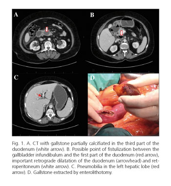
The patient was admitted to the emergency room complaining about an upper abdominal pain radiated to the back with nausea and vomiting. The laboratory tests revealed a serum amylase of 648 U/l and leucocytosis with neutrofilia. The abdominal radiography was completely normal. The patient was admitted to hospital with the diagnosis of acute lithiasic pancreatitis. During her stay she revealed a bad evolution with worsening of the abdominal pain and uncontrolled vomiting despite the placement of a nasogastric tube. Seventy two hours later, a CT was performed finding a big (4.2x2.8 cm) partially calcified gallstone, impacted in the third portion of the duodenum (Fig. 1A). This gallstone was producing an important gastroduodenal retrograde dilatation and a possible point of fistulization between the gallbladder and the first portion of the duodenum (Fig. 1B). In addition, pneumobilia was present (Fig. 1C), as well as free gas and fluid in the retroperitoneum (Fig. 1B), which was initially interpreted as a duodenal perforation in a different point of the cholecystoduodenal fistula.
With these findings and the patient hemodinamically stable, she was taken to the operating room. At subcostal laparotomy there was a large amount of gastrobilliary fluid in the supramesocolic space and the gallbladder was perforated with an important peritoneal mass. In the first part of the duodenum, there was a 2 cm perforation with retroperitoneal communication. A Kocher manoeuvre was performed doing an extensive debridement, fluid aspiration and retroperitoneal washing with no evidence of another point of duodenal perforation as the CT suggested. A big gallstone was impacted in the third portion of the duodenum which was extracted by enterotomy 15 cm further of the Treitz angle (Fig. 1D). The big size of the gallstone caused a partial loss of serous membrane of the small bowel which was repaired with simple sutures.
The orifice of the duodenal fistulization was repaired with double layer transverse suture using absorbable monofilament and an epiploplasty was added. Due to the peritoneal mass reaction and the gallbladder perforation, it was necessary to carried out a difficult cholecystectomy. Finally, after an exhaustive washing, two Penrose drains were placed in the retroperitoneum and near the zone of the duodenal reparation. The postoperative evolution was satisfactory, except for the infection of the surgical wound which required antibiotics and cures.
Discussion
Bouveret's syndrome was described for the first time by Leon Bouveret in 1896 (1). It is more usual in older women and it is an infrequent form of presentation of biliary ileum where the transiting gallstone gets impacted in a duodenal or pyloric point producing obstruction. The symptoms are nonspecific: abdominal pain, sickness, nausea and vomiting (2). The presence of a big size cholelithiasis during a long time produces the formation of a cholecystogastric or cholecystoduodenal fistula, which allows the transit of the obstructive gallstones (3). The cholecystoenteric fistulas appear in 0.3-0.5% of the patients with cholelithiasis, the majority as cholecystoduodenal (60%). Also we can found cholecystocolic (17%), cholecystogastrics (5%) or choledocoduodenal (5%) fistulas. Only the 6% of the transiting gallstones origin a small bowel obstruction due to its size and it occurs in the terminal ileum (50-90%), the proximal ileum and the jejunum (20-40%), an in the duodenum (less than 5% of the cases) (4). The formation of the fistula between the gallbladder and the lumen of the digestive tract is originated in the context of a chronic inflammatory reaction, which stops the free perforation and allows the transit of the gallstone. In our case this connection was established in a small amount of time because of the big size of the stone, which had originated the disruption point and the fistula resulting in a Bouveret's syndrome plus duodeno-biliar retroperitoneal perforation.
The diagnosis of biliary ileum is clinical (intestinal obstruction) and radiological (abdomen Rx, US and CT) (5,6), showing biliary pathology and small bowel obstruction because of an impacted gallstone in some point of the digestive tract. The abdomen Rx can show the Rigler's triad (gastric distension, pneumobilia and ectopic gallstone) (7), only present in a third part of the cases (8). In our case the duodenal free perforation carried to the intervention. Generally, for a less acute case, the final diagnosis is endoscopic (5) which can show the damage of the duodenal wall and sometimes the orifice of the fistula (6).
The treatment in patients with high surgical risk like elderly people or those with a lot of comorbidities can be endoscopic. The disadvantage of this approach is the low rate of success (5,6), for most of the patients the treatment is surgical (5,6). The extraction of the gallstone can be performed by gastrostomy or enterotomy according to the situation and it is necessary to check all the digestive tract because in the 16% of the cases there are more gallstone loss in the bowels (6,7,10).
To our knowledge, there are not published any cases like ours until the present, a Bouveret's syndrome associated to free duodeno-biliar perforation to the retroperitoneum. This fact determines a substantial change in the way a biliary ileum should be managed. First, the duodenal free perforation to retroperitoneum adds gravity to the syndrome and forces to a meticulous closure of the duodenum in addition to performing an exhaustive debridement and drainage. Second, the big size of the gallbladder perforation requires a difficult cholecystectomy (despite of the recommendations in the usual cases of biliary ileum (6,10-12). The global mortality of the case without free duodenal perforation is established in a 25% of the cases and the prognosis is determined by the comorbidities of the patient, the advanced age and the delay in the diagnosis (9).
In conclusion, the Bouveret's syndrome should be suspected in those patients with symptoms of small bowel obstruction and personal history of biliary pathology with big gallstones. The early diagnosis is important because of the high rate of mortality, mainly in the elderly patients and those with a lot of comorbidities. The treatment is principally surgical, although the endoscopic treatment is taking advantage in selected patients. Nevertheless, in our case it was contraindicated because of the presence of duodenal free perforation. Therefore the surgical way was the only treatment resolving the obstruction with enterolithotomy, adding cholecystectomy, closure of the duodenal default and drainage of the retroperitoneum.
References
1. Bouveret L. Stenose du pylore adherent a la vesicule. Rev Med (Paris) 1896;16:1-16. [ Links ]
2. Chick JFB, Chauhan NR, Mandell JC, et al. Traffic jam in the duodenum: Imaging and pathogenesis of Bouveret syndrome. J Emerg Med 2013;45:e135-7. DOI: 10.1016/j.jemermed.2013.04.058. [ Links ]
3. Lowe AS, Stephenson S, Kay CL, et al. Duodenal obstruction by gallstone (Bouveret's syndrome): A review of the literature. Endoscopy 2005;37:82-7. DOI: 10.1055/s-2004-826100. [ Links ]
4. Liew V, Layani L, Speakman D. Bouveret's syndrome in Melbourne. ANZ J Surg 2002;72:161-3. DOI: 10.1046/j.1445-2197.2002.02319.x. [ Links ]
5. Rivera R, Ubiña E, García G, et al. Síndrome de Bouveret resuelto mediante litotricia mecánica endoscópica. Rev Esp Enferm Dig 2006;98:790-2. [ Links ]
6. Cappell MS, Davis M. Characterization of Bouveret's syndrome: A comprehensive review of 128 cases. Am J Gastroenterol 2006;101:2139-46. DOI: 10.1111/j.1572-0241.2006.00645.x. [ Links ]
7. Rigler LG, Borman CN, Noble JF. Gallstone obstruction: Pathogenesis and roentgen manifestations. JAMA 1941;117:1753-9. DOI: 10.1001/jama.1941.02820470001001. [ Links ]
8. Marschall J, Hayton S. Bouveret's syndrome. Am J Surg 2004;187:547-8. DOI: 10.1016/j.amjsurg.2003.12.031. [ Links ]
9. Crespo-Pérez L, Angueira-Lapeña T, Defarges-Pons V, et al. Una causa infrecuente de obstrucción gástrica: síndrome de Bouveret. Gastroenterol Hepatol 2008;31:646-51. DOI: 10.1016/S0210-5705(08)75813-8. [ Links ]
10. Vidal O, Seco JL, Álvarez A, et al. Síndrome de Bouveret: cinco casos. Rev Esp Enferm Dig 1994;86:839-44. [ Links ]
11. Reisner RM, Cohen JR. Gallstone ileus: A review of 1001 reported cases. Am Surg 1994;60:441. [ Links ]
12. Iñíguez A, Butte JM, Zúñiga JM, et al. Síndrome de Bouveret. Resolución endóscopica y quirúrgica de cuatro casos clínicos. Rev Méd Chile 2008;136:163-8. DOI: 10.4067/S0034-98872008000200004. [ Links ]
![]() Correspondence:
Correspondence:
María Victoria Vieiro-Medina.
Department of General Surgery.
Hospital Universitario 12 de Octubre.
Avenida de Córdoba, s/n.
28041 Madrid, Spain
e-mail: vickyvieiro@gmail.com
Received: 20/03/2015
Accepted: 13/05/2015
