


Meu SciELO
 Serviços customizados
Serviços Personalizados
Journal
Artigo
 Inglês (pdf)
 Artigo em XML
 Referências do artigo
 Enviar este artigo por email
Indicadores
-
 Citado por SciELO
 -
 Acessos
Links relacionados
-
 Citado por Google
 -
 Similares em
SciELO
Similares em
SciELO -
 Similares em Google
Compartilhar

 Permalink
Revista Española de Enfermedades Digestivas
versão impressa ISSN 1130-0108
Rev. esp. enferm. dig. vol.108 no.12 Madrid Dez. 2016
PICTURES IN DIGESTIVE PATHOLOGY.
A recurrence of pancreatic non-functioning neuroendocrine tumor mimicking splenosis.
Yeon-Ji Kim and Chang-Nyol Paik
Department of Internal Medicine. St. Vincent's Hospital. College of Medicine. The Catholic University of Korea. Korea.
Case report
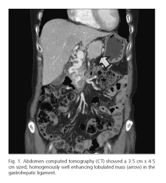
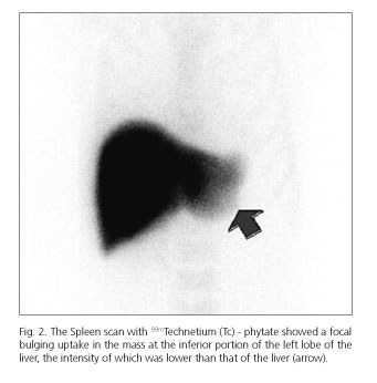
A 63-year-old woman presented with three-month history of epigastric pain. She underwent distal pancreatectomy with splenectomy for pancreatic non-functioning neuroendocrine tumor (NET) 19 years ago, which was completely resected without any evidence of recurrence for 5 years. Initial serologic tests and tumor markers including serum CEA and CA 19-9 were normal. Initial abdomen computed tomogram (CT) showed a 3.5 cm x 4.5 cm sized, homogenously well enhancing lobulated mass in the gastrohepatic ligament (Fig. 1). The scan with 99m Technetium (Tc)-phytate showed a focal bulging uptake in the mass at the inferior portion of the left lobe of the liver, the intensity of which was lower than that of the liver (Fig. 2), suspected to be the intraperitoneal splenosis. Endoscopic ultrasonography with biopsy sampling was not done, because the lesion was located on the periceliac area and the severity of pain was gradually exacerbated despite conservative management. Therefore, surgical resection of the mass was performed, but being partially removed as the mass was firmly encased with the celiac axis. The lesion was consistent with the pancreatic NET.
Discussion
In our case, initially, a long standing history of operation and radioactive uptake of mass were suspected to be the intraperitoneal splenosis. A definitive diagnosis of splenosis can be difficult because radiologic images from sonography, CT or magnetic resonance image are very similar to those found in hypervascular pancreatic tumors such as islet cell tumor and acinar cell carcinoma. Although a nuclear scintigraphy is a useful method to differentiate between abdominal splenosis and metastatic cancer (1), histopathological confirmation should be considered. To the best of our knowledge, this is the first report of a Tc-99m phytate scintigraphy study in the English literature showing the uptake in the metastatic mass of pancreatic NET which has been resected in 19 years. Herein, we present a case of recurrence of pancreatic NET mimicking intraperitoneal splenosis.
References
1. Short NJ, Hayes TG, Bhargava P. Intra-abdominal splenosis mimicking metastatic cancer. Am J Med Sci 2011;341(3):246-9. DOI: 10.1097/MAJ.0b013e318202893f. [ Links ]
