


Mi SciELO
 Servicios personalizados
Servicios Personalizados
Revista
Articulo
 texto en
  Inglés (pdf)
 Articulo en XML
 Referencias del artículo
 Enviar articulo por email
Indicadores
-
 Citado por SciELO
 -
 Accesos
Links relacionados
-
 Citado por Google
 -
 Similares en
SciELO
Similares en
SciELO -
 Similares en Google
Compartir

 Permalink
Revista Española de Enfermedades Digestivas
versión impresa ISSN 1130-0108
Rev. esp. enferm. dig. vol.108 no.12 Madrid dic. 2016
PICTURES IN DIGESTIVE PATHOLOGY
Gastric schwannoma presenting as a casual ultrasonographic finding
Schwannoma gástrico como hallazgo ecográfico casual
Francisco Javier Álvarez-Higueras1, Ana Pereñíguez-López1, Esther Estrella-Díez1, María Muñoz-Tornero1, Juan Egea-Valenzuela1, Águeda Bas-Bernal2, Carmen Garre-Sánchez1, Ángel Vargas-Acosta1, Eduardo Sánchez- Velasco1 and Luis Fernando Carballo-Álvarez1
Departments of 1Digestive Diseases and 2Pathology. Hospital Clínico Universitario Virgen de la Arrixaca. Murcia, Spain
Introducción
Gastric schwannoma is an extremely rare neoplasm (0.2% of all gastric tumors), with neural origin and rising from the nerve plexus of the gastrointestinal tract. It is usually a benign tumor, presenting as solitary o multiple lesions and sometimes associated to neurofibromatosis (1).
Case report
An 80-year-old woman with past history of elevated liver enzymes presented with bilateral lower-extremity edema and ascites. In laboratory tests, it was to highlight the presence of cholestasis and positive antimitochondrial antibodies (AMA-M2).
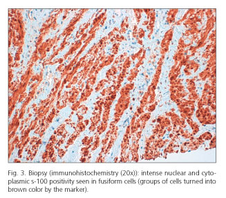
During ultrasonography, signs of chronic liver disease were observed, as well as portal hypertension and ascites. We also could see a heterogeneous hypoechoic mass (56 x 45 mm) located on the posterior wall of the stomach (Fig. 1). In an upper endoscopy this lesion was described as an extrinsic compression, and in a computed tomography as an extraluminal gastric tumor (Fig. 2). Endoscopic ultrasound could not be performed because of the clinical situation of the patient so an ultrasound-guided percutaneous biopsy was carried out. The pathologist observed a mesenchymal neoplasm with fusiform cells with intense S-100 protein expression, as well as negative actin, desmin, CD34 and CD117, compatible with a benign peripheral nerve sheath tumor (schwannoma). Conservative management was decided as surgical treatment was rejected.


Discussion
Diagnosis of gastric schwannoma is usually casual as clinical evolution of this tumor is silent. Exceptionally, compression or bleeding secondary to ulceration can be seen in those with endoluminal location (2). Endoscopic ultrasound is the election diagnostic tool (3) and differential diagnosis must be established with other gastric submucosal tumors: gastrointestinal stromal tumor (GIST), leiomyoma and leiomyosarcoma. Definite diagnosis requires biopsy and immunohistochemical analysis because its ultrasonographic characteristics and location do not differ from GIST (4). The main difference is that schwannoma presents intense S-100 expression and is negative for CD117 and CD34 (5). In symptomatic patients or cases of malignization surgery is the preferred treatment.
References
1. Prévot S, Bienvenu L, Vaillant JC, et al. Benign schwannoma of the digestive tract: A clinicopathological e immunohistochemical study of five cases, including a case of esophageal tumor. Am J Surg Pathol 1999;23:431-6. DOI: 10.1097/00000478-199904000-00007. [ Links ]
2. Yoon W, Paulson K, Mazzara P, et al. Gastric schwannoma: A rare but important differential diagnosis of a gastric submucosal mass. Case Rep Surg 2012;2:123-8. DOI: 10.1155/2012/280982. [ Links ]
3. Martínez-Ares D, Souto-Ruzo J, Yáñez López J, et al. Usefulness of endoscopic ultrasonography in the preoperative diagnosis of submucosal digestive tumors. Rev Esp Enferm Dig 2005;97:416-26. DOI: 10.4321/S1130-01082005000600004. [ Links ]
4. Voltaggio L, Murray R, Lasota J, et al. Gastric schwannoma: A clinicopathologic study of 51 cases and critical review of the literature. Hum Pathol 2012;43:650-9. DOI: 10.1016/j.humpath.2011.07.006. [ Links ]
5. Karamchandani JR. Sox10 and S100 in the diagnosis of soft-tissue neoplasms. Appl Immunohistochem Mol Morphol 2012;20:445-50. DOI: 10.1097/PAI.0b013e318244ff4b. [ Links ]
