


Mi SciELO
 Servicios personalizados
Servicios Personalizados
Revista
Articulo
 texto en
  Inglés (pdf)
 Articulo en XML
 Referencias del artículo
 Enviar articulo por email
Indicadores
-
 Citado por SciELO
 -
 Accesos
Links relacionados
-
 Citado por Google
 -
 Similares en
SciELO
Similares en
SciELO -
 Similares en Google
Compartir

 Permalink
Revista Española de Enfermedades Digestivas
versión impresa ISSN 1130-0108
Rev. esp. enferm. dig. vol.109 no.1 Madrid ene. 2017
PICTURES IN DIGESTIVE PATHOLOGY
Pneumatosis cystoides intestinalis
Neumatosis quística intestinal
Adriana F. Romano-Munive and Rafael Barreto-Zuñiga
Department of Endoscopy. National Institute of Medical Sciences and Nutrition Salvador Zubirán. Mexico City, Mexico
Case report
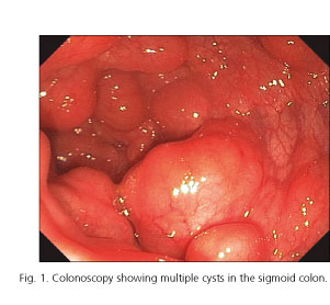
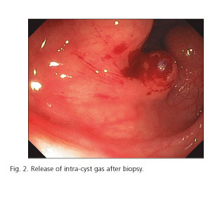
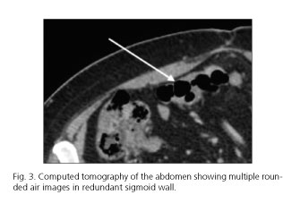
A 54-year-old woman underwent colonoscopy for colon cancer screening. Colonoscopy showed multiple cysts in the sigmoid colon, the largest being 4 cm in diameter. One of the cysts was biopsied. Cyst walls were observed; during biopsy, the gas was released and the cyst collapsed (Figs. 1 and 2). Computed tomography of the abdomen confirmed a diagnosis of pneumatosis cystoides intestinalis (Fig. 3).
Discussion
Pneumatosis cystoides intestinalis is a rare disease characterized by the presence in the intestinal submucosa or subserosa of multiple cysts filled with gas (nitrogen, oxygen, carbon dioxide and hydrogen) (1). This condition occurs more often in males than in females, with cysts most frequently located in the colon. Causes may include elevated intraluminal pressure, pulmonary diseases, bacterial gas production, malnutrition, chemotherapy, and connective tissue diseases, among others. Symptoms of pneumatosis cystoides intestinalis include abdominal pain, diarrhea, bloating and gastrointestinal bleeding. This condition is diagnosed by endoscopy or computed tomography of the abdomen (2,3).
Conservative treatment is successful in 93% of patients. However, 3% of patients develop complications such as intestinal obstruction or perforation.
References
1. Rodríguez-Sánchez D, Sáez-Martínez ME, Sánchez-Jiménez RM, et al. Pneumatosis cystoides, CT colonoscopy and endoscopic correlation. Rev Esp Enferm Dig 2013;105:486-7. DOI: 10.4321/S1130-01082013000800007. [ Links ]
2. Wu LL, Yang YS, Dou Y, et al. A systematic analysis of pneumatosis cystoides intestinalis. World J Gastroenterol 2013;19:4973-8. DOI: 10.3748/wjg.v19.i30.4973. [ Links ]
3. Rivera Vaquerizo PA, Caramuto Martins A, Lorente García MA, et al. Pneumatosis cystoides intestinalis. Rev Esp Enferm Dig 2006;98:959-61. DOI: 10.4321/S1130-01082006001200007. [ Links ]
