


Mi SciELO
 Servicios personalizados
Servicios Personalizados
Revista
Articulo
 texto en
  Inglés (pdf)
 Articulo en XML
 Referencias del artículo
 Enviar articulo por email
Indicadores
-
 Citado por SciELO
 -
 Accesos
Links relacionados
-
 Citado por Google
 -
 Similares en
SciELO
Similares en
SciELO -
 Similares en Google
Compartir

 Permalink
Revista Española de Enfermedades Digestivas
versión impresa ISSN 1130-0108
Rev. esp. enferm. dig. vol.109 no.6 Madrid jun. 2017
PICTURES IN DIGESTIVE PATHOLOGY
Diverticulitis of the cecal appendix: a case report
Diverticulitis del apéndice cecal: presentación de un caso
Marta Alberich1, Carla Bettonica1, Mario Huete2 and Juan Azcarate3
Departments of 1General and Gastrointestinal Surgery, 2Radiology and 3Pathological Anatomy. Hospital Universitario de Bellvitge. Hospitalet de Llobregat, Barcelona. Spain
Case report
A 51-year-old man who presented to the Emergency Room due to a skin rash on the chest and lower limbs and a fever of 39 oC, with associated shivering without any other symptoms, was admitted to the Infectious Disease service due to E. coli bacteremia with no apparent cause. The urine culture was negative and the abdominal ultrasound was unremarkable.
Thirty-six hours after admission he presented with right iliac fossa pain with vomiting, diarrhea and fever, despite undergoing antibiotic therapy. Physical examination revealed tenderness in the right iliac fossa, associated with leukocytosis. The abdominal CT report was compatible with acute appendicitis.
During surgery, an appendix with diverticula along its entire length was observed (Fig. 1). Appendicectomy was performed, without postoperative complications. The patient was discharged on the fourth day.

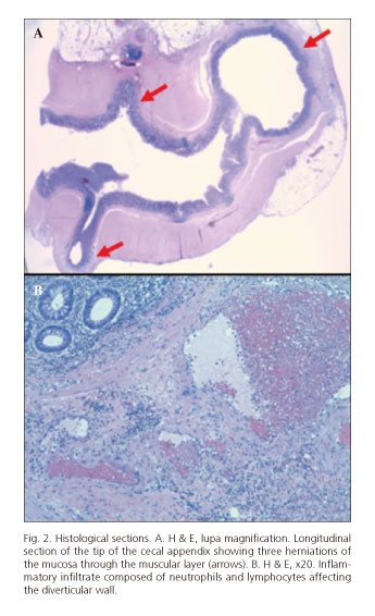
The anatomopathological study reported diverticulosis and non-perforated appendicular diverticulitis with no signs of appendicitis (Fig. 2).
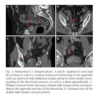
A subsequent revision of the abdominal CT with the radiologists led to a clear identification of the appendicular diverticula (Fig. 3).
Discussion
Appendicular diverticulosis is a rare disease, and complications lead to appendicectomy. Preoperative diagnosis is uncommon in acute forms of presentation as they are usually mistaken for acute appendicitis (1). Abdominal CT could be useful in promptly diagnosing insidious presentations, allowing an early treatment and a decrease in morbidity and mortality (up to a 30% higher than in appendicitis) (1). Moreover, in cases of incidental findings, prophylactic appendicectomy (2,3) will be performed in order to prevent complications and neoplasm development (7.1% incidence) (3).
References
1. Lobo I, Delgado L, Hernández I, et al. Appendiceal diverticulitis and acute appendicitis: Differences and similarities. Rev Esp Enferm Dig 2014;106:452-8. [ Links ]
2. Escobar F, Vega NV, Valbuena E, et al. Diverticulitis apendicular, revisión de la literatura y presentación de dos casos. Rev Colomb Cir 2013;28:223-8. [ Links ]
3. Marcacuzco A, Manrique A, Calvo J, et al. Clinical implications of diverticular disease of the appendix. Experience over the past 10 years. Cir Esp 2016;94(1):44-7. DOI: 10.1016/j.ciresp.2014.05.003. [ Links ]
