


Mi SciELO
 Servicios personalizados
Servicios Personalizados
Revista
Articulo
 texto en
  Inglés (pdf)
 Articulo en XML
 Referencias del artículo
 Enviar articulo por email
Indicadores
-
 Citado por SciELO
 -
 Accesos
Links relacionados
-
 Citado por Google
 -
 Similares en
SciELO
Similares en
SciELO -
 Similares en Google
Compartir

 Permalink
Revista Española de Enfermedades Digestivas
versión impresa ISSN 1130-0108
Rev. esp. enferm. dig. vol.109 no.7 Madrid jul. 2017
PICTURES IN DIGESTIVE PATHOLOGY
Multiseptate gallbladder: a rare ultrasonographic finding
Vesícula biliar multiseptada: un raro hallazgo ecográfico
Raúl Honrubia-López, Joaquín Poza-Cordón, Silvia Gómez-Senent and Pedro Mora-Sanz
Department of Digestive Diseases. Hospital Universitario La Paz. Madrid, Spain
Case report
A 28-year-old male with an unremarkable history was referred from the outpatient clinic due to right-flank abdominal pain during the last three months. On physical examination the patient had right-flank tenderness and was negative for Murphy's sign. The laboratory tests were normal.
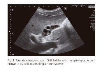
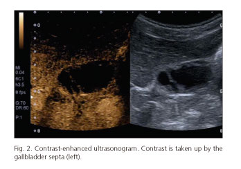
An abdominal ultrasound was performed, revealing a normal sized gallbladder with thin walls and no stones, although there were signs of multiple thin intraluminal and hyperechogenic septa. The bile duct had a normal caliber with no inner contents. An intravenous contrast (Sonovue®) was used which revealed B-mode septal uptake, thus confirming the diagnosis with multiseptate gallbladder. Due to the low symptom burden, which consisted of only mild episodic dull pain that subsided with standard painkillers, a conservative management was selected with no further episodes during a two year follow-up. A repeat abdominal ultrasound after six months showed no changes when compared with the index scan (Figs. 1 and 2).
Discussion
Multiseptate gallbladder is a congenital abnormality categorized as a gallbladder shape variant with some 20 cases reported thus far in the literature. Clinical presentation may be highly variable, ranging from asymptomatic to chronic pain in the right upper quadrant, cholecystitis, and even pancreatitis. It may be associated with other bile duct abnormalities such as choledochal cyst, ectopic gallbladder or anomalous biliopancreatic junction (1).
Ultrasonographic findings include multiple thin hyperechogenic septa within the gallbladder, perpendicular to the gallbladder's walls delineating hypoechogenic cyst-like cavities resembling a "honeycomb" (2). Differential diagnosis includes conditions such as adenomyomatosis, necrotizing cholecystitis, cholesterolosis or hydatid cyst (3).
None of the cases reported in the literature so far include an intravenous contrast medium for the diagnosis of this condition. We believe that ultrasound, particularly contrast-enhanced sonography, should be considered as the "gold standard" technique for the diagnosis of gallbladder conditions. Given that its outcome is usually benign, multiseptate gallbladder should not be considered as an indication for cholecystectomy except when associated with cholecystopancreatitis, diagnostic doubt or disabling chronic pain.
References
1. Wanaguru D, Jiwane A, Day AS, et al. Multiseptate gallbladder in an asymptomatic child. Case Rep Gastrointest Med 2011;2011:1-4. DOI: 10.1155/2011/470658. [ Links ]
2. Nakazawa T, Ohara H, Sano H, et al. Multiseptate gallbladder: Diagnostic value of MR cholangiography and ultrasonography. Abdom Imaging 2004;29:691-3. DOI: 10.1007/s00261-004-0184-5. [ Links ]
3. Demirpolat M, Duygulu G, Tamsel D. Multiseptate gallbladder in a child with recurrent abdominal pain. Diagn Interv Radiol 2010;16:306-7. [ Links ]
