


Meu SciELO
 Serviços customizados
Serviços Personalizados
Journal
Artigo
 texto em
  Inglês (pdf)
 Artigo em XML
 Referências do artigo
 Enviar este artigo por email
Indicadores
-
 Citado por SciELO
 -
 Acessos
Links relacionados
-
 Citado por Google
 -
 Similares em
SciELO
Similares em
SciELO -
 Similares em Google
Compartilhar

 Permalink
Revista Española de Cirugía Oral y Maxilofacial
versão On-line ISSN 2173-9161versão impressa ISSN 1130-0558
Rev Esp Cirug Oral y Maxilofac vol.29 no.3 Madrid Mai./Jun. 2007
CARTA AL DIRECTOR
Massons pseudotumor of the tongue: a possible false positive
Pseudotumor de Masson lingual: un posible falso positivo
Dear Sir,
It is well known that certain oral lesions are very complex for clinicians to evaluate and classify as benign or malignant so that the most appropriate therapeutic attitude can be adopted in each case. Without doubt, experience is a plus in this task, but anyone who has been confronted by these lesions in this location will be conscious of the importance of histological confirmation, and more so when malignancy is suspected. There is, nevertheless, a very large group of tumors and pseudotumors with uncommon oral locations that represent a diagnostic challenge not only for the clinician or surgeon but also for the pathologist, as there is a tendency to over estimate lesions that are sent with a suspected malignancy diagnosis, providing the context is adequate for these to develop.
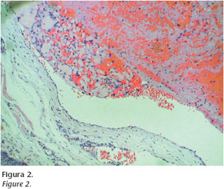
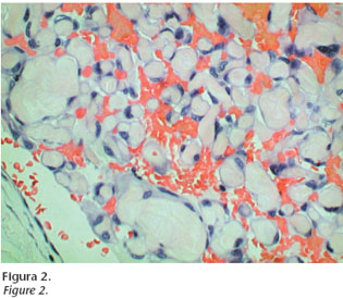
A review of the literature is interesting in order to understand that the oral cavity in not only one of the doors into the organism, but that it forms an important part of the digestive system and of the organism in general. For this reason lesions may be found in this area that we are accustomed to find elsewhere. This is the case of our patient, aged 45, who underwent excision of a lesion on the edge of her tongue, which was slightly ulcerated and hemorrhaging, nodular and with a soft consistency. Its maximum dimension was 2cm. There was no personal history of interest and the patient reported the rapid appearance of the lesion, which had grown slowly over a few weeks. The lesion was sent to the Department of Pathological Anatomy with certain urgency, as there were more than enough signs to suspect malignancy. The histological study of the lesion showed a relatively well-circumscribed vascular lesion that was made up of a fibrin-rich vascular lake that was adhered to the walls. These were covered by endothelium showing no disturbance, although a third of the circumference of the lesion displayed fibrous papillary hyperplasia of the wall, which was also covered with endothelium with no abnormality. The overlying epidermis showed general acanthosis and there was a strip of untouched dermis separating it from the vascular lesion. Given these findings, a diagnosis of vascular endothelial hyperplasia, more commonly known as Massons vascular pseudotumor was given.
With the presentation of this case we would like, not only to increase the casuistry of uncommon oral lesions, but to warn that, given the continuous trauma to which the oral mucosa is subjected (including frequent bites), there is a more than sufficient stimulus for a vessel (lingual in our case) to clot, giving rise to a lesion which can rearrange itself and simulate a neoplasm. Appropriate histological examination is necessary in order to guarantee that, in each case, patients have the most appropriate treatment.

F.J. Torres Gómez1, M. Díaz Delgado1, Silvia Gallana Álvarez2
1Servicio de Anatomía Patológica. Hospital Universitario Virgen Macarena. Sevilla, España
2Servicio de Cirugía Oral y Maxilofacial. Hospital Universitario Virgen Macarena. Sevilla, España

