


 Servicios personalizados
 Inglés (pdf)
 Articulo en XML
 Referencias del artículo
 Enviar articulo por email
 Citado por SciELO
  Citado por Google
  Similares en
SciELO
Similares en
SciELO  Similares en Google
 Permalink
INTRODUCTION
Facial asymmetry can have lasting psychological effects, often resulting from mandibular dysmorphology. The dysmorphology may be caused by various factors, including uncoordinated growth, trauma, developmental abnormalities, and tumors1. Unilateral temporomandibular joint (TMJ) ankylosis is a significant cause of mandibular dysmorphology in growing individuals, which, if left untreated, may cause significant changes to the mandible's morphological characteristics and underlying soft tissue structures. Surgical procedures like genioplasty, distraction osteogenesis, orthomorphic, and orthognathic surgery have been shown to correct mandibular dysmorphology. Orthomorphic surgery corrects morphology, while traditional orthognathic surgery corrects jaw function. In this report, four patients with mandibular dysmorphology secondary to unilateral TMJ ankylosis were surgically treated with orthomorphic correction to restore facial symmetry. The patients were evaluated for various outcomes, including mental nerve paraesthesia, pre- and post-surgical mouth opening, post-operative mandibular symmetry, and complications.
These cases have been described in accordance with the CARE reporting checklist.
CASE DESCRIPTION
Case 1: A 30-year-old male presented to the Department of Maxillofacial Surgery with a flattened right side of the face since childhood. The patient underwent interpositional gap arthroplasty with bilateral coronoidectomy for right TMJ ankylosis 1 year back. On extraoral examination, clinically evident asymmetry was noted in the inferior mandibular border (Figure 1). The patient had a convex profile with incompetent lips and was diagnosed with mandibular asymmetry. Posteroanterior cephalogram and Grummon's analysis confirmed the diagnosis.

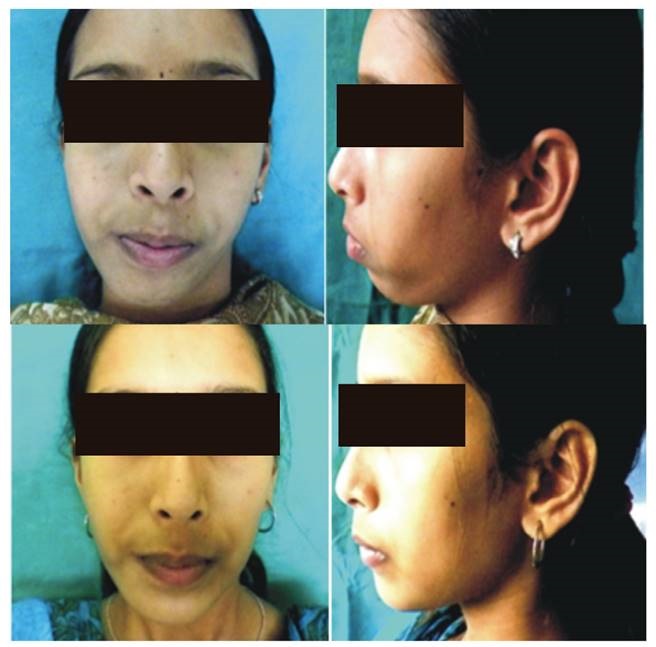
Case 2: A 20-year-old female reported to the department with a complaint of facial disfigurement over the left side of her face since birth. The patient provided us with a history of childhood trauma that led to left temporomandibular joint ankylosis and underwent interposition gap arthroplasty using temporalis muscle for the same, a month back. On extraoral examination, the patient had a convex profile with evident asymmetry with respect to the inferior border of the mandible (Figure 2). The diagnosis of mandibular asymmetry was arrived upon, which was confirmed with pre-operative CT scans. The DICOM images were transferred to 3D Dolphin software for virtual patient modelling (Figure 3).


Case 3: A 28-year-old female with the complaint of a flattened left side of the face since childhood reported to our department. The patient had a successful release of TMJ ankylosis on the left side 4 years back with interposition gap arthroplasty. On examination, the patient had an appreciable asymmetry of the mandible with a convex profile and incompetent lips (Figure 4). A preoperative Grummon's analysis was performed to confirm the diagnosis of mandibular asymmetry (Figure 5).


Case 4: A 16-year-old female presented to us with a complaint of left-sided facial disfigurement. She gave a history of undergoing resection of right TMJ ankylotic mass followed by interposition arthroplasty using temporalis muscle two years back. On examination patient presented with a convex facial profile, asymmetry of the mandible, and incompetent lips (Figure 6).
Written consent was obtained from the patients for undergoing the aforementioned procedure and for the publication of the case report and the accompanying images.
Surgical Procedure (Figure 7)

Figure 7. Intraoperative photographs (A) Incision design (B) Corpus osteotomy (C) Mobilization of the anterior segment (D) The overlapping edges sandwiched with autogenous corticocancellous anterior iliac crest graft (E) Final position and rigid fixation with Titanium miniplates.
All surgeries were performed under general anaesthesia, with local anaesthetic infiltration (1:80,000 adrenaline) at the proposed incision site. Two lower vestibular incisions were used to access the ramal and mental regions, and a mucoperiosteal flap was raised to the inferior border of the mandible while carefully protecting the mental nerve. The surgery involved an eccentric genioplasty continuing along the corpus to a ramus lateral cortical osteotomy on the non-ankylotic aspect of the mandible. Full-thickness sectioning of the inferior border was performed adjacent to the area of eccentric fusion using a combination of rotary instruments and reciprocating saws. The use of piezoelectric scalpel is a potential improvement to this technique. The piezoelectric devices have demonstrated favourable outcomes in a recent systematic review and meta-analysis and have been found to reduce operative blood loss and minimize neurosensory disturbances, while also being less traumatic in comparison to traditional saws2. The anterior portion was mobilized and oriented to rectify the midline and reinstate the proposed vertical and anteroposterior relationship based on the preoperative analysis. Titanium mini plates were used for fixation, and an autogenous corticocancellous iliac bone graft was sandwiched along the overlying margins. Postoperative intravenous antibiotics and analgesics were administered, and the patients were assessed for mental nerve paraesthesia, mouth opening, and postoperative mandibular asymmetry at 3rd day, 3 weeks and 3 months postoperatively. The surgical team assessed post-operative facial symmetry using a 3 point scale (good, moderate, poor). Neurosensory changes were evaluated using Light touch sensation test and Brush directional strokes test. Interincisal clearance was measured using a Willis's gauge. During the follow-up period of 1 year, there were no reported cases of relapse. The patients expressed contentment with the results of the surgical intervention.
Employing Dolphin Imaging software 11.8 (Dolphin Imaging and Management Solutions, Chatsworth, USA), a 3-D virtual model of patient 2 was constructed, followed by marking the virtual mandibular osteotomies in two segments. The virtual treatment involved lateral mobilization of the osteotomized segments to correct frontal asymmetry (Figure 3).
The lack of financial resources among the remaining patients prevented the utilization of 3-D virtual planning. Consequently, cephalometric analysis was used for assessment purposes.
DISCUSSION
Salins and colleagues1 described the orthomorphic correction as "an eccentric genioplasty from the body reaching to the buccal cortex of the mandible". The section transitions from the full thickness of the body to a sagittally oriented buccal cortical osteotomy in the ramal region. After that, the fragment is positioned to address the anteroposterior and midline deficit. Patients with a normal ramus dimension, functional occlusion, insufficient/ standard posterior airway space, or unilateral ankylosis may benefit from orthomorphic osteotomies3.
The study authors noted that mental nerve paraesthesia is a common complication associated with orthomorphic correction for mandibular dysmorphology. In a previous study, the authors reported that two out of five patients (40 %) undergoing orthomorphic correction experienced mental nerve paresthesia. Similarly, Lindquist et al reported alteration in lower lip and chin sensation in 28.5 % of patients following sagittal split osteotomy when performed concurrently with genioplasty. Furthermore, isolated genioplasty resulted in neurosensory changes in 10 % of patients4. In contrast, the current study found that only one patient experienced mental nerve paresthesia. The study authors suggested that the damage to the mental nerve in this case may be due to the shortened height of the body of the ramus, and that leaving a fringe of soft tissue in the premolar region during incision-making could help avoid such damage.
Postoperative mouth opening reduction was due to discomfort and edema, resolving at 3 months. The assessment of postoperative symmetry was deemed satisfactory in three cases, indicating good symmetry, while in one case the assessment showed a moderate level of symmetry (Figure 8). The results were similar to the authors' previous results with this technique i.e., 60 % of cases showed good mandibular symmetry while 40 % showed moderate symmetry5. No postoperative complications were observed, but the follow-up period was deemed insufficient to assess graft resorption (Table I). The technique's limitations include imperfect symmetry and deficient mandibular body in some patients. The preoperative and postoperative remained unaltered as orthomorphic correction focuses on correcting the shape and contour-related deformities without impacting the occlusal status1.

Figure 8. The image displays a PA Cephalogram depicting the radiographic changes that occurred before and after orthomorphic surgery.

Since its inception, very little literature exists regarding its use in the correction of mandibular dysmorphology, mostly in the form of isolated case reports.
Pipalia et al.6) used 3D virtual patient modelling for orthomorphic surgical simulation in a 23-year-old female with facial asymmetry, diagnosed as left condylar hypoplasia. Rudagi et al.7) demonstrated the successful correction of facial asymmetry in a 27-year-old Marfan syndrome patient presenting unilateral TMJ joint ankylosis, obstructive sleep apnoea, and unesthetic contour to correct the residual facial deformity.
With the advent of virtual surgical planning, most surgeons have forgone the extensive laboratory preparations synonymous with the conventional surgical work-up8. Three-dimensional imaging has expanded our stability to assess and plan orthomorphic surgery by eliminating the ambiguity associated with 2D imaging9.
Orthomorphic surgery effectively corrects bony tissues, producing significant transformations and correcting soft tissue deficiencies in various conditions.
In the reported cases, the patients exhibited unwillingness to undergo orthodontic correction and expressed a strong desire for aesthetic improvement. Therefore, a single stage surgery for correction of mandibular asymmetry was undertaken instead of conventional orthognathic surgery.
Compared to the conventional orthognathic surgery and more recent chin augmentation procedures using PEEK or polyethylene (Medpor) implants, orthomorphic surgery has certain limitations. Firstly, orthomorphic correction confers less predictable and proportional changes to the facial contour than orthognathic surgery. Secondly, it is incapable to correct the occlusal cant, a common finding in patients with unilateral temporomandibular joint ankylosis. Thirdly, the need for autologous bone grafts increases the operative time and adds morbidity compared to alloplastic chin augmentation procedures. PEEK, a durable and malleable biomaterial, is conducive to intraoperative modifications. In addition, the ability to prefabricate an implant based on the patient's anatomy can decrease operating time and result in more precise custom fit, in contrast to orthomorphic surgery10.
CONCLUSION
Orthomorphic surgery is more than just a necessary complement to orthognathic surgery. It gives the surgeon a reliable single-stage treatment alternative for complex mandibular dysmorphic structures. The described technique can modify the entire region of the contour defect as a single unit, in any desired dimension.

