Mi SciELO
Servicios Personalizados
Revista
Articulo
Indicadores
-
 Citado por SciELO
 -
 Accesos
Links relacionados
-
 Citado por Google
 -
 Similares en
SciELO
 -
 Similares en Google
Compartir
Anales del Sistema Sanitario de Navarra
versión impresa ISSN 1137-6627
Anales Sis San Navarra vol.32 no.1 Pamplona ene./abr. 2009
Unilateral non-traumatic radiocarpal volar dislocation in a child. A long-term evolution
Luxación radiocarpiana volar atraumática unilateral en una niña. Evolución a largo plazo
S. García-Mata, A.M. Hidalgo-Ovejero
Pediatric Orthopaedic Section. Department of Trauma and Orthopaedic Surgery. Virgen del Camino Hospital. Pamplona. Spain.
Dirección para correspondencia
ABSTRACT
We report an eight year-old female with trisomy 21 referred to our clinic for limitation of wrist mobility. The patient had been diagnosed and treated by polyarticular juvenile chronic arthritis for six months. Clinical and radiological study revealed volar radiocarpal dislocation of the left wrist.
She was treated surgically by open reduction, temporary K-w fixation with six weeks of immobilization. The dislocation relapsed but the joint remained painless. One year later she had 5º of dorsal flexion and 25º of volar flexion and absence of pain. In spite of this situation the family said that the girl lived normal life without any limitation because of her condition, refusing further treatment.
Eight years later the girl remains asymptomatic and does a normal life without limitation because of her condition. The patient and her family refused any treatment.
Based in the failure of surgical treatment performed in our case we think that abstention could be the most reasonable option, mainly in the immature patients.
Key words. Volar radiocarpal dislocation. Trisomy 21. Polyarticular juvenile chronic arthritis.
RESUMEN
Presentamos el caso de una niña de ocho años con trisomía 21 que fue remitida a nuestra consulta por limitación de la movilidad de la muñeca izquierda. La paciente había sido diagnosticada y tratada por artritis crónica juvenil poliarticular durante seis meses. El estudio clínico y radiológico mostraba una luxación radiocarpiana volar de la muñeca izquierda.
Se le efectuó reducción abierta y fijación temporal mediante agujas de Kirschner, con seis semanas de inmovilización. La luxación recidivó, pero la articulación permanecía indolora. Un año más tarde presentaba 5º de flexión dorsal y 25º de flexión volar con ausencia de dolor. A pesar de esa situación, la familia manifestaba que la niña realizaba una vida normal, sin ninguna limitación para su condición, rehusando cualquier tratamiento posterior.
Ocho años más tarde, la paciente permanecía asintomática, realizando una vida normal sin limitaciones para su condición. La paciente y su familia han rehusado otro tipo de tratamiento.
Basándonos en el fracaso del tratamiento quirúrgico realizado en el caso de la paciente que presentamos, pensamos que la abstención puede ser la opción de tratamiento más razonable, principalmente en pacientes con inmadurez en el desarrollo
Palabras clave. Luxación volar radiocarpiana. Trisomía 21. Artritis crónica juvenil poliarticular.
Introduction
In juvenile arthritis, the wrist is the commonest joint in the hand to be involved and, after the knee, the commonest in the body. Wrist is involved at the onset of juvenile rheumatoid arthritis in about 30% of the patients and eventually involved in the disease process in about 60% of the patients1.
The inflammation in the wrist can spread easily because there are communications among the compartments except for the carpometacarpal thumb compartment in the 30-60% of the normal people, increasing with age1.
Chaplin et al2, in 1969, gave the first detailed account of the natural history of juvenile arthritis affecting the wrist.
The habitual evolution of the wrist in juvenile rheumatoid arthritis is progressive. The first signs of wrist involvement are soft-tissue swelling and loss of extension. Later narrowing of the intercarpal joint spaces. The ulna is short and there is progressive ulnar translocation of the carpus. The lunate moves away from the radial articular surface and may lose contact with it. The ulna becomes progressively shorter in relation to the radius. In addition, there is a change in shape of the distal end of the ulna. In progressive cases, the lunate and the ulnar end of the radial epiphysis shows signs of damage. Finally, changes and erosions may be found in the bones forming the wrist-joint.
Trisomy 21 (Down’s syndrome patients) is a well known genetic disorder with mental retardation, cardiac and respiratory problems and shorter life expectancy, associated with ligamentous laxity. This condition facilitates the presence of orthopaedic manifestations.
Theoretically, the association of the two conditions makes the development of orthopaedic lesions easier.
The facts that dorsal and ulnar collateral ligaments are much thinner and weaker than volar and radial collateral ligaments of the carpal bones and that the articular surface is volarly oriented (usually 9º), explain the possibility of volar dislocation of the wrist1.
The mechanical factors contributing to wrist subluxation are: inclination of articular surface of the radius, the distal insertion of the muscles (the strongest are the flexors), contractures in palmar structures and destruction of dorsal ligaments.
Only two cases have been reported of volar non-traumatic radiocarpal dislocation. One case in an 11-year-old girl with juvenile rheumatoid arthritis1 and one case of bilateral wrist dislocation in a 14-year-old female with trisomy 213.
We report a case of one patient with both trisomy 21 and chronic juvenile rheumatoid arthritis who developed unilateral volar radio-carpal dislocation, our management and, the more importantly, the long-term evolution.
Case report
An eight year-old female with trisomy 21 was referred to our Orthopaedic Service by the paediatric rheumatology department in order to perform wrist infiltration with triamcinolone acetate as local treatment of the “swollen” wrist. The patient had been diagnosed and treated as polyarticular juvenile chronic arthritis for six months. The girl presented clear blumb in the left wrist during the previous three months. She presented as synovial inflammation and pannus but had complete volar radiocarpal dislocation.
On physical examination the left wrist was swollen and the metacarpophalangeal joints were also swollen. The joint was painless both at rest and movements. The motion of the wrist was severely limited, mainly the dorsiflexion. The wrist mobility was: volar flexion 15º and 0º of dorsal flexion. The motion of the elbow and shoulder was normal. The motion of the metacarpophalangeal and interphalangeal joints was normal.
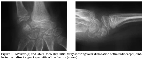
Hand strength was determined by manual dynamometer to assess grip strength. The grip strength was 9.5 Kg for the right hand and 1 Kg for the left (Grip strength dynamometer T.K.K. 5401, Takei scientific instruments, CO., LTD. Japan). The radiographs demonstrated volar radiocarpal dislocation of the left wrist (Figs. 1A and 1B).
We carried out surgical treatment in order to reduce the dislocation and so to achieve higher range of motion. We found complete destruction of the dorsal radiocarpal ligaments and hypertrophic synovium. In the surgery positive drawer sign of the radiocarpal joint was checked with audible “clunk”, which could not have been demonstrated previously with the patient awake. We also found evident attenuation of the dorsal radiocarpal ligaments (these ligaments and the dorsal capsule were very thin and elongated) and therefore it was impossible to differentiate between them. Easy reduction of the radiocarpal joint was performed. Once reduction was achieved we carried out fixation with two Kirschner wires, synovectomy and tightening of the dorsal capsule-ligamentous structures also was performed (Fig. 2). A plaster cast was applied for six weeks.
Both the Kirschner-wires and the plaster cast were removed sixth weeks after surgery and immobilization was continued with a moulded splint at night for three months. Three months later X-rays showed recurrence of the dislocation but the wrist remained painless (Fig. 3). She did not interrupt her school activities. The family agreeded with us not do any further surgery because of the lack of pain and ability of the girl to do daily living activities. We planned that in case of pain or aggravation of the deformity to perform a wrist arthrodesis in older age.
One year later she had 5º of dorsal flexion and 25º of volar flexion (due to the midcarpal motion) and was painless. The grip strength was 10 Kg for the right hand and 3 Kg for the left. In spite of this situation the family said that the girl lived normal life without any limitation because of for her condition, refusing further treatment (Figs. 4A and 4B).
Eight years later the girl remained asymptomatic and did a normal life without limitations because of her condition, with grip strength of 12.2 Kg for the right hand and 9.2 for the left. The patient and her family refused any treatment (wrist arthrodesis) because she is asymptomatic and performed a normal life without any limitation. X-rays showed a SLAC wrist (scapholunate advanced collapse), with collapse and partial destruction of the first carpal row. She had neutral position of the wrist with limitation of the active dorsal and volar flexion.
Discussion
Patients with Down’s syndrome have every day more social integration and more social and sports activities. The mental stimulation and the better management of the cardiac derangements have increased the expectancy of the patients and their families. For this reason their families are more concerned for their possible orthopaedics manifestations.
It is well known the occurrence of ligamentous laxity in Down’s syndrome patients and the frequent association with several orthopaedics disorders such as: atlanto-axial subluxation, hip subluxation and dislocation with acetabular dysplasia, patella instability, genu valgum, flatfoot and metatarsus varus. The abnormal collagenous composition is also responsible of the broad and non-aesthetic scars that they made.
Additionally, in the rheumatic disease, wrist is involved at the onset of the juvenile rheumatoid arthritis in about 30% of the patients and is eventually involved in the disease process about 60% of the patients1.
In most cases, as the children grows up, the disease subsides and some healing occurs, although there is usually residual damage. In the untreated patients the disease leads to ulnar translocation of the carpus, with eventual dislocation leading to the bayonet deformity described by Chaplin et al2. In early management the splintage maintains the alignment.
Wrist deformity is causative factor in the development of distal deformities. Flexion deformity of the wrist leads to progressive hyperextension of the metacarpo-phalangeal joints and flexion of the proximal interphalangeal joints.
The treatment at the presentation of deformity is the general management of the disease (drug therapy) and splintage. Some local preventive measures such as steroid injection (triamcinolone acetate) and surgery (synovectomy) are indicated. But surgery is mainly indicated in case of deformity and/or established functional disturbance.
Arthrodesis is the treatment of choice for uncorrectable ulnar translocation but in adult joints. As Evans et al4 stated, indications for prophylactic treatment during childhood are infrequent: steroid injection, synovectomy, ulnar lengthening, proximal row carpectomy or release of volar capsule for resistant flexion contracture.
Soft-tissue release of volar capsule for flexion deformity is indicated if the radiocarpal joint is well preserved and resistant to reduction. It is an occasional procedure that frequently relapses4.
The patient reported in this paper has the infrequent concomitance of trisomy 21 and chronic juvenile rheumatoid arthritis. This is the first report of a non-traumatic volar radiocarpal joint dislocation in a patient with such combination of conditions in a child.
Findley1 reported volar dislocation of the radio-carpal joint in an otherwise normal 11-year-old girl with juvenile rheumatoid arthritis who was treated with an inflatable sphygmomanometer bag installed in a splint but he did not reported the result of the treatment with this splinting.
Probably this type of splinting can be useful in the initial acute and subacute phases of the juvenile rheumatoid arthritis in addition with rest and medication, at the onset of the dislocation, but no so useful when the deformity has been installed.
Only one case of bilateral wrist dislocation has been reported in a 14-year-old female with trisomy 21, by Jan et al3.
Up to date no one case has been reported in a 21 trisomy with juvenile arthritis affected of volar dislocation of the radiocarpal joint.
The case here reported is similar to that of by Jan et al3, in spite of the mistaken “dorsal wrist dislocation” that he referred, and differed with the previous described by these authors in the pathogenesis. The bilaterality of their case evidences the congenital predisposition for the ligamentous laxity4. But in our case the unilateral occurrence and the coincidence with the wrist affected with chronic arthritis makes us to think that the pathogenesis was the destruction of the dorsal ligaments due to the pannus and inflammation of the radio-carpal synovial on an predisposed congenital abnormal ligamentous laxity because of the trisomy.
In the case reported the affected wrist was the left. This fact makes easy the normal life in a right-handed person.
Jan et al3 did not perform any surgical treatment but an aggressive physical therapy programme was employed without success. The two previous cases reported: one child with trisomy 213, and one with juvenile rheumatoid arthritis1, received non-surgical treatment without success. To date the management and evolution of the radiocarpal volar dislocation in a patient with trisomy 21 and juvenile rheumatoid arthritis has not been reported. According with the evolution of the case here reported, it seems that surgical reduction and tightening of the dorsal capsule-ligamentous structures and temporary fixation with K-wire are not the solution to maintain the reduction. The evolution at eight years of follow-up is a painless wrist with limited range of motion, without functional limitation for this kind of children.
Based in the failure of surgical treatment performed in our case we think that abstention could be the most reasonable option, mainly in an eight year old girl. Radiocarpal arthrodesis could be an option of treatment in case of presence of pain and/or severe volar and ulnar deformity in the adult age, which is not the case of our patient.
As conclusion we recommend close observation in children with rheumatic diseases and Down’ syndrome because the possibility of progressive dislocation of the wrist, that can only be prevented in early stages. In spite of the presence of this lesion, the “normal” life of these children can remain without disabilities. Open reduction with temporary kirschner-wire fixation and tightening of the dorsal capsule-ligamentous structures does not resolve the problem, unless in late diagnosed and structural cases.
The natural history of this dislocation is toward collapse of the first carpal row and clinical arthrodesis due to the scarce but painless active movement.
References
1. Findley TW, Halpern D, Easton JKM. Wrist subluxation in juvenile rheumatoid arthritis: pathophysiology and management. Arch Phys Med Rehabil 1983; 64: 69-74. [ Links ]
2. Chaplin D, Pulkki T, Saarimaa A, Vainio K. Wrist and finger deformities in juvenile rheumatoid arthritis. Acta Rheumatologica Scandinavica 1969; 15: 206-223. [ Links ]
3. Jan WM, Kennedy JG, Dowling FE, Fogarty EE, Moore D. Bilateral wrist dislocation in trisomy 21: A case report. J Pediatr Orthop B 2001; 10: 349-351. [ Links ]
4. Evans DM, Ansell BM, Hall MA. The wrist in juvenile arthritis. J Hand Surg Br 1991; 16: 293-304. [ Links ]
![]() Correspondence:
Correspondence:
Serafín García-Mata
Servicio de Cirugía Ortopédica y Traumatología
Hospital Virgen del Camino
31008 Pamplona. Spain
E-mail: sgarcima@cfnavarra.es
Recepción el 18 de septiembre de 2008
Aceptación provisional el 13 de octubre de 2008
Aceptación definitiva el 16 de octubre de 2008


