


 Serviços customizados
 Inglês (pdf)
 Artigo em XML
 Referências do artigo
 Enviar este artigo por email
 Citado por SciELO
  Citado por Google
  Similares em
SciELO
Similares em
SciELO  Similares em Google
 Permalink
Blue toe syndrome (or sign) is a condition characterised by the development of a blue or violaceous coloration in one or more toes in the absence of any evident trauma, cold-induced lesion or disorders that induce generalised cyanosis1. The location can be unilateral or bilateral and can also affect the upper extremities; it may be accompanied by pain and hypersensitivity when touched, and in most cases is accompanied by peripheral pulses2.
There are many causes for this syndrome (Table 1)1-5. It is essential to carry out an early diagnosis and start a therapy that prevents the natural evolution of the disease towards necrosis, amputation or the patient’s death2,4.
Table 1. Aetiology of signs of blue toe syndrome.
| 1. Reduced arterial flow: |
| 1.1. Embolism: |
| Atheroembolism. |
| Arterial aneurysms. |
| Cardiac or aortic tumour. |
| Myxoma, intimal angiosarcoma of the aorta. |
| Cardiac vegetation. |
| Infectious endocarditis. |
| Non-infectious thrombotic endocarditis. |
| 1.2. Thrombosis: |
| Antiphospholipid syndrome. |
| Neoplasia (acral vascular paraneoplastic syndrome). |
| Thrombotic Thrombocytopenic purpura. |
| Disseminated intravascular coagulation. |
| Cutaneous necrosis caused by anticoagulants. |
| 1.3. Disorders with vasoconstriction: |
| Acrocyanosis. |
| Chilblains. |
| Lupus pernio erythematosus. |
| Medication that causes vasoconstriction. |
| Drugs (cocaine, amphetamines). |
| 1.4. Infectious and non-infectious inflammation: |
| Syphilis, SARS-Cov-2. |
| Purulent infections. |
| Behçet disease. |
| Thromboangiitis obliterans. |
| Other forms of vasculitis. |
| 1.5. Other vascular obstructions: |
| Calcified Vascular disease. Calciphylaxis |
| 2. Reduction of venous return: |
| 2.1. Extensive venous thrombosis: |
| Phlegmasia cerulea dolens and venous gangrene. |
| 3. Changes in blood circulation: |
| 3.1. Hyperviscosity induced by paraproteinemia. |
| 3.2. Ferative myeloproliferative syndromes (polycythaemia vera, essential thrombocythemia). |
| 3.3. Cryofibrinogenemia. |
| 3.4. Cryoglobulinemia. |
| 3.5. Cold agglutinins. |
The case described here is a male subject of 46 years of age, with blood pressure within normal limits and of normal weight. He smoked 20 cigarettes a day and was a polysubstance user (cocaine, heroin and benzodiazepines). He was treated with venlafaxine, olanzapine, quetiapine and gabapentin; with a record of treated syphilis, hepatitis A and B with a positive Mantoux test, with no radiological signs of tuberculosis.
The patient visited the doctor for the first time on 29/11/2022 complaining of a pain in the 4th toe of the left foot, with a blueish coloration, and unrelated to any trauma. He was treated with analgesics and anti-inflammatory drugs. A necrotic patch subsequently appeared, which was treated with dressings, after which the ulcer was completely healed on 02/01/2023.
On 04/01/2023, the patient visited the doctor again, presenting intense pain and cyanosis of the first toe of the left foot that was unrelated to any trauma. The toes evolved to form an abscess in the distal region, for which cloxacillin and ibuprofren were prescribed. Two bullous lesions also appeared on the internal lateral side of the toe that later changed into necrotic patches. The patient was referred to the dermatologist, who gave a diagnosis of blue toe syndrome on 03/03/23 and referred the patient to vascular surgery to rule out the possibility of thromboangiitis obliterans (non-atherosclerotic inflammatory disorder that usually affects small and medium arteries, and is associated with the formation of inflammatory occlusive thrombi that cause ischaemia of extremities and digital necrosis; it often occurs in young men between 20 and 40 years of age, and is closely related to tobacco consumption4).
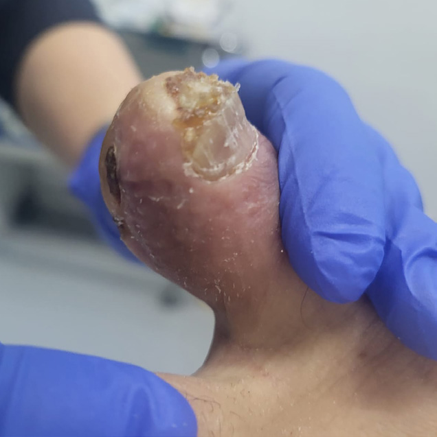
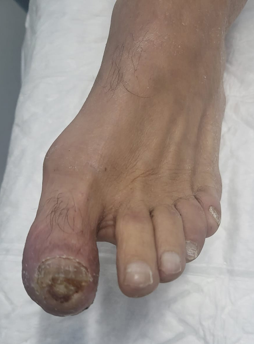
On 11/03/23, the assessment from vascular surgery was still pending, and the inmate was transferred to our centre to attend a trial. On admission, we found a dry ulcer in the distal region of the first toe of the left foot (Figures 1 and 2) along with two necrotic patches on the internal lateral side of the same toe (Figure 1), accompanied by intense pain. The pedis pulse was present.
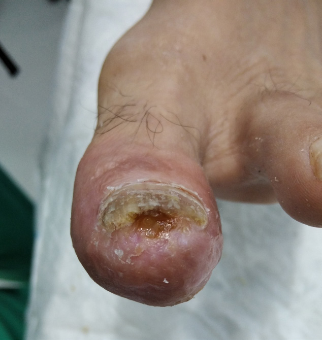
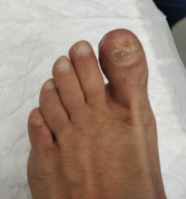
We commenced treatment with hydrogel (Purilon®) and silicon dressings (Mepitel®), on the lateral necrotic patches and in the distal region of the toe. This therapy continued until the patches were completely healed. Given the possibility of thromboangiitis obliterans, an individual smoking cessation intervention was applied, which led to the patient completely giving up smoking, accelerating the favourable evolution of the ulcers (Figure 3), which were finally healed (Figure 4).
The patient was discharged at the treatment room on 09/05/2023, and is awaiting an assessment of his condition by the vascular surgery service.

