


Mi SciELO
 Servicios personalizados
Servicios Personalizados
Revista
Articulo
 texto en
  Inglés (pdf)
 Articulo en XML
 Referencias del artículo
 Enviar articulo por email
Indicadores
-
 Citado por SciELO
 -
 Accesos
Links relacionados
-
 Citado por Google
 -
 Similares en
SciELO
Similares en
SciELO -
 Similares en Google
Compartir

 Permalink
Medicina Oral, Patología Oral y Cirugía Bucal (Internet)
versión On-line ISSN 1698-6946
Med. oral patol. oral cir.bucal (Internet) vol.11 no.1 ene./feb. 2006
ORAL MEDICINE AND PATHOLOGY
Clinical evaluation of dental and periodontal status in a group
of oncological patients before chemotherapy
Mónica Paula López Galindo 1, José V. Bagán 2, Yolanda Jiménez Soriano 3,
Francisco Alpiste 4, Carlos Camps 5
(1) Dentist. University of Valencia.
(2) Chairman of Oral Medicine, Valencia University Medical and Dental School.
Head of the Service of Odontology, Valencia University General Hospital.
(3) Associate Professor of Oral Medicine, Valencia University Medical and Dental School.Valencia
(4) Associate Professor of Periodontology, Valencia University.
(5) Head of the Service of Oncology, Valencia University
General Hospital. (Spain)
ABSTRACT
Objective: To evaluate the dental status of 88 cancer patients before chemotherapy.
Material and methods: Eighty-eight patients with cancer in different body locations were studied and compared with a control group. Dental plaque was assessed by means of the Silness and Löe index, dental status with the DMFT index, and periodontal status with the modified CPI index.
Results: In the oncological patients the mean Silness and Löe index was 1.28±0.11. Patients showed multiple missing teeth (mean number 7.55±0.80); the mean number of decayed teeth was 2.10±0.36; and the mean number of filled teeth was 2.27±0.37. As to periodontal status, the mean modified CPI index was 1.45±0.11.
In the control group, the mean Silness and Löe index was 0.94±0.00. The mean number of decayed teeth was 1.21±0.25; the mean number of missing teeth was 4.97±0.67; and the mean number of filled teeth was 4.82±0.44. The mean modified CPI index was 1.29±0.10.
Conclusions: Oncological patients in our study showed more dental plaque versus healthy patients and more decayed and missing teeth. However, patients in the control group showed more filled teeth than cancer patients. Periodontal status as determined by the modified CPI index was similar in both patient groups.
Key words: Chemotherapy, dental and periodontal status.
RESUMEN
Objetivos:
Valorar el estado bucodental en 88 pacientes con cánceres corporales, previo al
inicio de su tratamiento quimioterápico.
Diseño del estudio: Estudiamos 88 pacientes con cánceres de diferentes
localizaciones corporales y los comparamos con un grupo control. Analizamos la
placa dental (mediante el índice de Silness y Löe), el estado dental (mediante
el índice CAO.D) y el estado periodontal (índice CPI modificado).
Resultados: En el grupo de pacientes oncológicos, la media del índice
de placa de Silnness y Löe fue de 1,28±0,11. Los pacientes presentaban múltiples
ausencias dentarias, siendo la media de dientes ausentes por caries de 7,55±0,80.
También se observó que la media de caries por paciente era de 2,10±0,36 y de
dientes obturados de 2,27±0,37; por lo que respecta al estado periodontal, el
valor del índice CPI modificado fue de 1,45±0,11.
En el grupo control, la media del índice de placa de Silnness y Löe fue de
0,94±0,00. Por lo que respecta a los dientes cariados, la media era de 1,21±0,25;
la media de dientes ausentes por caries era de 4,97±0,67 y el valor de la media
de los dientes obturados era de 4,82±0,44. La media del índice periodontal CPI
modificado, en el grupo control, fue de 1,29±0,10.
Conclusiones: Los pacientes oncológicos de nuestro estudio
presentaron mayor cantidad de placa dental que los pacientes sanos. Además tenían
más dientes cariados y ausentes que los individuos sanos. En cambio, los
pacientes del grupo control presentaron más dientes obturados que los pacientes
afectos de cáncer. El estado periodontal estudiado en ambos grupos de
pacientes, mediante el índice CPI modificado, demostró que era similar en los
individuos sanos y en los oncológicos.
Palabras clave: Quimioterapia, estado dental y periodontal.
Introduction
Antineoplastic therapy includes surgery, radiotherapy and chemotherapy alone or in combination, depending on the nature and extent of the tumor (1).
Antineoplastic chemotherapy presently consists of the use of drugs (cytostatic agents) that destroy or hinder the proliferation of tumor cells. Treatment is followed by tumor cell necrosis, after which a neoplastic cell recovery phase may be observed. The problem of these treatments is that in most cases their action is not selectively target to tumor cells. In effect, anticancer drugs affect not only neoplastic cells but also other similarly rapidly dividing normal cells such as bone marrow, hair follicle cells and the orodigestive epithelium (2). Chemotherapy is characterized by a narrow borderline between its antitumor effects and toxicity (which may even prove fatal) (3). Due to the side effects upon the oral cavity, patient oral status prior to chemotherapy is important for the quality of life of these patients, because the possibilities for intervention after chemotherapy are limited.
The present study explores oral and dental status in a group of patients with cancer before chemotherapy, with the evaluation of possible prior dental intervention, taking into account aspects such as tumor stage and location, and patient dental hygiene and motivation.
Material and Methods
Oral and dental status was explored in 88 patients in the Service of Odontology (Valencia University General Hospital; Valencia, Spain), between October 2000 and January 2004.
Inclusion criteria were:
1. A diagnosis of cancer in any location, except oral cancer.
2. Patients programmed for systemic chemotherapy.
3. Presence of teeth to allow the evaluation of dental and periodontal status. Edentulous patients were excluded from the study.
The mean patient age was 56.75 years (standard deviation (SD) = 14.16 years). There were 38 males (43.2%) and 50 females (56.8%). The patients were examined in the mentioned Service of Odontology prior to chemotherapy. A clinical history was compiled, and certain data were recorded, such as: toxic habits (smoking, alcohol abuse) and tumor diagnosis, location and stage. Depending on their oral hygiene status, the patients were classified into three groups: excellent oral hygiene (tooth brushing 3 times a day); good oral hygiene (brushing 1-2 times a day); poor oral hygiene (failure to brush daily).
To conduct a comparative study versus healthy individuals, 90 controls were included, based on the following inclusion criteria: absence of systemic disorders; absence of medication of any kind at the time of the study.
There were no significant differences between the two groups in terms of age and sex. Mean age in the control group was 55.51 years (SD = 15.18). There were 41 males (45.6%) and 49 females (54.4%).
After compiling the clinical history, the following explorations were performed in both groups:
(a) Evaluation of dental plaque. Buccal and lingual/palatal examination of the dental arches was performed with a plane mouth mirror (number 5) and a dental probe (number 23). Dental plaque was assessed in the following teeth: 1.6, 1.1, 2.4, 3.6, 3.1 and 4.6, according to the Silness and Löe scale (4). After the teeth were examined, the arithmetic mean was calculated for all scores obtained in each patient.
(b) Evaluation of the DMFT index (4). Using a plane mouth mirror (number 5) and a dental probe (number 23), we recorded the number of decayed (D), missing (M) and filled (F) permanent teeth. The sum of these three values yielded the corresponding DMFT index (T = permanent teeth). The caries criteria used were those of the World Health Organization (WHO), which defines caries when "... a lesion in a pit or fissure, or on a smooth tooth surface, has an unmistakable cavity, undetermined enamel, or a detectably softened floor or wall" (5). A crown was considered filled, with decay, when it had one or more permanent restorations and one or more areas that were seen to be decayed. Third molars were excluded from the study.
(c) Evaluation of the modified Community Periodontal Index (modified CPI)(4,6-8). Instead of dividing the mouth into sextants, we took the following tooth numbers: 1.7 or 1.6, 1.1, 2.6 or 2.7, 3.6 or 3.7, 3.1 and 4.6 or 4.7. Periodontal probing was carried out with a plane mouth mirror (number 5) and a dental probe. Each tooth was examined in the buccal and lingual/palatal surfaces at three points (mesial, medial and distal); the greatest probe depth was registered in mm. The mean value of the pocket depth was obtained by calculating the arithmetic mean of the greatest values obtained in the explored teeth. Periodontal status was scored as follows: 0 = health; 1 = bleeding; 2 = supra- or subgingival calculus, excessive fillings; 3 = pocket depth 4-5 mm; 4 = pocket depth 6 mm or more; X = excluded sextant.
The data obtained were subjected to descriptive and comparative statistical analysis. The Student t-test was used for comparing the means of quantitative variables, while analysis of variance (ANOVA) was performed to compare the means of more than two groups of quantitative variables. Statistical significance was considered for p≤ 0.05.
Results
Thirty-six patients (40.9%) were diagnosed with adenocarcinoma, 22 (25.0%) with infiltrating ductal carcinoma, 8 (9.1%) squamous cell carcinoma, and 22 patients (25.0%) with some other type of cancer (Table 1).

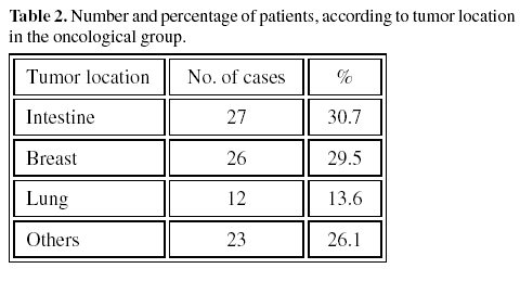
The distribution of tumor location was as follows: 27 patients (30.7%) had intestinal cancer, 26 (29.5%) had breast cancer, 12 patients (13.6%) had lung cancer, and 23 (26.1%) presented multiple locations (Table 2).
Tumor staging according to the TNM classification showed 13 patients (14.8%) to be in stage I, 19 (21.6%) in stage II, 22 (25.0%) in stage III, and 34 (38.6%) in stage IV. The data relating to oral hygiene on both groups are reported in Table 3.

The results relating to dental and periodontal evaluation (including plaque index, mean carious decayed and filled teeth, DMFT index and modified CPI index) are described in Table 4.
Discussion
While chemotherapy offers important positive results in the management of cancer, it also has a series of undesirable effects. In this context, the creation of healthy oral conditions before chemotherapy is administered can afford patient benefits, since the negative effects of chemotherapy upon the oral cavity are less pronounced in the presence of a healthy mouth without dental or periodontal disorders (4,9-12).
To assess oral hygiene, the Silness and Löe index was measured yielding a mean score of 1.28±0.11 in the 88 oncological patients, and 0.94±0.00 in the control group. The only study similar to our own found in the literature was published by Jankovic et al. (13). In this study, 20 healthy patients were compared with 30 oncological patients before the administration of chemotherapy; in both groups the age and sex distributions were similar to our own. Based on the Silness and Löe index, the authors recorded a value of 1.39±0.65 in the 20 healthy subjects and 1.57±0.90 in the 30 oncological patients. The study failed to mention whether the intergroup differences were statistically significant, however. The values recorded in both groups were slightly greater than our own.
Our study reflects a high DMFT index in the oncological group, with a mean value of 11.89±0.88. The magnitude of this score was not due to the decayed (mean 2.10±0.36) or filled teeth (mean 2.27±0.37), but to the missing teeth (mean 7.55±0.80). The mean DMFT score for the control group was 10.97±0.71, and was largely attributable to missing and filled teeth.
In a study of 736 healthy adults aged between 19-64 years, Athanassouli et al (14) in 1990 reported a DMFT index of 8.99±5.98 for the 19-24 years age interval, versus 17.05±6.58 for the 55-64 years interval. These values are slightly greater than our own.
As regards periodontal status assessed by the modified CPI index, the mean value was found to be higher among the oncological patients (mean 1.45±0.11) than in the healthy group (mean 1.29±0.10) though the difference failed to reach statistical significance (t=1.08; p=0.28).
Diamanti-Kipioti et al. (15), in 169 healthy Athenian farmers aged 25-64 years, found the CPITN (Community Periodontal Index of Treatment Needs) to be 1±1.4 (i.e., similar to the score recorded in our groups of patients).
On relating different parameters to periodontal status, statistically significant correlations were observed. In effect, a positive relation was found between the Silness and Löe plaque index and the modified CPI periodontal index (R=0.55; p=0.00), i.e., the higher the plaque index, the higher the periodontal index. The presence of dental plaque is related to poorer periodontal status (16,17).
A positive relationship was found between carious teeth and the modified CPI index (R=0.24; p=0.03).
Patients initially presenting a poor periodontal and/or carious status who are treated before chemotherapy and receive intense oral care during treatment show a significant reduction in the frequency of the oral complications associated with chemotherapy (18-20). Many authors consider it necessary to provide dental treatment before chemotherapy, eliminating any possible foci of dental disease with the aim of minimizing or eradicating the effects of chemotherapy upon the oral cavity (21-33). Moderate dental plaque levels may contribute to local infection in immunocompromised patients (18).
Trauma-inducing prostheses, and sharp-edged or broken teeth imply an increased risk of ulcerations and mucositis (34). Hence the importance of correcting these possible alterations before chemotherapy is administered.
In conclusion, our oncological patients showed more dental plaque, and more decayed and missing teeth than the controls. However, no differences in modified CPI periodontal index were observed between the two groups.
![]() Correspondence
Correspondence
Dr. José Vicente Bagán Sebastián
Hospital General Universitario de Valencia
Servicio de Estomatología
Av/ Tres Cruces s/n
46.014 Valencia
E-mail: bagan@uv.es
Received: 25-01-2005
Accepted: 4-12-2005
References
1. Feliu J, Artal A, Garrido P. Tratamientos oncológicos. En: Ordóñez A, García de Paredes ML, Feliu J, Zamora P, eds. Oncología clínica. Madrid: Interamericana McGraw-Hill; 1992. p. 261-5. [ Links ]
2. Oñate RE, Bermejo A. Asistencia odontológica a pacientes oncológicos. En: Bullón P, Machuca G, eds. La atención odontológica en pacientes médicamente comprometidos. Madrid: Laboratorios Normon; 1996. p. 387-414. [ Links ]
3. Holmes S. The oral complications of specific anticancer therapy. Int J Nurs Stud 1991;28:343-60. [ Links ]
4. Cuenca E. La identificación de problemas en Odontología Comunitaria. En: Cuenca E., Manau C, Serra LL eds. Manual de Odontología Preventiva y Comunitaria. Barcelona: Ed. Masson, S.A. 1991. p. 226-42. [ Links ]
5. WHO.Oral Health Surveis Basic Methos. 2ª Edition. World Health Organization. Geneva; 1977. [ Links ]
6. Ainamo J. Epidemiología de la enfermedad periodontal. En: Lindhe J ed. Periodontología Clínica. Buenos Aires: Editorial Médica Panamericana; 1989. p. 70-86. [ Links ]
7. Ainamo J, Barmes D, Beagrie G, Cutress T, Martin J, Sardo-Infirri J. Development of the World Health Organization (WHO) Community Periodontal Index of Treatment Needs (CPITN). Int Dent J 1982;32:281-91. [ Links ]
8. Cutress T., Ainamo J., Sardo-Infirri J. The Community Periodontal Index of Treatment Needs (CPITN) procedure for population groups and individual. Int Dent J 1987;37:223-33. [ Links ]
9. Milián MA, Silvestre J, Jiménez Y. Patología infecciosa oral en enfermos con procesos sistémicos. En: Liébana J, Bagán JV eds. Terapéutica antimicrobiana en odontoestomatología. Madrid: IM & C; 1996. p. 369-82. [ Links ]
10. Toht BB, Martin JW, Fleming TJ. Complicaciones orales asociadas a la terapia oncológica. La experiencia del Centro Oncológico MD Anderson. Arch Odontoestomatol 1991;7: 87-96. [ Links ]
11. Lockhart PB, Clark J. Pretherapy dental status of patients with malignant conditions of the head and neck. Oral Surg Oral Med Oral Pathol 1994;76:236-41. [ Links ]
12. Scully C, Epstein JB. Oral health care for the cancer patient. Oral Oncol Eur J Cancer 1996;23B:281-92. [ Links ]
13. Jankovic L, Jelic S, Filipovic-Ljeskovic I, Ristovic. Salivary immunoglobulins in cancer patients with chemotherapy-related oral mucosa damage. Oral Oncol, Eur J Cancer 1995;31B: 160-5. [ Links ]
14. Athanassouli T, Koletsi-Kounari H, Mamai-Homata H, Panagopoulos H. Oral health status of adult population in Athens, greece. Community Dent Oral Epidemiol 1990;17:82-4. [ Links ]
15. Diamanti-Kipioti A, Papapanou PN, Moraitaki-Tsami A, Lindhe J, Mitsis F. Comparative estimation of periodontal conditions by means of different index systems. J Clin Periodontol 1993;20:656-61. [ Links ]
16. Baca P, Llodra JC, Bravo M. Caries dental. Etiopatogenia. Clínica. Diagnóstico. Control y tratamiento. En: Terapéutica antimicrobiana en Odontoestomatología. Madrid: IM&C, 1996. p. 219-32. [ Links ]
17. Martínez P. Enfermedad periodontal. En: Bagán JV, Ceballos A, Bermejo et al. Medicina Oral. 1ª ed. Barcelona: Masson SA, 1995. p. 103-17. [ Links ]
18. DePaola LG, Minah GE, Peterson DE, Williams LT, Overholser CD, Stansbury DM et al. Dental care for patiens receiving chemotherapy. JADA 1986;112:198-203. [ Links ]
19. Guggenheimer J, Verbin RS, Appel BN, Schmitz J. Clinicopathologic effects of cancer chemotherapeutic agents on human buccal mucosa. Oral Surg 1977;44:58-63. [ Links ]
20. Lockart PB, Sonis ST. Alterations in the oral mucosa caused by chemotherapeutic agents. J Dermatol Surg Oncol 1981;7:1019-25. [ Links ]
21. Laine PO, Lindquist JC, Pyrhönen SO, Strand-Pettinen M, Teerenhovi LM, Meurman JH. Oral infection as a reason for febrile episodes in lymphoma patients receiving cytostatic drugs. Oral Oncol, Eur J Cancer 1992;28B:103-7. [ Links ]
22. Bagán Sebastián JV, Peñarrocha Diago M. Patología oral inducida por fármacos y sustancias químicas. En: Esplugues J, Morcillo EJ, de Andrés-Trelles F, eds. Farmacología en clínica dental. Barcelona: J. R. Prous Editores; 1993. p. 438-9. [ Links ]
23. Rutkauskas JS, Davis JW. Effects of chlorhexidine during immunosuppressive chemotherapy. Oral Surg Oral Med Oral Pathol 1993;76:441-8. [ Links ]
24. Graham KM, Pecoraro DA, Ventura M, Meyer CC. Reducing the incidence of stomatitis using a quality assesment and improvement approach. Cancer Nurs 1993;16:117-22. [ Links ]
25. Lindquist SF, Hickey AJ, Drane JB. Effect of oral hygiene on stomatitis in patients receiving cancer chemotherapy J Prosthet Dent 1978;40:312-4. [ Links ]
26. Heimdal A, Mattson T, Dahlöf G, Lönnquist B, Ringden O. The oral cavity as a port of entry for early infections in patients treated with bone marrow transplantation. Oral Surg Oral Med Oral Pathol 1989;68:711-6. [ Links ]
27. National Institutes of Health. National institutes of health consensus development conference statement: oral complications of cancer therapies: diagnosis, prevention and treatment. JADA 1989;119:179-83. [ Links ]
28. Hickey AJ, Toth BB, Lindquist SB. Effect of intravenous hyperalimentation and oral care on the developement of oral stomatitis during cancer chemotherapy. J Prosthet Dent 1982; 47:188-93. [ Links ]
29. Carl W, Higby DJ. Oral manifestations of bone marrow transplantation. Am J Clin Oncol 1985;8:81-7. [ Links ]
30. Öhrn KO, Wahlin YB, Sjödén PO, Wahlin ACE. Indications for and referrals to oral care for cancer patients in a county hospital. Acta Oncológica 1996;35:743-8. [ Links ]
31. Ellegard B, Bergman OJ, Ellegard J. Effect of plaque removal on patients with acute leukemia. J Oral Pathol Med 1989;18:54-8. [ Links ]
32. Peterson DE, Overholser CD. Dental management of leukemic patients. Oral Surg 1979;47:40-2. [ Links ]
33. Sonis S, Kunz A. Impact of improved dental services on the frequency of oral complications of cancer therapy for patients with non-head-and-neck malignancies. Oral Surg Oral Med Oral Pathol 1988;65:19-22. [ Links ]
34. Carl W. Local Radiation and systemic chemotherapy: preventing and managing the oral complications. JADA 1993:124:119-23. [ Links ]

