Meu SciELO
Serviços Personalizados
Journal
Artigo
Indicadores
-
 Citado por SciELO
 -
 Acessos
Links relacionados
-
 Citado por Google
 -
 Similares em
SciELO
 -
 Similares em Google
Compartilhar
Medicina Oral, Patología Oral y Cirugía Bucal (Internet)
versão On-line ISSN 1698-6946
Med. oral patol. oral cir.bucal (Internet) vol.12 no.1 Jan. 2007
Clinical and radiological course in apicoectomies with the Erbium: YAG LASER
María Isabel Leco Berrocal 1, José María Martínez González 2, Manuel Donado Rodríguez 3
(1) Associate Professor of Integrated Adult Odontology, European University of Madrid. Collaborating Professor of the Master of Oral surgery, Madrid Complutense University
(2) Assistant Professor of Surgery, Madrid Complutense University
(3) Chairman of Oral and Maxillofacial Surgery, Madrid Complutense University. Madrid (Spain)
ABSTRACT
Objective. A study is made of the efficacy of the Erbium:YAG laser in granulomatous periapical lesions, based on clinical and radiographic controls.
Material and methods. The study comprised a series of 45 patients amenable to periapical surgical treatment of incisors, canines and premolars. A conventional surgical technique was used, with silver amalgam retrograde filling and irradiation of the bone defect and remnant root cement with the Erbium:YAG laser. Clinical and radiographic controls were made during 24 months, assessing the absence of symptoms and the presence of pain, swelling or fistula and ossification of the lesions, respectively.
Results. The clinical course proved asymptomatic in 95.5% of the cases. As regards remodeling of the bone cavity, 77.7% had completed ossification after 24 months, 13.3% were in an advanced stage of ossification and 4.5% in an intermediate stage, while 4.5% showed treatment failure.
Conclusions. The combination of silver amalgam and irradiation with the Erbium:YAG laser in periapical surgery showed a very high clinical success rate, with a 77.7% bone cavity remodeling rate after 24 months.
Key words: Erbium:YAG laser, periapical surgery, silver amalgam, retrograde filling.
RESUMEN
Objetivo. Valorar la eficacia del láser de Erbium:YAG en lesiones granulomatosas periapicales, mediante controles clínicos y radiográficos.
Material y método. Estudio clínico en el que participó una muestra de 45 pacientes susceptibles de tratamiento quirúrgico periapical en dientes incisivos, caninos y premolares. Realizándose una técnica quirúrgica convencional con relleno retrógrado de amalgama de plata e irradiación del defecto óseo y cemento radicular remanente con láser de Erbium:YAG. Se realizaron controles clínicos y radiográficos durante 24 meses, valorando la ausencia de síntomas o la presencia de dolor, inflamación o fístula y osificación de las lesiones respectivamente.
Resultados. La evolución clínica de los pacientes en un 95,5% de los casos fue asintomática. En cuanto a la remodelación de la cavidad ósea el 77,7% terminaron su osificación a los 24 meses, el 13,3% se encontraban en un estadio avanzado, el 4,5% en un estadio intermedio y en otro 4,5% fracasó el tratamiento.
Conclusiones. La combinación de amalgama de plata e irradiación con láser de Erbium:YAG en cirugía periapical supuso un éxito clínico muy elevado y una remodelación de la cavidad ósea del 77,7% a los 24 meses.
Palabras clave: Láser Erbium:YAG, cirugía periapical, amalgama de plata retrógrada.
Introduction
Periapical surgery comprises a surgical technique applied to both periapical and apical tissues of the teeth, in order to seal the root canal and clean the affected tissue (1). At present, and since it is increasingly common to prescribe conservative management, periapical surgery is increasingly focusing on the achievement of good sealing and apical sterilization. In this context, new techniques have been developed, and the use of the surgical microscope, microinstrumentation and ultrasound preparation allow lesser apical resection and smaller obturation cavities - transforming periapical surgery into a minimally invasive procedure (2).
The more recent introduction of the Erbium:YAG laser paved the way for bactericidal and sterilizing action in periapical surgery. In addition, the absence of secondary heat damage defines the laser as an instrument of choice in these treatments (3-6).
The present study was designed to evaluate the efficacy of the Erbium:YAG laser in periapical lesions, based on clinical and radiographic controls.
Material and methods
The study comprised a series of 45 healthy patients with periapical lesions in incisors, canines and premolars previously diagnosed on the basis of the clinical and radiological findings. Oral and written informed consent was obtained from all patients.
Surgery started with local anesthesia, followed by incision, flap raising, osteotomy and location of the lesion using a handpiece with a rounded tungsten carbide drill. The lesion was then cleaned and subjected to curettage, followed by apical sectioning with the handpiece and fissuring drill. The apical cavity was prepared with a micro-handpiece and rounded and truncoconal drills. Retrograde filling with silver amalgam was then carried out.
Posteriorly, the bone defect left by the osteotomy and curettage of the cavity was irradiated with the Erbium:YAG laser, along with the remnant root cement. To this effect we used the Movil Key Laser 2 (Kavo®), which is a class 4 laser with a wavelength of 2940 nm, operating in pulse mode and at a power rating of 50-500 mJ. The instrument is moreover equipped with a He-Ne guiding beam. Application of the laser was carried out in a focalized manner at a power of 350 mJ and a frequency of 10 Hz.
Finally, conventional suturing was carried out with triple-0 suture. Antibiotic, antiinflammatory and analgesic treatment was prescribed, and the patient was instructed on measures of oral hygiene.
Clinical and radiographic controls were made 3, 6, 12 and 24 months after the intervention, with the help of periapical X-rays and orthopantomography (Figure 1).
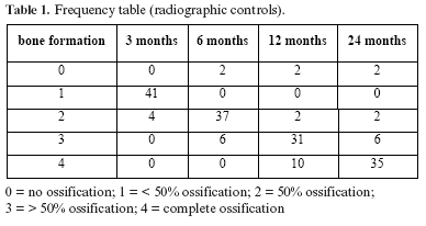
The radiographic controls were used to assess lesion ossification on the following scale: 0 = no ossification; 1 = less than 50% ossification; 2 = 50% ossification; 3 = over 50% ossification; and 4 = complete ossification.
The clinical controls evaluated the presence of symptoms, considering a score of (+) in the presence of a favorable course without clinical manifestations, and (-) when the patient presented swelling, pain or fistulization.
The results were analyzed using the SPSS version 11.5 statistical package. Descriptive statistics comprised the absolute and relative percentage frequencies, with frequency tables to describe the response of each study variable. The Wilcoxon nonparametric test was in turn used to assess the course of lesion ossification.
Results
The results obtained at the clinical control after three months proved positive (no symptoms) in 100% of the study series, i.e., no patient presented pain, swelling, or fistula three months after surgery. However, after 6 months two patients showed symptoms (fistula), requiring repeat intervention. On occasion of the subsequent controls at 12 and 24 months, the rest of the patients exhibited a positive course, i.e., 95.5% of the series showed no clinical manifestations (Figure 2).
As regards the patients with symptoms after 6 months, a radiological evaluation was made and in both cases repeat surgery was decided. In one case we identified a vertical root fracture, and the decision was taken to extract the tooth. In the second case all the contaminated tissue was eliminated, revealing good apical sealing with no apparent cause of failure. The course following repeat surgical management proved positive.
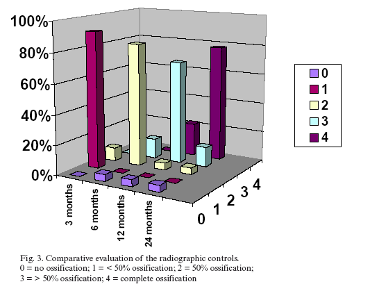
The results obtained in terms of remodeling of the bone lesion showed as most common finding an increase in ossification over time, with complete ossification at the last control after 24 months – the trend being statistically significant (p<0.001)(Wilcoxon nonparametric test)(Table 1 and Figure 3).
Specifically, at the first radiographic control three months after surgery, most of the patients (91.1%) showed initial stage ossification (score = 1). The rest (8.9%) were in intermediate stage ossification (score = 2), i.e., 50%. After 6 months, lesion size in most of the patients (82.2%) had decreased 50% (intermediate ossification). On the other hand, at this point 13.3% of the patients had already exceeded 50% ossification (score = 3), while two patients (4.5% of the study series) presented treatment failure as commented above (score = 0).
At the third radiographic control after 12 months, over half of the series (68.8%) were in advanced stages of ossification (score = 3), i.e., the size of the bone cavity had decreased by more than 50%. In turn, 10 patients (22.2%) had completed ossification at this control (score = 4), versus two patients (4.5%) with intermediate stage ossification.
Lastly, at the final control 24 months after surgery, and as expected, most patients (77.7%) had completed ossification (score = 4). The rest (33.3%) comprised treatment failure (4.5%) and individuals who had not completed the ossification process. This latter group comprised two patients with intermediate ossification (4,5%) and 6 patients (13,3%) with advanced stage (over 50%) ossification.
Discussion
On comparing our results with those published in the literature, no studies of periapical surgery with the Erbium:YAG laser assessing success and lesion ossification under the conditions of our study were identified.
A comparison between our findings in relation to the clinical course of the patients and the data published by other authors involving conventional periapical surgery shows that 95.5% of the operated subjects in our series presented no signs or symptoms at any of the controls made. These results coincide with those published by Rapp et al. (7) and Marti-Bowen et al. (8), with a 95% success rate in periapical surgery of anterior teeth. According to Amagasa et al. (9), the success rate is in the range of 85%. Baca et al. (10) published an 85-90% success rate for anterior teeth. In contrast, in posterior sectors (premolars and molars) the figure barely reaches 44%. These comparatively poor results seem to be related to the existence of isthmuses in the canals of multiple-root teeth, and coincide with the findings of Friedman et al. (11) and Ioannides et al. (12), who likewise reported an increased incidence of failure in apicoectomized premolars and molars – with a success rate of between 44-73%, However, Peñarrocha et al. (13) obtained a 90.4% success rate in application to molars.
Thus, according to the literature, the majority of authors coincide in citing a success rate of 60-90%. In this context, in 2002, Vallecillo et al. (14) published a study on 29 patients subjected to periapical surgery, with a clinical success rate of 58.6%. According to Balandron (15), the success of periapical surgery can range from 60-90%, depending on the authors consulted. Fernandez-Vazquez et al. (16), after a period of 6 years and 180 apicoectomies, recorded a success rate of just over 80%, while Donado et al. (17) indicated that the success rate is always greater than the failure rate or the proportion of doubtful cases – with figures of 70-90%, depending on the author.
Nevertheless, most authors agree that the results of periapical surgery are better after conventional retreatment of the canals, without differences in terms of patient age, sex, location of the tooth, length of the sectioned root, or type of lesion (granuloma or cyst). Likewise, there is general agreement that surgery does not correct deficient endodontic management (17-19).
Gay-Escoda et al. (20) coincide with the above and consider that periapical surgery is not the solution if canal treatment is not correct. In this sense, the percentage success rate for periapical surgery, according to these authors, is in the range of 90-95%.
The results obtained in the evaluation of bone cavity remodeling show ossification to increase over time after treatment. This appears logical in the light of the results obtained at the clinical controls.
Nevertheless, it should be pointed out that the criteria for periapical surgery success or failure are not the same for all authors. According to Molven et al. (21), success is achieved one year after the operation, defined not only as complete disappearance of the radiographically evidenced bone lesion but also of incomplete healing (scar tissue) in the radiotransparent zone within the apicoectomized area. These authors take the persistence of the latter radiographic image one year after treatment to constitute failure, while a decreased image would be indicative of uncertain treatment outcome. Balandron (15) considers that 45% of all periapical lesions require 1-10 years for complete radiological resolution, while 30% require more than 10 years. Orstavik (22) reported that 6 months after treatment, radiological evidence of repair becomes apparent. However, if these signs are not seen after one year, the healing prognosis is poor.
In any case, the complete healing process varies greatly among individuals. Thus, healing becomes radiologically visible in some patients within a matter of months, while in others it may take a year or more for the normal trabecular pattern to become established. There are even cases of periapical lesions that require 10 years or more for complete radiological resolution. On the other hand, it must also be taken into account that the in vivo healing process lies several steps ahead of radiological healing (20).
According to August (23), the criteria for success are the absence of signs and symptoms, and reduction of the radiotransparency to 1 mm or less. Baca et al. (10) in turn defined criteria for success or failure largely coincident with those of Zetterqvist et al. (24). These authors defined complete healing as the absence of clinical signs and symptoms, accompanied by radiographically manifest normal bone trabeculation in the operated zone – accepting persistent bone defects of no more than 1 mm, and a slightly widened periodontal ligament.
Doubtful cases are those situations without clinical manifestations but with a radiological picture that has not experienced changes since the surgical intervention.
Thus, on the basis of our results, it can be concluded that combination of the Erbium:YAG laser and retrograde filling with silver amalgam in periapical surgery affords an important success rate.
References
1. Martínez-González JM, Donado M. Láser en Cirugía Bucal. En: Donado M. Cirugía Bucal. Patología y Técnica. Madrid: Ed. Masson;1990.p.799-816. [ Links ]
2. Gay C, Méndez UM, Berini L. Nuevas aportaciones en cirugía periapical. RCOE 1996;1:405-14.
3. Keller U, Hibst R. Proceedings of medical applications of laser. SPIE 1994; 2327:146-54.
4. Ando Y, Aoki A. Bactericidal effect of Er:YAG laser on perdontopathic bacteria. Lasers Surg Med 1996;19:190-200.
5. España AJ. Láser de Er:YAG en Odontología. Rev ANEO 1997;37:8-13.
6. Leco MI, Martínez-González JM, Donado M, López C. Sterilizing effects of the Erbium:Yag laser upon dental structures: an in vitro study. Med Oral Patol Oral Cir Bucal 2006;11:158-61.
7. Rapp EL, Brown CE, Newton CW. An analysis of success and failure of apicoectomies. J Endod 1991;17:508-12.
8. Marti-Bowen E, Penarrocha M, García B. Periapical surgery using ultrasound technique and silver amalgam retrograde filling. A study of 71 teeth with 100 canals. Med Oral Med Patol Cir Bucal 2005;10:67-73.
9. Amagasa T, Nagase M, Sato T. Apicoectomy with retrograde gutappercha root filling. J Oral Surg 1989;68:339-74.
10. Baca R, Alobera MA, Sirvent F. La cirugía periapical del nuevo milenio (2a parte). Prof Dent 2002;5:103-11.
11. Friedman S, Lustmann J, Sharabany V. Treatment results of apical surgery in premolar and molar teeth. J Endod 1991;17:30-3.
12. Ioannides C, Bortslap WA. Apicoectomy on molars: a clinical and radiologic study. Int J Oral Surg 1983;12:73-9.
13. Peñarrocha M, Sanchís JM, Gay C. Periapical surgery of 31 lower molars based on the ultrasound technique and retrograde filling with silver amalgam. Med Oral 2001; 6:376-82.
14. Vallecillo M, Muñoz E, Reyes C, Prados E, Olmedo V. Cirugía periapical de 29 dientes. Comparación entre técnica convencional, microsierra y uso de ultrasonidos. Med Oral 2002;7:46-53.
15. Balandrón J. Atlas de Cirugía Oral. Madrid: Ed. Ergón; 1997. p.168-85.
16. Fernández J, Garrón G. Apicectomía: otro complemento endodóncico. Rev Actual Estomatolog Esp 1988;48:39-55.
17. Donado A, Gomeza A, Sirvent F, Martínez-González JM, Donado M. Cirugía, ¿una solución en endodoncia? Perspectivas futuras. Parte II. Endodoncia 2001;19:195-07.
18. Molven O, Halsen A, Grung B. Surgical management of endodontic failures: indications and treatment results. Int Dent J 1991;41:33-41.
19. Allen RK, Newton CW, Brown CE. A statistical analysis of surgical and non surgical endodontic retreatment cases. J Endod 1989; 15: 261-6.
20. Gay C, Peñarrocha M, Berini L. Lesiones periapicales. En: Gay C, Berini L, eds. Cirugía Bucal. Madrid: Ed.Ergon;1999. p. 749-80.
21. Molven O, Halsen A, Grung B. Incomplete healing (scar tissue) after periapical surgery-radiographic finding 8 to 12 years after treatment. J Endod 1996;22:264-8.
22. Orstavik D. Time-course and risk analyses of the development and healing of chronic apical periodontitis in man. Int Endod J 1996; 29:84-92.
23. August DS. Long-term postsurgical results on teeth with periapical radiolucencies. J Endod 1996;22:380-3.
24. Zetterqvist L, Hall G, Holmlund A. Apicoectomy: a comparative clinical study of amalgam and glass ionomer cement as apical sealants. Oral Surg Oral Med Oral Pathol 1991;71:489-91.
![]() Correspondence:
Correspondence:
Dra. Ma Isabel Leco Berrocal
C/ Clara del Rey, 44, 5°D
28002 Madrid
E-mail: maria.leco@uem.es
Received: 8-02-2006
Accepted: 24-06-2006


