


Mi SciELO
 Servicios personalizados
Servicios Personalizados
Revista
Articulo
 Inglés (pdf)
 Articulo en XML
 Referencias del artículo
 Enviar articulo por email
Indicadores
-
 Citado por SciELO
 -
 Accesos
Links relacionados
-
 Citado por Google
 -
 Similares en
SciELO
Similares en
SciELO -
 Similares en Google
Compartir

 Permalink
Medicina Oral, Patología Oral y Cirugía Bucal (Internet)
versión On-line ISSN 1698-6946
Med. oral patol. oral cir.bucal (Internet) vol.12 no.2 mar. 2007
Facial nerve injury following surgery for the treatment of ankylosis of the temporomandibular joint
Ricardo Viana Bessa Nogueira1, Belmiro Cavalcanti do Egito Vasconcelos2
(1) Postgraduation student - PhD, Oral and Maxillofacial Surgery
(2) Head of the program of PhD, Oral and Maxillofacial Surgery, University of Pernambuco – Recife - Brasil
ABSTRACT
Objective: The purpose of the present paper was to carry out a longitudinal study of a series of cases in which injury of the facial nerve was observed following surgery for the treatment of temporomandibular ankylosis.
Study design: The sample was composed of 13 patients, both male and female, in whom 18 surgical approaches were made. A postoperative assessment of the motor function of the facial nerve was made in accordance with the House-Brackmann grading system. All the patients were photographed and assessed at the following postoperative times: 24 hours, one week, one month and three months.
Results: The results showed that injury of the facial nerve occurred in 31% of the cases. An increase in the frequency of nerve injury was observed in the cases in which the interpositional arthroplasty technique was employed, as well as the fact that 75% of the patients had undergone at least one surgical intervention prior to the study. After three months all the patients displayed normal function of the facial nerve.
Conclusion: The frequency of facial nerve injury is related to the degree of difficulty involved in the surgery determined by the type of ankylosis. The nerve lesions were shown to be of a temporary nature.
Key words: Ankylosis, nerve injury, facial nerve, facial palsy, arthroplasty.
RESUMEN
Objetivo: El propósito de este trabajo fue realizar un estudio longitudinal de una serie de casos, en los cuales se observó disfunción del nervio facial subsecuente a cirugía para el tratamiento de anquilosis temporomandibular.
Diseño del estudio: La muestra estuvo compuesta por 13 pacientes de ambos géneros, en los cuales fueron realizados 18 abordajes quirúrgicos. Fue realizada una evaluación postoperatoria de la actividad motora del nervio facial, de acuerdo con la escala de House-Brackmann. Todos los pacientes fueron fotografiados y evaluados para los periodos de tiempo: 24 horas, 1 semana, 1 mes y 3 meses.
Resultados: Los resultados mostraron que el porcentaje de casos de disfunción del nervio facial fue de 31%. Fue observado un aumento en la frecuencia de lesión nerviosa en los casos donde fue usada la técnica de artroplastia interposicional, asi como también que el 75% de los pacientes habían sido sometidos al menos a una intervención quirúrgica previa al estudio. Después de 3 meses todos los pacientes presentaban actividad normal del nervio facial.
Conclusión: La frecuencia de lesión del nervio facial se relaciona al grado de dificultad quirúrgica determinda por el tipo de anquilosis. Las lesiones nerviosas mostraron un caracter temporal.
Palabras clave: Anquilosis, lesión nerviosa, nervio facial, parálisis facial, artroplastía.
Introduction
The facial nerve is the main anatomical structure that the surgeon should consider in performing a surgical approach to the temporomandibular joint (TMJ). Skill in preserving its functional integrity is a critical point when this type of surgery is performed. Despite the care taken during the procedure and the surgeons skill in the approach to the TMJ, the facial nerve may be damaged, resulting in injury to the frontal and/or orbicular muscles of the eye. Nerve injury is a major complication from this type of technique (1).
Weinberg and Kryshtalskyj (1), in 1992, analyzed the function of the facial nerve alter performing a preauricular approach in 68 patients. They found a 10.84% occurrence of injury and presented a review of the literature in which the incidence ranged from 1% to 55% among different authors.
Dolwick and Kretzschmar (2) when comparing a perimeatal approach with the preauricular one, reported an incidence of disorder of facial nerve function of 14% and 32%, respectively. In another article, Hall, Brown and Lebowitz (3) modified the dissection technique in the preauricular approach, thereby managing to reduce the incidence of facial nerve injury from 25% to 1.7%.
In cases of facial nerve injury it is necessary to assess the degree and type of nerve lesion. The methods of assessment of the function of the facial nerve may be classified in two major groups: clinical and electrophysiological (4, 5). The House Brackmann grading system, among the methods of clinical evaluation, is quite comprehensive and includes important ítems such as the appearance of the frontal, periorbital and peribuccal musculature, both at rest and in motion, and is used in several countries (6, 7, 8). The literature shows rates of agreement of 93% among the different evaluators using the House-Brackmann grading system to assess the function of the facial nerve (9).
Thus the purpose of this investigation was to carry out a longitudinal study of a series of cases on the frequency of facial nerve injury following surgery for the treatment of temporomandibular ankylosis.
Patients and method
The sample consisted of 13 patients (18 joints) with varying degrees of temporomandibular ankylosis who were operated on using different techniques. The study was conducted between July 2000 and July 2002 in the Department of Oral and Maxillofacial Surgery of Oswaldo Cruz University Hospital of the University of Pernambuco in Recife, Brazil.
The House-Brackmann grading system (Graph 1) was used to assess motor function of the facial nerve. Photographs were taken of the patients operated on who presented nerve disorders at the following times after surgery: 24 hours, one week, one month and threee months. The photographs were obtained using the same camera and the same magnitude. The patients were photographed facing the camera in the following positions: at rest, closing the eyes with a minimum of effort and tightly, raising the eyebrows and with maximum mouth opening (6, 10).
A descriptive method using tables and graphs was employed to elucidate the data. The tables show absolute distributions and the graphs percentage data. The statistical analysis was performed by means of the software program MS Excel Version 2.000.
Results
Table 1 shows the distribution of patients according to gender, surgical approach and the presence of facial nerve injury. Five cases of ankylosis are seen in males and eight in females. It may be observed that four of these 13 patients (31%) presented signs of facial nerve injury, amounting to 22% of the 18 joints operated on.

Table 2 presents the different types of surgical techniques employed and their correlation with the presence of facial nerve injury, which is seen to be found predominantly among the cases operated on using the interpositional arthroplasty technique.

In Figure 1 one may observe the distribution of the sample in relation to the number of operations performed prior to the study and the presence of facial nerve injury. It may be seen that 75% of the patients with dysfunction of the facial nerve had undergone at least one operation prior to the study.

In Table 3 it may be observed that all the patients presented grade 4 on the House-Brackmann grading system at 24 hours.
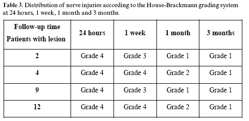
At one week there was an improvement in 50% of the cases. At one month half the patients presented normal function of the facial nerve and finally, at three months, all the patients had recovered.
Discussion
Surgery for the treatment of temporomandibular ankylosis requires elaborate planning and a refined surgical technique on the part of the surgeon (11). In addition, the preparation of a surgical approach that allows a suitable exposure of the surgical field with a minimum risk of damage to the facial nerve represents a great challenge for the performing of surgery of the TMJ in general (12).
The potential complications of this type of surgery include injury to the facial nerve. For Weinberg and Kryshtalskyj (1) nerve lesions may manifest themselves to a greater or lesser extent, and this is in agreement with our results, for in the series presented here the cases of nerve injury had varied classifications on the House-Brackmann grading system
McBride (13) found a frequency of 3% of complications of the marginal nerve of the mandible, a branch of the facial nerve in 140 joints operated on for the placement of prosthesis for reconstruction of the TMJ. The author does not describe the reason for these complications or the distribution to the other branches or main trunk of the facial nerve. On the other hand, Roychoudhury, Parkash and Trikha (14) reported that of the 50 cases of ankylosis operated on, 20% presented postoperative injury to the facial nerve. Our results showed that there were four cases of facial nerve injury, amounting to 31%. The percentage distribution of facial nerve lesion in relation to gender showed that the males presented a larger number of nerve injuries (40%). Thus, facial nerve injury in surgery for correction of temporomandibular ankylosis is a significant finding and should be identified, evaluated and followed up.
Kaban, Perrott and Fisher (11) on analyzing the distribution of the types of ankylosis in a series of patients, observed that 64% were fibro-osseous and 22% osseous in 18 surgical approaches and the type of ankylosis may influence the grade of the injury. From an evaluation of our results it is noted that facial nerve injury occurred in 75% of the cases of osseous ankylosis and in 25% of those of fibro-osseous ankylosis, thus enabling us to realize that the degree of surgical difficulty was an important factor in the appearance of facial nerve lesion.
Al-Kayat and Bramley (15) carried out a cadaveric study of the relationship of the facial nerve and its branches with the region of the TMJ. The zygomatic branch of the facial nerve crosses the region of the zygomatic arch at a distance of 2.0 ± 0.5 cm from the anterior wall of the external auditory canal. The bifurcation of the main trunk of the facial nerve occurs 3.0 ± 0.31 cm from the postglenoidal tubercule and at a mean distance of 2.3 ± 0.28 cm from the inferior concavity of the external acoustic meatus. Roychoudhury, Parkash and Trikha (14) reported that, on using the the measurements of the study conducted by Al-Kayat and Bramley (15) as a parameter in their operations, they achieved a decrease in the number of facial nerve injuries in a series of surgical approaches for the treatment of temporomandibular ankylosis. In the series of cases presented in our study, it was observed that 22% of the surgical approaches resulted in facial nerve injury.
The incisions that provide access to the TMJ are established in accordance with the requirements of the anatomical region and its function. It is considered that the preauricular approach produces a less satisfactory cosmetic result, but provides an excellent retraction and exposure of the surgical field (12). In this connection, Hall, Brown and Lebowitz (3) showed a reduction from 25% to 1.7% in facial nerve injuries on comparing two dissection techniques, one with the making of a skin flap and the other without such a flap. Their conclusions ascribed the decreased incidence of facial nerve in jury to three factors: the elimination of the skin flap before starting the dissection of the deep anatomical planes, where performing this maneuver renders the skin more susceptible to trauma during the operation; a change in direction in the dissection of the deep planes; and not performing the dissection underneath the lateral ligament and the articular capsule. From an evaluation of our results it may be observed that the number of cases of nerve injury was proportionally greater when the technique employed was interpositional arthroplasty, in which the length of the surgical flap is greater and the temporal region is approached for rotation of the temporal muscle.
The increased facial nerve injury resulting from a previous operation is a fact and this may be attributed to the presence of a cicatricial contraction and fibrosis of the region, thereby causing a distortion of the anatomic planes and making an accurate surgical dissection more difficult at a second intervention (1). On assessment of such parameters in our study, it was found that 75% of the patients with nerve injury had undergone at least one previous surgical intervention.
Nerve injury resulting from surgical approaches may also be related to the following: excessive dissection of the tissues; localized edema and hematomas; presence of inflammation and/or infection; trauma with electrocoagulation; and application of anesthetic in the area (1). In these cases facial nerve injury could be classified as being a neurapraxia and presents a favorable prognosis. When the recovery of the patients with facial nerve injury was evaluated, it was observed that at three months they had all recovered and were classified as grade 1 on the House-Brackmann grading system.
Failure to fulfil the protocols for TMJ ankylosis surgery might result in serious facial nerve injury, which could be resolved only by means of microsurgical techniques for repairing the damaged nerve. The ones most commonly described in the literature are epi- or perineural neurorrhaphies, autogenous nerve grafts and alloplastic artificial conduits (4, 5).
References
1. Weinberg S, Kryshtalskyj B. Facial nerve function following temporomandibular joint surgery using the preauricular approach. J Oral Maxillofac Surg 1992;50:1048-51. [ Links ]
2. Dolwick MF, Kretzschmar DP. Morbidity associated with the preauricular and perimeatal approaches to the temporomandibular joint. J Oral Maxillofac Surg 1982;40:699-700.
3. Hall MB, Brown RW, Lebowitz MS. Facial nerve injury during surgery of the temporomandibular joint: a comparison of two dissection techniques. J Oral Maxillofac Surg 1985;43:20-3.
4. Mackinnon SE, Dellon AL. Surgery of peripheral nerve. 1 ed. New York: Thieme Medical; 1988. p. 638.
5. Vasconcelos BC, Gay-Escoda C. Facial nerve repair with expanded polytetrafluoroethylene and collagen conduits: an experimental study in the rabbit. J Oral Maxillofac Surg 2000;58:1257-62.
6. Yen T L, Driscoll C L W, Lalwani A K. Significance of House-Brackmann Facial Nerve Grading Global Score in the Setting of Differential Facial Nerve Function. Otol Neurotol 2003;24:118-22.
7. Satoh Y, Kanzaki J, Yoshihara S. A comparison and conversion table of the House-Brackmann facial nerve grading system and the Yanagihara grading system. Auris Nasus Larynx 2000;27:207-12.
8. Kang TS, Vrabec JT, Giddings N, Terris DJ. Facial nerve grading systems (1985-2002): beyond the House-Brackmann scale. Otol Neurotol 2002;23:767-71.
9. Evans RA, Harries ML, Baguley DM, Moffat DA. Reliability of the House and Brackmann grading system for facial palsy. J Laryngol Otol 1989;103:1045-6.
10. Ellis E 3rd, McFadden D, Simon P, Throckmorton G. Surgical complications with open treatment of mandibular condylar process fractures. J Oral Maxillofac Surg 2000;58:950-8.
11. Kaban LB, Perrott DH, Fisher K. A protocol for management of temporomandibular joint ankylosis. J Oral Maxillofac Surg 1990; 48:1145-51.
12. Kreutziger KL. Surgery of the temporomandibular joint. I. Surgical anatomy and surgical incisions. Oral Surg Oral Med Oral Pathol 1984; 58:637-46.
13. Mcbride, K. L. Total Temporomandibular Joint Reconstruction. En: Bell, W. H. Modern Practice in Orthognathic and Reconstructive Surgery, Philadelphia: WB Saunders; 1992. p. 1115-6.
14. Roychoudhury A, Parkash H, Trikha A. Functional restoration by gap arthroplasty in temporomandibular joint ankylosis: a report of 50 cases. Oral Surg Oral Med Oral Pathol Oral Radiol Endod 1999;87:166-9.
15. Al-Kayat A, Bramley P. A modified pre-auricular approach to the temporomandibular joint and malar arch. Br J Oral Surg 1979;17:91-103.
![]() Correspondence:
Correspondence:
Prof. Dr. Belmiro Cavalcanti do Egito Vasconcelos
Facultad de Odontología de Pernambuco
Av. General Newton Cavalcanti, 1650CE
Camaragibe – Pernambuco – Brasil.
CEP: 54.753-220
E-mail: belmiro@pesquisador.cnpq.br
Received: 25-03-2005
Accepted: 5-01-2007

