


My SciELO
 Custom services
Services on Demand
Journal
Article
 English (pdf)
 Article in xml format
 Article references
 Send this article by e-mail
Indicators
-
 Cited by SciELO
 -
 Access statistics
Related links
-
 Cited by Google
 -
 Similars in
SciELO
Similars in
SciELO -
 Similars in Google
Share

 Permalink
Medicina Oral, Patología Oral y Cirugía Bucal (Internet)
On-line version ISSN 1698-6946
Med. oral patol. oral cir.bucal (Internet) vol.12 n.3 May. 2007
Clinical justification of dental radiology in adult patients: A review of the literature
Yolanda Martínez Beneyto1, Miguel Alcaráz Baños2, Leonor Pérez Lajarín3, Vivian E. Rushton4
(1) Profesor Ayudante Doctor Odontología Preventiva y Comunitaria. Universidad de Murcia, España
(2) Profesor Titular Radiología y Medicina Física. Universidad de Murcia, España
(3) Profesor Titular Odontología Preventiva y Comunitaria, Universidad de Murcia, España
(4) Profesor Titular Radiología Oral y Maxilofacial, Universidad de Manchester, Reino Unido
ABSTRACT
Although the radiological doses used by dentists are low individually, patients are often exposured to many repeat dental radiographic examinations. The routine use of dental radiography, such as screening of all patients using dental panoramic radiography (DPRs) or a random decision to take a dental radiograph, will inevitable lead to unnecessary patient exposure.
The use of Radiographic Referral Criteria has now become a legal requirement for all practitioners following the adoption of European Legislation. All exposures to x-rays should be clinically justified and each exposure should be expected to give the patient a positive net benefit.
Recently the European Commission has published guidelines (1) on radiation protection in dental radiology. Guidelines have previously been available in a number of European countries (2,3) and also within the United States (4,5). At the present time, no specific guidelines have been published within Spain.
The aim of this review article is to provide the Spanish dentist with guidance as to the appropriateness of different radiographic techniques for a variety of clinical conditions and also the frequency with which they should be taken. It is hoped that this document will act as a useful work tool in daily dental practice.
Key words: Radiation protection, dentistry, European guidelines, dental radiology, regulations.
RESUMEN
Aunque generalmente las dosis de radiación empleadas por los odontoestomatólogos no suelen ser altas, consideradas individualmente, las exposiciones a las que algunos y determinados pacientes están sometidos pueden ser excesivas.
En este sentido, hay que evitar radiaciones rutinarias innecesarias para no incrementar la dosis de irradiación recibidas por los pacientes.
El empleo de criterios de selección radiográfica se ha convertido en requerimientos legales europeos a cumplir por todos los dentistas. Todas las exposiciones a los rayos X deberían de estar clínicamente justificadas y, a la vez, proporcionar un beneficio neto para el paciente.
Recientemente, la Comisión Europea ha publicado unas Guías (1) sobre Protección Radiológica Dental, existentes ya en numerosos países de la Unión Europea (2, 3) y en Estados Unidos (4, 5); sin embargo, en España no se ha publicado ningún documento propio específico que permita difundir un protocolo de actuación en radiología dental.
El objetivo de este artículo es proporcionar al odonto-estomatólogo general español guías de actuación en radiología dental apropiadas para cada situación clínica y, además, recomienda la frecuencia con la que se deben de realizar dichas exploraciones.
Palabras clave: Protección radiológica, odontología, Guías europeas, radiología dental, legislación.
Introduction
The use of x-rays requires the adoption of measures to limit the exposure to both the patient and the clinian. One of the basic tenets of radiation safety is to ensure that all exposures to ionising radiation are clinically justified.
All radiation exposures must be kept as low as reasonably achievable (ALARA principle). This is achieved in three ways, using physical methods of minimising dose (i.e. equipment and film factors), the application of Selection Criteria when choosing whether or not to use a radiographic examination and, finally by Quality Assurance Programmes. In the latter, efforts are made to ensure the consistent production of high quality radiographs, thereby avoiding repeat exposure and maximising the benefit to the patient.
The dentist is the person who takes responsibily for the necessity of a radiographic examination and its frequency. The clinician must review any previous radiographs as these may well provide useful information on the patients present symptoms. If these radiographs are not useful, then further radiography is may well be justified. Selection criteria aid the clinician in choosing the appropriate radiographic examination to maximise the diagnostic yield while limiting the dose to the patient. The use of Selection Criteria is well established within many countries of the European Union. However, Spain has not adopted the routine use of selection criteria although several published papers have highlighted the importance of implementing radiographic guidelines within dental practice (6-8).
The main objetive of this study was to review some of the more relevant aspects of the guidelines that appear in the recent publication of the European Commission (1) on dental radiographic selection criteria in adult patients.
European and Spanish regulations
Within the European Union, the most recent ICRP recommendations were incorporated into several Euratom Directives (9-11). These directives outline the best radiographic practice and also include many recommendations to ensure high quality radiography. These recommendations include: justification and optimization of the radiographic examination; measures on quality control of radiolographic equipment; procedures for the annual evaluation of the doses received by patients in the most frequently conducted radiographic examinations; the evaluation of the image quality and also an assessment of annual dose levels. These directives were implemented within Spanish legislation by two Royal Decrees 1976/1999 (12) and 783/2001(13).
The control of the doses to patients combined with the production of high quality images constitutes the first assessment of the state of the radiological equipment used and, also, of the training of personnel involved with ionising radiation.
The necessity of selection criteria in dental radiology: frequency of radiographic examinations
The Scientific Committee on the Atomic Effects of Radiation within the United Nations noted that dental radiography was the most frequent radiographic technique in medical practice. Dental radiography accounts for nearly one third of all the total number of radiological examinations conducted within the European Union (14) (see Table 1).
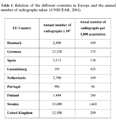
Within Spain, there are believed to be approximately 440 dental sets per million of the population, although this figure has not been reliably confirmed. This figure represents 57% of the total medical X-ray units in clinical pracrice. Throughout the European Union, the number of dental x-ray sets per million of population varies enormously with 1534 sets in Sweden, 975 to Denmark, 667 sets in Greece, 631 to France and 350 within the UK (14).
Vaño and colleagues (15) in a recent study assessed the number of dental sets within Spanish dental practices and the number of dental radiographs taken per annum compared with medical exposures. The study found that the number of medical and dental x-ray machines in clinical practice totalled 14,411, of which 7,327 (50.8%) were dental x-rays sets. The annual number of medical radiolographic examinations has been assessed as 25,058,622, representing an annual rate of 629 examinations per 1000 habitants. Corresponding figures for the 5,226,823 dental x-ray examinations undertaken in Spain translates into 131 dental x-ray exposures per 1,000 inhabitants.
In the last 20 years within England and Wales, dental panoramic radiography (DPR) has become well-stablished in general dental practice, as evidenced by a seven-fold increase when compared with intra-oral radiography over the same period (2). Between 1998 and 1999, approximately 2,05 million panoramic radiographs were taken in the general dental service in England and Wales (16).This increasing use of panoramic radiography has been observed in other countries. Within the USA, it was estimated 20 years ago that 60% of all practioners had access to panoramic equipment (17).
Panoramic equipment often delivers a wide range of doses to patients and these can vary by a factor of 200. In Spain, the published data of a recent study illustrates that in the region of 3,1% of the panoramic equipment fail to reach the manufacturers nominal kilovoltage and machines also display timer inaccuracies. These innacuries have decreased from 12% of all equipment for the year 1997 to 3% in 2001 following the adoption of compulsory annual quality control assessment of x-ray equipment (18).
Although a large number of dental radiographs are exposed within primary dental care, a large proportion of these exhibit poor image quality. These films represent a cumulative increase in dose to the exposed population without benefit as often these films are essentially non-diagnostic because of faults in technique and/or processing. Research has shown that 42% of dental practitioners in the United Kingdom practise routine screening of new adult patients using panoramic radiography without any clinical findings to support such a radiographic examination (19). Of these screening panoramic films, when the yield from posterior bitewing radiographs and the radiological findingsof no relevance to treatment were excluded, 57% of patients received no benefit from these panoramic films.
Several research studies have shown that the frequency of unacceptable panoramic films ranges from 18% to 33% of the total panoramic radiographs taken. These unacceptable panoramic films limit the diagnostic yield that the practitioner can obtain from the radiographic image. The faults range from inadequate processing to technical technical faults, such as movement of the patient. More often inadequate panoramic films exhibit a combination of both technical and processing errors (20). Films faults are not confined solely to panoramic radiography as a recent study has reported levels of unacceptable intraoral films ranging between 45.2-56.4% (21).
Similarly, in the USA, it has been estimated that the elimination of non-productive examinations could lead to the reduction of the collective population dose from medical radiography by 30% (4)
The method proposed to eliminate unnecessary x-ray examinations is by the adoption of selection criteria in radiography. Selection criteria have been defined as "descriptions of clinical conditions observed from patient signs, symptoms and history that identify those patients who are likely to benefit from a particular radiographic examination" (22).
Radiation dose and risks
The biological effects of ionizing radiation can be extremely damaging. Somatic deterministic effects predominate with high doses of radiation, while somatic stochastic effects predominate with low doses. Dental radiology employs low doses and the risk of stochastic effects is very small (23). The estimated risk of a fatal cancer developing from two intraoral bitewing exposures, or from a dental panoramic tomography, is of the order of one tumour for every 2 million exposures (24).
In the case of panoramic radiology, the weighted dose equivalent from a panoramic examination was calculated to be 3,85-30 μSv , corresponding to a lifetime risk of fatal cancer (per million) of 0,21-1,9 (25,26). For an intra-oral radiograh the effective dose is 1-8.3 μSv and the risk of cancer is 0,02-0,6 (26, 27). These figures assume best practice is employed. A panoramic radiograph may be associated with an effective dose the same as 1-5 days additional background radiation, while two bitewing radiographs would be equivalent to about one day.
However lower levels of risk are associated with newer equipment and techniques. Recent studies have showed that the 72,79% of dental x-ray sets in Spain operate at 70 kVp, 88,02% employ a 20 cm of focus-to-film distance (PID) and the majority of this equipment employ a 6 cm diameter round beam. Ekta-speed dental film was used in the 10,24 % of the cases and intraoral digital imaging was used by 11,95% of practitioners (28-30)
A particular problem arises from the inclusion or exclusion of the salivary glands in the calculation of dose. The salivary glands have previously not been included as an organ in effective dose calculations (31). However, the most recent document from the International Commission on Radiation Protection (ICRP) has recognised this omission in view of the apparent relationship between dental radiography and increased risk of salivary gland tumours (32). The most recent ICRP document has included salivary tissue as a remainder organ and their inclusion in dose calculations increases the rate of risk of inducing tumours by a factor of two.
Methodology
A review of the literature relating to European guidelines and the protocols of performance of selection criteria in dental radiology was undertaken.
This study was specially related to the European Guidelines on Radiation Protection in Dental Radiology (1) which had been developped by a Committe of European Experts in Radiation Protection. It has been designed to be used as a guide for both general dental practitioners and dental specialists. This document (1) has been developed using a methodology supported in a critical review of the literature following an evidence-based practice. Depending on the available evidence, the recommendations given were graded to reflect their relevance. A similar document was produced by the Royal College of Surgeons, London using identical techniques leading to the production of "Selection Criteria for Dental Radiography [2nd edition, The Faculty of General Dental Practitioners, The Royal College of Surgeons, London WC2A 3PE] (33).
New adult patients
In some centres, it has become routine to take a panoramic film or full-mouth intraoral radiography of all new patients and this routine practice is not aceptable (34, 35).
A high proportion of practitioners (57%) continue to rely on panoramic radiography alone to assess common dental pathosis (34). Research has confirmed that intra-oral (bitewing and periapical) radiography is superior to panoramic radiography for the diagnosis of common dental pathology (i.e.caries, periodontal and periapical pathology). It is posible that anecdotal evidence of identifying a cyst or other uncommon lesion in a patient may reinforce this attitude. However, this standpoint ignores the low prevalence of the asymtomatic pathology and routine radiography without the presence of clinical signs or symptoms cannot be justified (35, 36). A panoramic radiograph may be apropriate for the patient in certain cases such as one whom presents with a grossly neglected mouth with significant numbers of clinically-determined carious lesions and periapical pathology, along with established periodontal disease (35). In these cases, it may be expeditious to use panoramic radiography as a means of identifying teeth requiring a more detailed (intra-oral) radiographic examination or, when limited to a hospital setting, prior to dental surgery under general anaesthesia.
Full-mouth periapical radiography can be criticised in the same way as routine panoramic radiography.
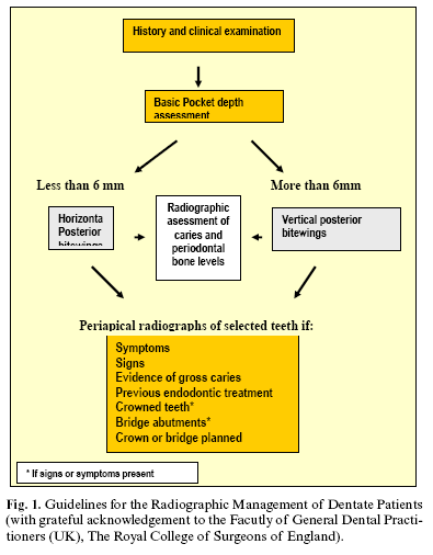
For a new adult dentate patient, the choice of radiography should be based upon history, clinical examination and an individualised prescription as illustrated in Figure 1.
Screening with panoramic radiography
Forty-two percent of practitoners were found to use panoramic radiography routinely to "screen" the jaws for clinically unsuspected pathology and 77.4% of these do so for no specific reason. Approximately, 65,3% of screening panoramic radiographs have no relevance to treatment rising to 71% in the screened asymptomatic attender. Some dentists defend routine screening on the basis of detecting of large cyst and tumours. These lesions are very rare and often have signs or symptoms, which would alert the practitioner to the need for radiography. The detection of a small number of lesions, which are completely asymptomatic, does not justify the routine screening of the population.
Radiography in endodontics
Radiographs are essential for the mechanical aspects of endodontic treatment allowing evaluation of the root canal configuration and also for confirmation that treatment goals have been achieved. Radiographs should have optimum geometry obtained by using the paralleling technique and a beam-aiming device (37).
The radiographs recommendated for endodontic treatment are shown within Table 2.
The edentulous patient
In the absense of any clinical signs or symptoms, there is no justification for any radiographic examinations unless implant treatment is planned (38). If implant treatment is extensive, other more advanced imaging techniques, such as Computed Tomography (CT) imaging, may well be appropriate. Where the clinical examination identifies the posible presence of an abnormality, such as a possible retained root, then an intra-oral radiograph of the site is the apropriate radiographic examination.
Referral criteria for dental radiology: prior to third molars exodontia, simple extractions and surgery
The panoramic radiograph is commonly used to assess third molars prior to their surgical renoval but this examination does not need to be carried out at the initial examination (3). Routine radiography of unerupted third molars is not recommended.
The techniques recommended in the extractions of the third molar vary depending on the geographic situation of the tooth:
Lower third molar: a panoramic radiograph provides information about the tooth position, the relationship to the inferior dental canal and the distance to the lower border. A periapical radiograph is indicated where there is any question of a complex root formation or an intimate relation between the molar roots and the ID canal (3).
Upper third molar: If the tooth is erupted fully then a periapical radiograph should be requested in the first instance, and remember to always check previous radiographs before requesting new films. Previous radiographs may show that there are no contralateral third molars present and thus avoid the need to take a full panoramic radiograph.
In other surgical situacions, such as apicectomy, root renoval or enucleation of small cysts, an intra-oral radiograph may be all that is required for treatment planning.
There is no convincing evidence to support the need for radiography prior to uncomplicated routine extrabtions in adults; however, where a radiograph already exists, this should be referred to before commencing the procedure.
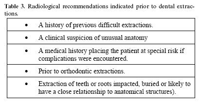
Radiological recomendations for dental extractions are shown at Table 3 and clinical situations that indicate radiological examination at Table 4.

Trauma
For simple dental trauma, intraoral radiography will provide greater diagnostic detail.
A panoramic radiograph is indispensble when assessing madibular fractures (39); however, poor panoramic film quality has been shown to severely affect diagnosis (40). Panoramic radiography has been shown to necesítate supplementary radiography in order accurately to diagnose high condylar fractures (41).
If there is clinical evidence of a bony fracture, it is probably more appropriate for a dentist to refer the patient for a complete radiographic examination at the hospital where treatment wil be performed. Panoramic radigoraphy has a limited ability to detect mid-facial fractures.
Temporomandibular joint problems
The panoramic radiograph shows an image of the mandibular condyles and is often used as a first choice imaging technique for those patients with TMJ symptoms.
A recent study (42) of patients with TMJ symptoms found that panoramic radiography provided little or no information that influenced diagnosis or patient management in the majority of cases examined.
The overwhelming majority of patients with symptoms and signs related to the TMJ region are suffering from myofacial pain/disfunction or internal disc derangements.
Radiography is not recommended for patients with joint sounds (clicking) in the absence of other signs or symptoms (43). Radiographic examination is indicated where there is recent evidence of progressive pathology (recent trauma, change in occlusion, madibular shift, sensory or motor alterations or change in range of movement).
To assess disc position in cases of internal derangement in which simple treatments have been unsuccessful, it may be useful to use Magnetic Resonance Imaging.
In the situation where a clinical diagnosis of condylar hyperplasia is suspected, it should be necessary to use Computed Tomography (CT).
Periodontal disease
There is insufficient evidence from research studies to develop robust evidence-based radiographic selection criteria for periodontal disease. While the panoramic radiograph can offer a dose advantage over large numbers of intra-oral radiographs, it may be considered as an alternative imaging modality, if available. This may be the case when there are other concurrent problems for which radiography is indicated (i.e. symptomatic third molars, multiple existing crowns/heavily restored teeth, and/or multiple endodontically-treated teeth in a patient new to a practice). The use of radiography should be view as secondary to a detailed clinical examination in the diagnosis of periodontal diseases. Access to previous radiographs may be useful in assessing the rate of disease progression (5).
Guidelines for the use of dental radiography in periodontal disease are shown in Table 5.
Suggested selection criteria for panoramic radiography
The general recommendations are detailed below:
Where a bony lesion or unerupted tooth is of a size or position that precludes its complete demostration on intra-oral radigraphs.
In the case of a grossly neglected mouth, with significant numbers of clinically-determined carious lesions and periapical pathology, along with established periodontal disease (other than simple gingivitis) and where there is pocketting greater than 6 mm in depth.
For the assessment of wisdom teeth prior to planned surgical intervention. Routine radiography of unrerupted third molars is not recommended.
As a part of an orthodontic assessment where there is a clinical need to know the state of the dentition and the presence/absence of teeth. The use of clinical criteria to select patients rather than routine screening of patients is essential
Panoramic radiographs should only be taken in the presence of specific clinical signs and symptoms. There is no justification for review panoramic radiography at arbitrary time intervals
The European Recommendations have been shaped to reflect the most frequent radiographic practices within General Dentistry.
Conclusion
The main conclusion of this study was to emphasise that All patients must have a clinical history taken prior to any radiological examination and when radiographs are clinically indicated, intra-oral radiographs should be considered first because of their better detail and lower radiation dose".
Within Spain, it is necessary to change the dentists attitude to the use of ionising radiation. This requires a readjustment to the new regulations on radiological safety of the patient and also to reinforce the need for justification for all radiographic examinations used in dental radiological diagnosis.
Acknowledgement
This paper has been made thanks to a Post Doctoral Grant of the University of Murcia for the dissemination of the European Dental Guidelines on Radiation Protection in Dental Radiology. Acknowledgement is made to the staff of the Department of Dental and Maxillofacial Radiology, The School of Dentistry, The University of Manchester, M15 6FH, U.K. In addition, the authors would like to express their thanks to The Faculty of General Dental Practitioners (UK) and The Royal College of Surgeons of England for allowing the use of their material to be translated into Spanish and to be published within this document.
References
1. European Union European Commission. Radiation Protection 136. European guidelines on radiation protection in dental radiology. Office for Official Publications of the EC, Luxembourg; 2004 [ Links ]
2. Dental Practice Board. Guidelines for panoral radiography. Eastbourne: Dental Practice Board of England den Wales; 1983. [ Links ]
3. Scottish Intercollegiate Guidelines Network (SIGN). Management of Unerupted and Impacted Third Molar Teeth. SIGN publication No 43. Edimburgg: SIGN; 2000. [ Links ]
4. Brown FR, Shaver JW, LAmel DA. The selection of patients for x-ray examination. U.S. Department of Health, Education and Welfare. HEW Publicacion (FDA) 80-8104. Rockville MD: Bureau of Radiological Health; 1980. [ Links ]
5. White SC, Heslop EW, Hollender LG, Mosier KM et al. Parameters of radiologic care: AN official report of the AmericanAcademy or Oral and Maxillofacial Radiology. Oral Surg Oral Med Oral Pathol Oral Radiol Endod 2001;91:498-511. [ Links ]
6. Fernández R, González L, Vaño E, Villa A, Martínez JM, Ortega R et al. Criterios de calidad de imagen en radiodiagnóstico dental. Archivos de odontoestomatología 1996;9:501-7. [ Links ]
7. Finestres F, Miguel J, Cloquell DA, Rafael A, Chimenos E, Guix B. LA calidad en el servicio de radiología. Med Oral 2003; 8:311-21. [ Links ]
8. Alcaráz M, Martínez Y, Jódar S, Velasco E, García MC. Control de calidad en radiología dental intraoral: anomalías en el funcionamiento de los equipos radiológicos. Radioprotección 2004;41:22-9. [ Links ]
9. Council Directive 84/466 Euratom, laying down the basic measures for the radiation protection of persons undergoing medical examination or treatment. Official Journal of the European Communities No L 265, 5th October 1984. p. 1-3. [ Links ]
10. European Union. Council Directive 96/29 Euratom, on health protection of sanitary persona and persons undergoing ionizing radiation. Official Journal of the European Communities No L 159, 29th June; 1996. p. 1-114. [ Links ]
11. European Union. Council Directive 97/43 Euratom, on health protection of individuals against the danger of ionizing radiation in relation to medical exposure, and repealing Directiva 84/466 Euratom. Official Journal of the European Communities No L 180, 9th July; 1997. p. 22-7. [ Links ]
12. BOE. Royal Decree 1976/1999, from the Health and Consumer Affairs Department, establishing quality criteria in radiodiagnostic. In State Official Bulletin, January 29th 1999:45891-900.(In Spanish). [ Links ]
13. BOE. Royal Decree 783/2001, from the Health and Consumer Affairs Department, establishing the Regulation on Ionizing Radiation Protection In State Official Bulletin, July 26th 2001. [ Links ]
14. United Nations Scientific Committee on the Effects of Atomic Radiation UNSCEAR Report to the general assembly with scientific annex. 2001. [ Links ]
15. Vaño, E. Las exposiciones médicas en UNSCEAR 2000 y los datos del Comité Español. Radioprotección 2001;30:14-9. [ Links ]
16. Dental Practice Board. Personal Communication. Dental Data Services, Dental Practice Board for England and Wales. 1999. [ Links ]
17. Kaugars GE, Broga DW and Collett WK. Dental radiologic survey of Virginia and Florida. Oral Surg Oral Med Oral Pathol 1985;60:225-9. [ Links ]
18. Jodar S, Alcaraz M, Martínez Y, Perez L, Velasco E, López M.Manejo de las radiaciones ionizantes en instalaciones dentales españolas: intraorales y panorámicos. Avances en Odontoestomatología 2005;21: 361-70. [ Links ]
19. Rushton VE, Horner K, Worthington HM. Aspects of the use of panoramic radiography in genral dental practice. Br Dent J 1999;186:342-4. [ Links ]
20. Rushton VE, Horner K, Worthington HM. The quality of Panoramic Radiographs in General Dental Practice. Br Dent J 1999;186:630-3. [ Links ]
21. Helminen S, Vehkalahti M, Wolf J , Murtomaa H. Quality evaluation of young adults radiographs in Finnish public oral health service. Journal of Dentistry 2000;28:549-55. [ Links ]
22. U.S. Department of Health and Human Services. The selection of patients for x-ray examination: Dental radiographic examinations. HHS Publication (FDA) 1987;88:8273. [ Links ]
23. Whaites E. Essentials of Dental Radiography and Radiology.Churchill Livingstone, London 2005. [ Links ]
24. NCRP (National Council of Radiation Protection and Measurements). Quality Assurance for Diagnostic Imaging Equipment. Report Nº 99 (Bethesda, MD: NCRP); 1998. [ Links ]
25. Sanforth RA, Clark DE. Effective dose from radiation absorbed during a panoramic examination with a new generation machine. Oral Surg Oral Med Oral Pathol Oral Radiol Endod 2000;89:236-43. [ Links ]
26. Dula K, Mini R, van der Stelt PF, Buser D. The radiographic assessment of implant patients: decision-making criteria. Int J Oral Maxillofac Implant 2001;16:80-9. [ Links ]
27. Gijbels F, Jacobs R, Snaderink G, de smet E, Nowak B, van Dam J, et al. A comparison of the effective dose from scanography with periapical radiography. Dentomaxillofac Radiol 2002;31:159-63. [ Links ]
28. Martínez-Beneyto Y, Alcaraz M, Perez L, Jodar S, Saura AM. Radiation protection and quality assurance in dental radiology: I. Intraoral Raiography. In: International Atomic energy Agency, editors. Internacional conference of radiological protection of patients in diagnostic and interventional raddiology, nuclear medicine and radiotheraphy. Málaga; 2001. p. 110-3. [ Links ]
29. Alcaraz M, Martínez-Beneyto Y, Jodar S, Velasco E, García Vera M. Control de calidad en radiología dental intraoral: anomalías en el funcionamiento de los equipos radiológicos. Radioprotección 2004;41:22-30. [ Links ]
30. Alcaraz M, Martinez Y, Perez L, Jodar S, Velasco E, Canteras M.The estatus of Spain´s dental practices following the European Union directive concerning radiological installations. Oral Surg Oral Med Oral Pathol Oral Radiol Endod 2004;98:476-82. [ Links ]
31. ICPR Publication 60. Recommendations of the International Commission on Radiatiological Protectin. Annal of the ICRP 21. 1991. [ Links ]
32. Horn-Ross PL, Ljung BM, Morrow M. Environmental factors and the risk of salivary gland cancer. Epidemiology 1997;8:414-9. [ Links ]
33. Selection Criteria for Dental Radiography, 2nd Edition. The Faculty of General Dental Practitioners (UK). Royal College of Surgeons of England, London, 2004. [ Links ]
34. Rushton VE, Horner K, Worthington HV. Screening panoramic radiology of adults in general dental practice: radiological findings. Br Dent J 2001; 190:495-501. [ Links ]
35. Rushton VE, Horner K, Worthington HV. Screening panoramic radiology of a new adult patients in general dental practice: a measurement of diagnostic yield of relevance to treatment and identification of selection criteria. Oral Surgery, Oral Medicine Oral Pathology Oral Radiology and Endodontics 2002;93:488-95. [ Links ]
36. Richardson PS. Selective periapical radiology compared to panoramic screening. Prim Dent Care 1997;4:95-9. [ Links ]
37. Consensus report of the European Society of Endodontoloty on quality guidelines for endodontic treatment. Int Endod J 1996;29:150-5. [ Links ]
38. Bohay RN, Stephens RG, Kogon SL. A study of the impact of screening or selective radiography on the treatment and post delivery outcome for edentulous patients. Oral Surg Oral Med Oral Pathol Oral Radiol Endod 1998; 86:353-9. [ Links ]
39. Guss DA, Clark RF, Peitz T, Taub M. Pantomography vs mandibular series for the detection of mandibular fractures. Acad Emerg Med 2000; 7:141-5. [ Links ]
40. Markowitz BL, Sinow JD, Kawamoto HK, Shewmake K, Khoumehr F. Prospective comparison of axial computed tomography and standard and panoramic radiographs in the diagnosis of madibular fractures. Ann Plast Surg 1999;42:163-9. [ Links ]
41. Wilson IF, Lokeh a, Benjamin Cl, Hilger PA, Hamler DD, OndreyFG, et al. Contribution of conventional axial coputed tomography (nonhelical), in conjunction with panoramic tomography (zonography), in evaluation mandibular fractures. Ann Plast surg 2000;45:415-21. [ Links ]
42. Epstein JB, Caldwell J, Black G. The utility of panoramic imaging of the temporomandibular joint in patients with temporomandibular disorders. Oral Surg Oral Med Oral Pathol Oral Radiol Endod 2001;92:236-9. [ Links ]
43. Brooks SL, Brand JW, Gibbs SJ, Hollender L, Lurie AG, Ommell KA, et al. Imaging of the temporomandibular joint. A position paper of the American Academy of Oral and Maxillofacial Radiology. Oral Surg Oral Med Oral Pathol Oral Radiol Endod 1997;83:609-18. [ Links ]
![]() Correspondence:
Correspondence:
Prof. Yolanda Martínez Beneyto
Hospital Universitario Morales Meseguer
2ª Planta, Clínica Odontológica Universitaria
Marqués de los Vélez s/n, 30008 Murcia
E-mail: yolandam@um.es
Received: 15-02-2006
Accepted: 30-01-2007

