


Mi SciELO
 Servicios personalizados
Servicios Personalizados
Revista
Articulo
 Inglés (pdf)
 Articulo en XML
 Referencias del artículo
 Enviar articulo por email
Indicadores
-
 Citado por SciELO
 -
 Accesos
Links relacionados
-
 Citado por Google
 -
 Similares en
SciELO
Similares en
SciELO -
 Similares en Google
Compartir

 Permalink
Medicina Oral, Patología Oral y Cirugía Bucal (Internet)
versión On-line ISSN 1698-6946
Med. oral patol. oral cir.bucal (Internet) vol.12 no.5 sep. 2007
In vivo evaluation of the effects of 10% carbamide peroxide and 3.5% hydrogen peroxide on the enamel surface
Amparo Berga Caballero1, Leopoldo Forner Navarro2, José Amengual Lorenzo3
(1) Odontóloga. Diplomada en Técnicas de Blanqueamiento Dental. Universitat de València. Estudi General
(2) Médico Estomatólogo. Profesor Titular de Patología y Terapéutica Dentales, Co-Director del Máster en Endodoncia. Departamento de Estomatología
(3) Médico-Odontólogo. Dr. en Odontología. Co-Director del Diploma en Técnicas de Blanqueamiento Dental. Unidad Clínica de Blanqueamiento Dental. Clínica Odontológica. UVEG
ABSTRACT
Objectives: Bleaching of vital teeth performed at home by the patient under the dentists supervision, using low-concentration peroxides and custom-fitted trays specifically designed for this purpose, is one of several options for this type of dental treatment, whether alone or in combination with another in-office bleaching technique. The objective of this study is to analyse the effect on the enamel surface of two bleaching products recommended for this technique.
Materials & methods: Two bleaching products were used: VivaStyle (Vivadent), a 10% carbamide peroxide, and FKD (Kin), a 3.5% hydrogen peroxide. They were applied in trays to the anterior teeth of 20 patients (10 in each group). The application times were 2 and 3 hours a day respectively for 28-33 days. Replicas of the tooth surfaces before and after treatment were obtained. These were observed with a scanning electron microscope.
Results: The images obtained showed that the tooth surfaces remained entire and the enamel surface structures remained normal.
Conclusions: The results show that neither of the products affects the enamel surface: no post-operatory changes were observed.
Key words: At-home tray vital bleaching, carbamide peroxide, hydrogen peroxide, enamel, scanning electron microscope.
RESUMEN
Objetivos: El blanqueamiento de los dientes vitales que realiza el/la paciente en su domicilio bajo la supervisión del/de la dentista con férulas individualizadas especialmente diseñadas para ello y peróxidos de baja concentración, es una opción de entre las que integran esta terapéutica odontológica, ya sea, como tratamiento único o combinada con otra modalidad de blanqueamiento en la consulta. El objetivo del presente trabajo es analizar el efecto que producen sobre la superficie del esmalte dos productos blanqueadores indicados para esta técnica.
Diseño del estudio: Se emplearon dos productos blanqueadores, el VivaStyle (Vivadent), peróxido de carbamida al 10%, y el FKD (Kin), peróxido de hidrógeno al 3,5%, que se aplicaron mediante férulas sobre los dientes anteriores de 20 pacientes (10 en cada grupo). El tiempo de aplicación de cada producto fue de 2 y 3 horas al día respectivamente durante 28-33 días. Se obtuvieron réplicas de las superficies dentales antes y después del tratamiento, las cuales fueron observadas con un microscopio electrónico de barrido.
Resultados: Las imágenes obtenidas muestran la integridad de la superficie dental, con el mantenimiento de las estructuras normales de la superficie del esmalte.
Conclusiones: Los resultados obtenidos muestran que ninguno de los dos productos alteran la superficie del esmalte, no observándose, pues, cambios postoperatorios.
Palabras clave: Blanqueamiento vital domiciliario, peróxido de carbamida, peróxido de hidrógeno, esmalte, microscopía electrónica de barrido.
Introduction
A large proportion of patients attending dental clinics do so to seek treatments that will improve the appearance of their smile. Various types of dental treatment are available for this purpose: orthodontic, conservative, prosthetic, etc. However, one of the treatments that has had the greatest impact and aroused the highest expectations in recent years is dental bleaching (1); this has been associated with the appearance on the market of a large number of whitening products and the development of new dental bleaching techniques (2). If it is carried out in accordance with suitable procedures, the results are good and the side-effects that whitening agents can have are avoided. One of the possible side-effects that have aroused the greatest concern since whitening treatments began to be used is the damage that the bleaching agents can cause to dental tissues, particularly the enamel as it is the first on which they act (3, 4).
In 1991, Haywood and Heymann (5) reviewed the studies published to date on the safety and side-effects of at-home tray bleaching of vital teeth. They concluded that its safety is similar to that of other dental bleaching procedures, provided they are all carried out properly. At-home tray bleaching is now one of the most frequently used methods. It is suitable as a stand-alone treatment for mild to moderate discolouration or to complement an in-office bleaching technique, in which case the treatment is known as combined bleaching. Although 10% carbamide peroxide was initially used as the whitening agent, a considerable range of products at higher concentrations (between 15% and 30%) or which replace it with low concentrations of hydrogen peroxide (between 3.5% and 10%) is now available. The widespread use of this type of whitening treatment is mainly due to its simplicity, ease, safety and therapeutic effectiveness.
The objective of this work is to determine the effects on enamel surfaces in vivo of two low-concentration bleaching products used with the at-home tray bleaching technique described by Haywood and Heymann in 1989 (6).
Materials and methods
Two different products were used to carry out at-home tray bleaching in patients who attended the Dental Bleaching Clinical Unit at the University of Valencia Dental Clinic. One of the products contained 3.5% hydrogen peroxide (FKD® Kin, Barcelona, Spain) and the other 10% carbamide peroxide (Vivastyle® Vivadent, Schaan, Liechtenstein). The 20 patients selected for this study were assigned at random to each of the two treatment groups (n=10).
A thorough clinical and radiological examination and an accurate diagnosis was made of each patient to establish the indication of the treatment. After informing the patient about the characteristics of the whitening treatment to be carried out, he/she signed an informed consent document. The whitening product application trays were designed with a reservoir (7) and manufactured from 1 mm thick sheets of silicone (Soft Tray Sheets, Ultradent, UK).
The daily product application time was three hours in the case of the hydrogen peroxide, as recommended by the manufacturer. For the carbamide peroxide, however, the time was two hours instead of the one hour suggested by the manufacturer, as it was considered that after two hours treatment the product exhibited adequate activity to justify this move (8). At the beginning of treatment and at each check-up, the patients were given a card on which they had to write down the product application times each day.
Before starting and at the end of treatment, impressions of the teeth being treated were made with an addition cure silicone (Elite H-D®, Zhermack, Rovigo, Italy) so that replicas (9) could subsequently be obtained for microscopic study in order to assess any changes that occurred in the enamel surface during the treatment. The impressions were taken with the help of a lip separator after cleaning the tooth surfaces with a prophylaxis brush mounted in a counter-angle, used at low speed with refrigeration. The impressions were cast in an epoxy resin (Epofix® Struers, Copenhagen, Denmark) (9).
When selecting the samples for this work, the zones of each tooth that were easiest to study were taken into account in order to facilitate analysis of the images obtained from the pre and post-treatment replicas. Consequently, owing to their accessibility the vestibular faces of the anterior teeth were chosen. Using a hand piece with tungsten carbide burs, the replicas were divided into small fragments. Each fragment was cleaned with the aid of an ultrasound tank. The samples were placed in sealed bags, identifying each specimen with the patients clinical record number and the stage of treatment at which the impression was made.
After preparing the replicas in a metal-coating unit (SC500®, BIO-RAD, London, United Kingdom), they were observed with a scanning electron microscope (S-2500®, Hitachi, Tokyo, Japan) at 100x magnification.
Results
The images of an untreated tooth and of two treated teeth, each bleached with one of the two products, are shown in Figs. 1, 2 and 3. The bleached tooth surfaces of all the patients appeared clean and intact, showing a morphology compatible with that of the natural, untreated enamel; the structural arrangement was found to be characteristic of the enamel surface of the zone observed (vestibular face); no morphological irregularities or changes compared to the normal pattern of surface enamel were detected.
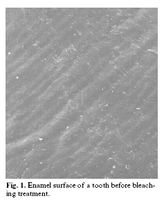
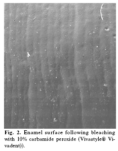
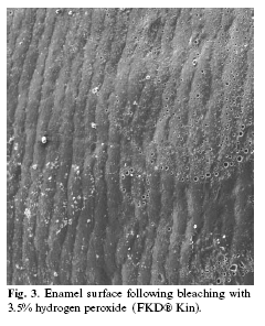
On comparing the pre-treatment images with those obtained after bleaching, no modifications were observed.
Discussion
This study was designed to assess the effect on the enamel surface caused by two whitening products for at-home tray vital bleaching, one of the bleaching procedures that is most often used. The product concentrations used were chosen as being equivalent, although with different formulations.
When determining the period for which the whitening products were to be used by each patient, it was decided to apply them for the as long as was needed to achieve the maximum whitening effect in each case.
The different response of each patient to the bleaching agents makes it difficult to establish a uniform time for all cases (10). The bleaching period lasted from 28 to 33 days.
Studies of the effects of at-home whitening agents on the enamel surface are usually conducted on extracted teeth or fragments of extracted teeth after the bleaching treatment has ended (11-14), as Bitter (15) did in a study of patients who needed exodontias for periodontal reasons and underwent prior at-home tray bleaching. In the present case, an in vivo study was conducted by making replicas of the teeth treated with the whitening technique. Although this method has not been much used for this purpose to date, it enables real changes in the enamel surface to be evaluated and contributes valuable information on this subject. In comparison to direct observation of the enamel surfaces, observation of replicas of the same surfaces shows the effects caused by the bleaching agents in greater detail, so this method is indicated for in vivo studies (9).
Scanning electron microscope observation of the replicas obtained for each group (hydrogen peroxide and carbamide peroxide) made it possible to see that the images of the enamel before treatment were very similar to those obtained after finishing the treatment. Typical enamel surface morphology was observed in all the images, with the usual perikymata pattern, which did not appear to be affected in any of the patients following the bleaching treatment. This leads to the conclusion that any changes caused by the whitening agents used in this technique are minimal or imperceptible, which agrees with the results presented by other authors (16-19).
As regards the ability to cause changes in the surface of the enamel, Bitter (15) found in an in vivo study that 10% carbamide peroxide can affect the tooth surface and underlined the importance of considering the effect of these changes on enamel integrity since, in the long term, they could be the cause of abrasions or cusp fractures, particularly in teeth that have been restored or weakened by other dental treatments. Llena et al. (20) showed the results of using scanning electron microscopy to analyse the surface of extracted teeth. They found that 10% carbamide peroxide did not result in modifications to the enamel surface, unlike 35% hydrogen peroxide, which led to severe destructuring of the surface and the appearance of aberrant crystals alongside the enamel prisms. In another laboratory study, Rotstein et al. (21) found that whitening agents, including those used in at-home tray bleaching, can affect the hard tissues of the teeth by altering their calcium concentration and recommended that they be used with caution. More recently, in another in vitro study, Goo et al. (22) encountered a slight loss of mineral content in the enamel on using 10% carbamide peroxide. A further experimental study by Hegedus et al. (12) also showed changes in the surface of the enamel when using products that contained 30% hydrogen peroxide and 10% carbamide peroxide. Lopes et al. (14) established in vitro that erosions in the enamel surface following dental bleaching did not present a uniform pattern and their intensity varied depending on the sample.
It has been asserted that the changes that have been observed in the surface of enamel which has undergone dental bleaching can modify the physical properties of the enamel, increasing its solubility and its susceptibility to caries (23). However, other studies have determined that the enamel did not present greater susceptibility to caries after dental bleaching (24, 25) and even that certain bleaching products can prevent demineralisation of the enamel by lactic acid (26).
The reason for the lack of unanimity concerning the effects that bleaching agents have on the enamel may be due to a variety of factors such as the use of non-standardised protocols in different studies (27), the origin of the enamel samples employed (teeth erupted or not) and their differing ages, the immediate remineralising effect of saliva after removal of the bleaching agent (13) and the pHs of the product employed and of some foods, which can alter the morphology of the enamel surface and of the dentine and cause alterations in their physical properties (28).
The results obtained coincide with those of other authors and enable at-home tray bleaching to be classed as a safe technique without adverse effects on dental tissues (2, 18, 20, 29).
Conclusions
In vivo microscope analysis using replicas of dental surfaces bleached with low-concentration products for use at home has established that no morphological changes took place in the enamel of the bleached teeth.
Acknowledgements
The authors wish to thank Mary Georgina Hardinge for assistance with the English text.
References
1. Lambert DL. Motivación estética y blanqueamiento dental vital. Sig Inter 2000;5:5-10. [ Links ]
2. Sarret DC. Tooth whitening today. J Am Dent Assoc 2002;133:1535-41. [ Links ]
3. Ernst CP, Marroquin BB, Willershausen-Zonnchen B. Effects of hydrogen peroxide-containing bleaching agents on the morphology of human enamel. Quintessence Int 1996;27:53-6. [ Links ]
4. Murchison DF,Charlton DG, Moore BK. Carbamide peroxide bleaching:effects on enamel surface hardness and bonding. Oper Dent 1992;17:181-5. [ Links ]
5. Haywood VB, Heymann HO. Nigthguard vital bleaching: how safe is it?. Quintessence Int 1991;22:515-23. [ Links ]
6. Haywood VB, Heymann HO. Nigthguard vital bleaching. Quintessence Int 1989;20:173-6. [ Links ]
7. Miller MB, Castellanos IR, Rieger MS. Efficacy of home bleaching systems with and without tray reservoirs. Pract Periodontics Aesthet Dent 2000;12:611-4. [ Links ]
8. Hannig C, Zech R, Henze E, Dreier S, Attin T. Peroxide release into saliva from five different home bleaching systems in vivo. Am J Dent 2005;18:13-8. [ Links ]
9. Turkun M, Sevgican F, Pehlivan Y, Aktener BO. Effects of 10% carbamide peroxide on the enamel surface morphology: a scanning electron microscopy study. J Esthet Restor Dent 2002;14:238-44. [ Links ]
10. Berga-Caballero A, Forner-Navarro L, Amengual-Lorenzo J. At-home vital bleaching: a comparison of hydrogen peroxide and carbamide peroxide treatments. Med Oral Patol Oral Cir Bucal 2006;11:E94-9. [ Links ]
11. McGuckin RS, Babin JF, Meyer BJ. Alterations in human enamel surface morphology following vital bleaching. J Prosthet Dent 1992;68:754-60. [ Links ]
12. Hegedus C, Bistey T, Flora-Nagy E, Keszthelyi G, Jenei A. An atomic force microscopy study on the effect of bleaching agents on enamel surface. J Dent 1999;27:509-15. [ Links ]
13. McCracken MS, Haywood VB. Effects of 10% carbamide peroxide on the subsurface hardness of enamel. Quintessence Int 1995; 26:21-4. [ Links ]
14. Lopes GC, Bonissoni L, Baratieri LN, Vieira L, Monteiro S. Effect of bleaching agents on the hardness and morphology of enamel. J Esthet Restor Dent 2002;14:24-30. [ Links ]
15. Bitter NC. A scanning electron microscope study of the long-term effect of bleaching agents on the enamel surface in vivo. Gen Dent 1998;46:84-8. [ Links ]
16. Scherer W, Cooper H, Ziegler B, Vijayaraghavan TV. At-home bleaching system: effects on enamel and cementum. J Esthet Dent 1991;3:54-6. [ Links ]
17. Haywood VB, Leech T, Heymann HO, Crumpler D, Bruggers K. Nightguard vital bleaching: effects on enamel surface texture and diffusion. Quintessence Int 1990;21:801-4. [ Links ]
18. Araujo EM, Baratieri LN, Vieira LC, Ritter AV. In situ effect of 10% carbamide peroxide on microhardness of human enamel: function of time. J Esthet Restor Dent 2003;15:166-74. [ Links ]
19. Cobankara FK, Unlu N, Altinoz HC, Fusun O. Effect of home bleaching agents on the roughness and surface morphology of human enamel and dentine. Int Dent J 2004;54:211-8. [ Links ]
20. Llena MC, Forner L, Faus VJ, Fernández A. Effet de deux agents pour blanchiment sur la surface de lémail. Etude in vitro. Bull Group Int Rech Sci Stomatol et Odontol 1992;35:117-20. [ Links ]
21. Rotstein I, Dankner E, Goldman A, Heling I, Stabholz A, Zalkind M. Histochemical analysis of dental hard tissues following bleaching. J Endod 1996;22:23-5. [ Links ]
22. Goo DH, Kwon TY, Nam SH, Kim HJ, Kim KH, Kim YJ. The efficency of 10% peroxide carbamide gel on dental enamel. Dent Mater J 2004;23:522-7. [ Links ]
23. Yeh ST, Su Y, Lu YC, Lee Sy. Surface changes and acid dissolution of enamel after carbamide peroxide bleach treatment. Oper Dent 2005;30:507-15. [ Links ]
24. Pretty IA, Edgar WM, Higham SM. The effect of bleaching on enamel susceptibility to acid erosion and demineralisation. Br Dent J 2005;198:285-90. [ Links ]
25. Al-Qunaian TA. The effect of whitening agents on caries susceptibility of human enamel. Oper Dent 2005;30:265-70. [ Links ]
26.- Nucci Marchionni S, Piana G, Mazzoni A, Prati C. Morphological evaluation of enamel surface after application of two `home´ whitening products. Oral Health Prev Dent 2004;2:221-9. [ Links ]
27. Spalding M, Taveira LA, Assis GF. Scanning electron microscopy study of dental enamel surface exposed to 35% hydrogen peroxide: alone, with saliva, and with 10% carbamide peroxide. J Esthet Restor Dent 2003;15:154-65. [ Links ]
28. Sulieman M, Addy M, Macdonald E, Rees JS. A safety study in vitro for the effects of an in-office bleaching system on the integrity of enamel and dentine. J Dent 2004;32:581-90. [ Links ]
29. Dadoun MP, Bartlett DW. Safety issues when using carbamide peroxide to bleach vital teeth -a review of the literature. Eur J Prosthodont Restor Dent 2003;11:9-13. [ Links ]
![]() Correspondence:
Correspondence:
Dr. José Amengual Lorenzo
Avda. Aragón nº 19, 1ª
Valencia
E-mail: amengual@mail.ono.es
Received: 5-11-2006
Accepted: 4-05-2007

