


Mi SciELO
 Servicios personalizados
Servicios Personalizados
Revista
Articulo
 Inglés (pdf)
 Articulo en XML
 Referencias del artículo
 Enviar articulo por email
Indicadores
-
 Citado por SciELO
 -
 Accesos
Links relacionados
-
 Citado por Google
 -
 Similares en
SciELO
Similares en
SciELO -
 Similares en Google
Compartir

 Permalink
Medicina Oral, Patología Oral y Cirugía Bucal (Internet)
versión On-line ISSN 1698-6946
Med. oral patol. oral cir.bucal (Internet) vol.12 no.8 dic. 2007
Spontaneous fracture of the mandibular genial tubercles. A case report
Lorena Gallego1, Luis Junquera2, Pedro Villarreal3, Juan Carlos de Vicente4
(1) Residente de Cirugía Oral y Maxilofacial
(2) Profesor Titular Vinculado
(3) Médico-adjunto. Servicio de Cirugía Oral y Maxilofacial
(4) Profesor Titular Vinculado. Jefe de Sección. Hospital Universitario Central de Asturias. Universidad de Oviedo
ABSTRACT
Fracture of the mandibular genial tubercles is an uncommon pathology affecting edentulous patients with severe maxillary atrophy. Usually occurs spontaneously which complicates the diagnosis. Their importance lies in the functional alterations, which occur as a consequence of the disinsertion of the genihyoid and genioglossus muscles. The treatment of fracture of the genial tubercles is controversial, including no surgical intervention, excision of the avulsed bone fragments, and muscular repositioning.
There have been only 11 cases reported in the literature of this fracture, most of them spontaneous. We present a difficult diagnosis situation of spontaneous fracture of the genial tubercles in an 86-year-old edentulous female with a painful sublingual and submental hematoma and anterior cervical echimosis. Computerized Tomography should be made to confirm the diagnosis. Surgical treatment was not necessary, and follow-up at 6 months revealed complete symptomatic recovery, and full return of function.
Key words: Mandibular fractures, symphysary fractures, genial tubercles fractures.
Introduction
The genial tubercles are a group of four bony extensions that surround the lingual foramen bilaterally on the lingual surface of the mandible, situated midway between the superior and inferior borders of the mandible (1,2). They serve as the insertion for the geniohyoid muscles (lower genial tubercles) and genioglossus muscles (upper genial tubercles). The action of these muscles is related to lingual mobility and deglutition, being important for two vital functions of capital importance: speech and feeding. Hypertrophy of the genial tubercles is common especially in atrophied mandibles.
The genial tubercles are affected in many mandibular fractures of the symphysis region (3), although the majority of cases described in the literature refer to spontaneous fractures in atrophic edentulous mandibles (1, 2, 4-8). The treatment is controversial, with the majority of authors arguing for a conservative treatment without the excision of the fractured tubercles from the floor of the mouth (3,5,6) or without replacement of the fractured tubercles and of the muscles inserted in them (1,3). This clinical report describes a situation of difficult diagnosis of this unusual pathology and reviews other documented cases.
Case report
An 86-year-old edentulous woman without important antecedents was referred to the Casualty department of our hospital for diagnosis and treatment of a sublingual hematoma. The patient complained of pain and swelling beneath the mandibular complete denture that started the previous day, without associated trauma.
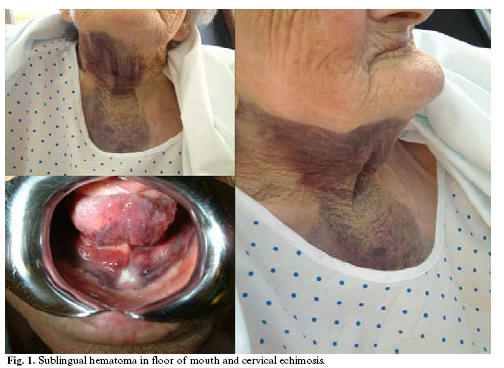
Clinical examination revealed a gross atrophy of the mandible, particularly in the incisor region. A painful sublingual and submental hematoma was noted (Fig. 1). She presented marked mandibular functional impotence, dysphagia with odinophagia, together with difficulty in lingual movement and speech. Furthermore, anterior cervical echimosis was observed (Fig. 1).
A panoramic radiograph revealed gross atrophy of the mandible without other disorders.
No hematological or sistemical pathologies were observed, so the pacient was controlled under the suspect of spontaneous trauma.
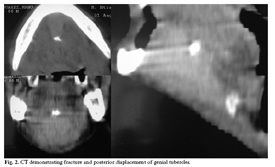
The Computerized Tomography (CT) showed a fracture of the genial tubercles with a marked posterior displacement of the genial segment , without simphysarial associated fractures (Fig. 2).
The patient was discharged from hospital at the fourth day without surgical treatment. Follow-up at 6 months revealed resolution of the hematoma, complete symptomatic recovery, and no loss of normal mobility of the tongue.
Discussion
Fracture of the genial tubercles without associated mandibular fractures is an uncommon pathology and there have been only 11 cases reported in the literature (1-11).
The majority of cases described refer to spontaneous fractures in elderly patients with atrophic edentulous mandibles, predominantly female (2,4,9).
It appears that fracture of the genial tubercles in an atrophied mandible can occur under normal masticatory forces, which are delivered through the mandibular denture (3).
The mandibular genial tubercles serve as the insertion for the geniohyoid and genioglossus muscles. The genioglossus muscle protrudes the tongue and raises the tip of the tongue, facilitating the passage of the food bolus to the pharynx (buccal phase of deglutition). Part of its middle fibers continue in the pharyngoglossus muscle, constituting Winslows geniopharyngeous muscle, which favors the passage of the food bolus through the pharynx (pharyngeal phase of deglutition). The geniohyoid muscle contributes to the laryngeal closure, as it lowers the epiglottis by raising the hyoideus (10).
The signs and symptoms associated with the fracture of the genial tubercles may include pain, swelling and hematoma in the floor of the mouth, limitation in tongue mobility and dysphagia (11). Although our case presented all these characteristics, the diagnosis of suspicion was difficulted without associated trauma. Panoramic radiographs could not resolve the diagnosis because of structures superposition or fractured tubercles demineralization (9). Occlusal radiographs, CT and craneal radiographs should be made to confirm the diagnosis and evaluate the severity of fragments displacement (5).
Treatment is controversial, some authors arguing for conservative treatment (1,3,7) and others for the removal of the fractured bone fragments and of the muscles inserted in them (2,6). These latter base their affirmation on the supposition that the intermittent muscular contraction will produce a chronic irritant effect on the floor of the mouth which will give rise to inflammatory phenomena which could be the origin of a malignant neoplasm (7).
The importance given to repositioning the muscular fascicles to their place of origin is supported by avoid the limitation on lingual protrusion, deglutition and speech (3). Although the function of these muscles is synergic with other muscle groups, the non-repositioning of these to a stable origin will generate at least a certain degree of discomfort. It seems reasonable to adopt a conservative treatment approach and postpone surgical treatment on the basis of functional restriction (6,11). In this clinical report with an elderly patient, a conservative approach to treatment was advised that resulted in a return to full function.
References
1. Goebel WM. Fractured genial tubercles. J Prosthet Dent. 1978 Jun;39(6):603-4. [ Links ]
2. Reifman S. Genial tubercle fracture. Report of a case. Oral Surg Oral Med Oral Pathol. 1969 May;27(5):595-7. [ Links ]
3. Maw RB, Lindsay JS. Conservative management of genial tubercle fractures. Oral Surg Oral Med Oral Pathol. 1970 Oct;30(4):445-9. [ Links ]
4. Carroll MJ. Spontaneous fracture of the genial tubercles. Br Dent J. 1983 Jan 22;154(2):47-8. [ Links ]
5. Santos-Oller JM, Junquera Gutierrez LM, De Vicente Rodriguez JC, Llorente Pendas S. Spontaneous fracture of hypertrophied genial tubercles. Oral Surg Oral Med Oral Pathol. 1992 Jul;74(1):28-9. [ Links ]
6. Shipman B. Genial tubercle fracture: a case. Va Dent J. 1976 Jun;53(3):7-8. [ Links ]
7. Smyd S. Fracture of the genial tubercles. J Am Dental Assoc. 1957; 55: 136-7. [ Links ]
8. Youngs R, Albert D. Fractured genial tubercles. J Laryngol Otol. 1984 Oct;98(10):1047-8. [ Links ]
9. Burnett CA, Clifford TJ. A case of fractured genial tubercles. Dent Update. 1993 Jun;20(5):219. [ Links ]
10. Shohat I, Shoshani Y, Taicher S. Fracture of the genial tubercles associated with a mandibular denture: a clinical report. J Prosthet Dent. 2003 Mar;89(3):232-3. [ Links ]
11. Yassutaka Faria Yaedú R, Regina Fisher Rubira-Bullen I, SantAna E. Spontaneous fracture of genial tubercles: case report. Quintessence Int. 2006 Oct;37(9):737-9. [ Links ]
![]() Correspondence:
Correspondence:
Dr. Luis Junquera
Universidad de Oviedo.
Escuela de Estomatología
C/ Catedrático José Serrano s/n.
Oviedo. España
E-mail: Junquera@uniovi.es
Received: 6-01-2007
Accepted: 9-10-2007

