


My SciELO
 Custom services
Services on Demand
Journal
Article
 text in
  English (pdf)
 Article in xml format
 Article references
 Send this article by e-mail
Indicators
-
 Cited by SciELO
 -
 Access statistics
Related links
-
 Cited by Google
 -
 Similars in
SciELO
Similars in
SciELO -
 Similars in Google
Share

 Permalink
Revista de Osteoporosis y Metabolismo Mineral
On-line version ISSN 2173-2345Print version ISSN 1889-836X
Rev Osteoporos Metab Miner vol.9 n.2 Madrid Apr./Jun. 2017
https://dx.doi.org/10.4321/s1889-836x2017000200002
Originales
Assessment of the predictive capacity of the Garvan calculator of 10 year risk of fracture in a Spanish population
1Instituto Universitario de Investigación Biomédicas y Sanitarias - Universidad de Las Palmas de Gran Canaria - Las Palmas de Gran Canaria (España)
2Departamento de Matemáticas - Universidad de Las Palmas de Gran Canaria - Las Palmas de Gran Canaria (España)
3Departamento de Medicina - Universidad de Sevilla - Sevilla (España)
4Servicio de Endocrinología - Hospital Quirón - Madrid (España)
5Unidad Metabólica Ósea - Hospital Universitario Insular - Las Palmas de Gran Canaria (España)
Introduction
Osteoporosis is a very prevalent disease, which produces the so-called "fragility fractures" as the only clinical complication [1]. In recent years, several calculation tools or scales have been published which, based on clinical data and with or without the aid of bone densitometry, estimate the risk of a fracture in the long term, up to 10 years [2,3,4,5,6].
Although these scales share many clinical data such as age or history of previous fractures, they also differ in the methodology and population in which they have been developed, as well as whether or not they include bone densitometry or other risk factors. For example, the more widely used FRAX® scale, published in more studies and sponsored by the World Health Organization (WHO) [3], apparently underestimates the risk of fracture in both patients with certain diseases [7,8,9,10,11,12] as well as globally in some countries, such as Spain [13], Argentina [14] or Canada [15].
The Garvan fracture risk calculator or Garvan scale was devised by Australian researchers at the Garvan Institute of Medical Research. It has been less widely used than the FRAX®, showing often divergent results in some studies which compared both scales [16,17,18]. It has not been validated in Spain, which led us to carry out this study, with the aim of observing its validity in a Canary Island population of both sexes. We have considered extending it to the Spanish population.
Material and methods
Design: This prospective study initially included 400 people of both sexes whose densitometries at the time of the first visit showed no osteoporotic values. The subjects had attended at least a second follow-up visit. Subsequently, those patients who were monitored over 10 years and who had not undergone pharmacological treatment for osteoporosis in those years were selected. The 121 who met this criterion were included in the follow-up study.
Fractures in the first 10 years of follow-up: All 121 individuals included in the study presented fragility fractures that occurred during the 10-year follow-up period.
Application of the Garvan calculator: All the patients included in our study were assessed for fracture risk due to long-term fragility using the Garvan calculator based on the data obtained during the first consultation. The tool considers a total of 5 calculation variables: sex, age, presence of fragility fractures beyond 50 years of age and falls in the last 12 months. The determination of bone mineral density by densitometry may be added if we have it. Otherwise, the calculation is also carried out, but the program requires including weight. In our study, all patients underwent bone densitometry screening at the first visit. This scale is freely available, without registration, on-line at:
https://www.garvan.org.au/promotions/bone-fracture-risk/calculator/
Once the data has been entered, the calculator shows the risk of frailty fracture for: a) any fragility fracture, and b) specifically hip fracture, and both at 5 and 10 years.
Statistical Study
Univariate analysis: Categorical variables were expressed as frequencies and percentages, and the continuous variables as means and standard deviations when the data followed a normal distribution, and as medians and interquartile ranges (percentiles 25-75) when the distribution followed was not normal. The percentages were compared using the chi-square test, the means with Student’s t test, and the medians with the Wilcoxon test for independent data.
Survival analysis: To explore the predictive ability of the fracture risk of the Garvan calculator, patients were classified according to the tertiles corresponding to this predictor. In each of these groups the survival curves were estimated up to the appearance of the first fracture using the Kaplan-Meier method. The difference between them was contrasted using the log-rank test.
Receiver Operating Characteristics (ROC) Curves: In order to evaluate the discriminatory capacity of any frailty fracture risk, the 121 patients who were monitored over 10 years were classified according to whether or not they suffered at least one fracture during this time period. For this classification, a ROC analysis was carried out, estimating the area under the corresponding ROC curve with a 95% confidence interval. The Garvan scale’s discriminatory optimal threshold was selected as the value associated with the point of the ROC curve that minimized the quantity:
(1 - sensitivity)2 + (1 - specificity)2
Sensitivity, specificity, positive predictive value (PPV) and negative predictive value (NPV) were estimated for this threshold with 95% confidence intervals.
A hypothesis test was considered statistically significant when the corresponding p value was less than 5%. Data were analyzed using the R program (version 3.1.0.).
Results
Table 1 shows the baseline characteristics of the 400 patients initially recruited for this study. It is observed that there is a greater proportion of women than men and that the mean age was 63 years, without obtaining statistically significant differences between both sexes. As expected, males were larger in size and weight than females, but body mass index (BMI) was similar in both groups, with an overweight average. The overall median risk of fracture fractures at 10 years when Garvan was applied was 15%, significantly higher in females than in males (p<0.001).
Table 1 General characteristics of the population recruited at the beginning of the study

tfn4Data expressed as #: means ± standard deviations; &: medians (interquartile ranges). *BMI: body mass index.
Table 2 shows the characteristics of the studied population over 10 years from the time of the Garvan estimation. The total number of patients was 121, of which 30 had at least one fracture due to fragility in this time frame. None of the patients received anti-osteoporotic treatment, although the patients with fractures were indicated after the fracture occurrence was reported. Of all the osteoporotic fractures (vertebral, hip, Colles, humerus, tibia, and ribs) only two were of the hip. At the outset of the study, the fractured patients had a mean risk of suffering any fragility fracture of 27%, compared to 13% of those who did not suffer a fracture (p <0.001). The same significant result was observed with the risk of hip fracture, since patients who suffered a new osteoporotic fracture (of any type) during follow-up showed an average value of 8% versus 3% of non-fractured ones.
Table 2 Characteristics of the studied population for 10 years from the time of the estimation of the Garvan value

tfn5*Fractures occurring within 10 years of follow-up. Data expressed as #: means ± standard deviations; &: medians (interquartile ranges).
Table 3 shows the statistical parameters used to assess the ability of the Garvan scale to predict any fracturing fracture within 10 years after its determination in the study population. The area under the corresponding ROC curve was 0.718 (CI-95% = 0.613 ; 0.824) (Figure 1). Considering this ROC curve, and looking for the value that offered the best statistical conditions to predict the risk of fracture, we set the optimum cutoff point at 18.5%. This value corresponds to a sensitivity of 0.67 (IC-95% = 0.47 ; 0.83), a specificity of 0.67 (CI -95% = 0.56, 0.77), a predictive value of 0.86 (CI-95% = 0.76 ; 0.93) and a positive predictive value of 0.40 (CI-95% = 0.26 ; 0.55).
Table 3 Capacity of the Garvan scale to predict an osteoporotic fracture within 10 years of being calculated
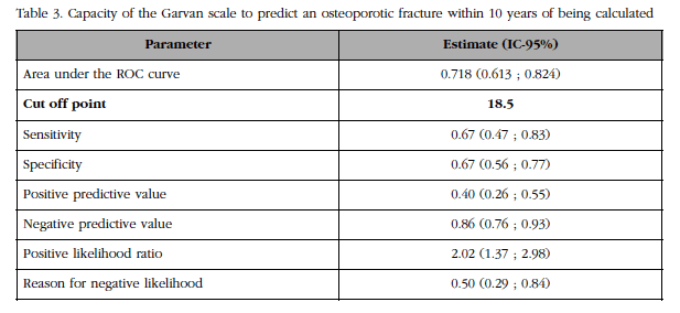

Figure 1 ROC curve for the risk of suffering any fracturing fracture calculated with the Garvan scale
Figure 2 shows the survival curves for the period between the estimation of the risk of frailty fracture and the first fragility fracture in each of the cohorts determined by the tertiles of the Garvan scale. According to these tertiles, the groups were divided according to whether the value obtained was less than 11% between 11 and 22%, and higher than 22%. The log-rank test showed statistically significant differences at 5 years (p<0.001).

Figure 2 Survival curves up to the first fracture according to the groups defined by the tertiles of the Garvan value for risk of any fragility fracture
The limited number of hip fractures (only 2) prevented an ROC analysis and one of survival for this type of fracture.
Discussion
In recent years, the diagnosis and treatment of patients with osteoporosis have changed, as a series of calculation tools or risk scales have been developed that allow us to estimate the probability of suffering a fracture due to fragility in the future, usually 10 years. This differs from the risk estimation offered by bone densitometry, which, in isolation, reports only a part of the fracture risk, which is clearly multifactorial [19,20]. Therefore, the combination of fracture risk factors and the results of densitometry have a greater specificity and sensitivity than each of them separately [21]. The FRAX® and Garvan scales, in contrast to QFracture®, include the value of bone mineral density per DXA in calculations for the likelihood of fracture risk.
The definitive role of these scales has not been established, although their presence is increasing in position papers and clinical guidelines.
Currently, FRAX® is the most accepted scale. It was the first to be published and is sponsored by the WHO [22]. It allows researchers to calculate fracture risk in a large number of countries. It is the tool with the greatest amount of literature published, with a treatment threshold of more than 20% for any fragility fracture and 3% for a hip fracture [23]. However, the FRAX® scale also has its limitations. On the one hand, it does not include falls, a very important risk factor in the production of most fragility fractures [24,25]. On the other hand, several authors have expressed their concern as it underestimates the risk of fracture in diabetic patients and in the Spanish population [12,13], because this scale has not yet been corrected for Spain. Finally, the formula with which the FRAX® calculator has been developed has not been published, a fact that has generated great controversy and suspicion in the scientific community.
Another fracture risk calculator is the QFracture® [5,26], developed by English authors, who added additional risk factors such as falls, diabetes mellitus and other diseases to variables already included in the FRAX® scale (http: //www.qfracture.org). In addition, the degree of alcohol and tobacco consumption was incorporated in more detail, and it has the novelty of making it possible to estimate fracture risk from 1 to 10 years, very useful for those individuals whose life expectancy is lower.
As for the limitations of QFracture® tool, it does not include calculations of bone densitometry and contains many variables [5,26], so the time required to complete the questionnaire is significantly longer. In addition, the QFracture® scale is not as widely used as FRAX®, which may be because it has not been validated outside the UK, and therefore there is less published material about this tool. On the other hand, the optimal cutoff points for the clinical management of patients with osteoporosis have not been established. Its website suggests a risk estimate for women of 11.1% in 10 years and for men, 2.6% over the same period of time.
Finally, there are few comparative studies between the QFracture® and FRAX® scales. We have been able to find only the work of Johansen et al. Who considered QFracture® better as a tool for estimating hip fracture risk, since it includes the history of falls [27]. On the other hand, Kanis et al published a review of the Scottish Intercollegiate Guidelines Network (SIGN), which concluded that the use of QFracture® should be used for estimating hip fracture risk and not for the risk of fragility fractures [19].
The Garvan fracture risk calculator was published by a group of Australian researchers from the Garvan Institute of Medical Research to predict in a given patient the absolute risk of having any osteoporotic fracture within 5 and 10 years. The study included a sample of more than 2,500 individuals, men and women, over 60 years of age from data collected by the Dubbo study [28]. They included the following four risk factors: age, number of previous fractures after 50 years of age, number of falls in the last year and the value of bone mineral density or weight (if bone densitometry is not available).
The Garvan scale, although apparently very practical and easy to use, is hampered by the limited relevant bibliography and that it has not been validated outside Australia.
In the main, existing publications compare the FRAX® scale with QFracture®, and FRAX® with the Garvan calculator [29]. Several studies have concluded that the FRAX® tool with bone mineral density (BMD) measurement underestimates the incidence of osteoporotic fractures, while both FRAX® without BMD and the Garvan scale overestimate the incidence of these fractures [6,30]. However, although the FRAX® and Garvan calculators include different risk factors, the therapeutic recommendation is the same [18].
As the Garvan scale has not yet been validated in Spain, the main contribution of our study is to give reliability to its predictive capacity in our population, which would allow its use in our patients, and with this the estimation of the risk of fracture due to fragility of A faster way than with the QFracture® scale, and a transparent methodology in its elaboration and with the inclusion of the falls, facts that the FRAX® does not offer.
With the FRAX® and QFracture® scales, an attempt has been made to identify a cutoff point from which we would consider the patient to be at high risk of fracture due to fragility and, therefore, it would be advisable to initiate some treatment. As we mentioned earlier, in the FRAX® scale, this value has been set at 20% for any fragility fracture and 3% for the hip, whereas in QFracture®, the authors recommend considering cut-off points for women and Men at 11.1% and 2.6%, respectively.
In the Garvan calculation tool this cut-off point has not yet been clearly established. According to our study results, an estimate of the risk of suffering any frailty fracture below 18.5% would be indicative of a very low risk, so starting treatment would not be necessary.
The main weakness of our study is the small sample size, due to the enormous difficulty found in our consultations of patients without densitometric osteoporosis and with a follow-up over so many years, besides not having received anti-osteoporotic treatment until the first fracture. The same reason has prevented us from performing the calculations for hip fracture risk, since the number of fractures incident at this location was insufficient to obtain a conclusive statistic. Despite this, the statistical study performed had enough robustness to be able to validate our findings.
In conclusion, according to the results of our study, the Garvan calculator can be used to a
ccess osteoporotic fracture risk in our population. Likewise, it could be used as a screening tool, since, according to the statistical calculations obtained, a value lower than 18.5% would allow us to establish in a given patient a very low risk of suffering any fragility fracture in the following 10 years.
Referencias
1 Kanis JA, Melton LJ, Christiansen C, Johnston CC, Khaltaev N. The diagnosis of osteoporosis. J Bone Miner Res. 2009;9:1137-41. [ Links ]
2 Black DM, Steinbuch M, Palermo L, Dargent-Molina P, Lindsay R, Hoseyni MS, et al. An assessment tool for predicting fracture risk in postmenopausal women. Osteoporos Int. 2001;12:519-28. [ Links ]
3 Kanis JA, Kanis JA. Assessment of fracture risk and its application to screening for postmenopausal osteoporosis: Synopsis of a WHO report. Osteoporos Int. 1994;4:368-81. [ Links ]
4 Kanis JA. Diagnosis of osteoporosis and assessment of fracture risk. The Lancet. 2002;359:1929-36. [ Links ]
5 Hippisley-Cox J, Coupland C. Derivation and validation of updated QFracture algorithm to predict risk of osteoporotic fracture in primary care in the United Kingdom: prospective open cohort study. BMJ. 2012;344:e3427. [ Links ]
6 Bolland MJ, Siu AT, Mason BH, Horne AM, Ames RW, Grey AB, et al. Evaluation of the FRAX and Garvan fracture risk calculators in older women. J Bone Miner Res. 2011;26:420-7. [ Links ]
7 Stephens KI, Rubinsztain L, Payan J, Rentsch C, Rimland D, Tangpricha V. Dual-energy X-Ray absorptiometry and calculated FRAX risk scores may underestimate osteoporotic fracture risk in Vitamin D deficient veterans with HIV infection. Endocr Pract. 2016;22:440-6. [ Links ]
8 Caffarelli C, Alessi C, Nuti R, Gonnelli S. Divergent effects of obesity on fragility fractures. Clin Interv Aging. 2014;9:1629-36. [ Links ]
9 FRAX from WHO underestimates fracture risk in diabetes. Bonekey Rep. 2012;1:69. [ Links ]
10 Dede AD, Tournis S, Dontas I, Trovas G. Type 2 diabetes mellitus and fracture risk. Metabolism. 2014;63:1480-90. [ Links ]
11 González Reimers E, Negrín A, Santolaria Fernández F, Martín González MC, Hernández Betancor I, Fernández Rodríguez CM, et al. Utilidad del FRAX® en el estudio de las fracturas en el paciente alcohólico. Rev Osteoporos Metab Miner. 2011;3:149-56. [ Links ]
12 Giangregorio LM, Leslie WD, Lix LM, Johansson H, Oden A, McCloskey E, et al. FRAX underestimates fracture risk in patients with diabetes. J Bone Miner Res. 2012;27:301-8. [ Links ]
13 González-Macías J, Marin F, Vila J, Díez-Pérez A. Probability of fractures predicted by FRAX® and observed incidence in the Spanish ECOSAP Study cohort. Bone. 2012;50:373-7. [ Links ]
14 Camporro F, Redondo L, Bulacio E, Gutiérrez Magaldi I, Chamale E, Sáenz F. Comparison of FRAX Score without bone mineral density determination and the criteria proposed by the Argentine Osteoporosis Society for the use of antiresorptive therapy in postmenopausal women. Medicina (Mex). 2015;75:155-8. [ Links ]
15 Roux S, Cabana F, Carrier N, Beaulieu M, April P-M, Beaulieu M-C, et al. The World Health Organization Fracture Risk Assessment Tool (FRAX) underestimates incident and recurrent fractures in consecutive patients with fragility fractures. J Clin Endocrinol Metab. 2014;99:2400-8. [ Links ]
16 Marques A, Ferreira RJO, Santos E, Loza E, Carmona L, da Silva JAP. The accuracy of osteoporotic fracture risk prediction tools: a systematic review and meta-analysis. Ann Rheum Dis. 2015;74:1958-67. [ Links ]
17 Crandall CJ. Risk assessment tools for osteoporosis screening in postmenopausal women: a systematic review. Curr Osteoporos Rep. 2015;13:287-301. [ Links ]
18 Billington EO, Gamble GD, Reid IR. Reasons for discrepancies in hip fracture risk estimates using FRAX and Garvan calculators. Maturitas. 2016;85:11-8. [ Links ]
19 Kanis JA, Compston J, Cooper C, Harvey NC, Johansson H, Odén A, et al. SIGN Guidelines for Scotland: BMD versus FRAX versus QFracture. Calcif Tissue Int. 2016;98:417-25. [ Links ]
20 Cummings SR, Bates D, Black DM. Clinical use of bone densitometry: scientific review. JAMA. 2002;288:1889-97. [ Links ]
21 Kanis JA, Oden A, Johnell O, Johansson H, De Laet C, Brown J, et al. The use of clinical risk factors enhances the performance of BMD in the prediction of hip and osteoporotic fractures in men and women. Osteoporos Int. 2007;18:1033-46. [ Links ]
22 Hillier TA, Cauley JA, Rizzo JH, Pedula KL, Ensrud KE, Bauer DC, et al. WHO absolute fracture risk models (FRAX): Do clinical risk factors improve fracture prediction in older women without osteoporosis? J Bone Miner Res. 2011;26:1774-82. [ Links ]
23 The Advisory Board of the National Osteoporosis Guideline Group, Kanis JA, Harvey NC, Cooper C, Johansson H, Odén A, et al. A systematic review of intervention thresholds based on FRAX: A report prepared for the National Osteoporosis Guideline Group and the International Osteoporosis Foundation. Arch Osteoporos [2016 Dec. consultado en: http://link. springer.com/10.1007/s11657-016-0278-z [ Links ]
24 Masud T, Binkley N, Boonen S, Hannan MT. Official positions for FRAX® clinical regarding falls and frailty: can falls and frailty be used in FRAX®? J Clin Densitom. 2011;14:194-204. [ Links ]
25 Grisso JA, Kelsey JL, Strom BL, Ghiu GY, Maislin G, O’Brien LA, et al. Risk factors for falls as a cause of hip fracture in women. N Engl J Med. 1991;324:1326-31. [ Links ]
26 Hippisley-Cox J, Coupland C. Predicting risk of osteoporotic fracture in men and women in England and Wales: prospective derivation and validation of QFracture scores. BMJ. 2009;339:b4229. [ Links ]
27 Johansen A. QFracture is better than FRAX tool in assessing risk of hip fracture. BMJ. 2012;345:e4988. [ Links ]
28 Nguyen ND, Frost SA, Center JR, Eisman JA, Nguyen TV. Development of prognostic nomograms for individualizing 5-year and 10-year fracture risks. Osteoporos Int. 2008;19:1431-44. [ Links ]
29 Leslie WD, Lix LM. Comparison between various fracture risk assessment tools. Osteoporos Int. 2014;25:1-21. [ Links ]
30 van Geel TACM, Eisman JA, Geusens PP, van den Bergh JPW, Center JR, Dinant G-J. The utility of absolute risk prediction using FRAX® and Garvan Fracture Risk Calculator in daily practice. Maturitas. 2014;77:174-9. [ Links ]
Received: January 13, 2017; Accepted: March 06, 2017
 Esta obra está bajo una Licencia Creative Commons Atribución-NoComercial 3.0 Internacional.
