INTRODUCTION
Coronary artery ectasia (CAE) is the dilatation of a coronary arterial segment to a diameter at least 1.5 times of the distal segment of the same coronary artery or adjacent coronary artery [1]. The incidence of CAE cases in angiographic examinations is 0.3% to 5.3%, and the presence of occlusive lesions has been rarely reported [2]. Damage to the coronary artery intima secondary to inflammation and cytokines released during the subsequent inflammation determine the clinical severity and prognosis of ectatic vessels [3]. Though the mechanism of CAE is unclear, it is known that inflammation, endothelial dysfunction, and vasculitides play a major role in the pathogenesis [4]. However, a complete consensus has not been reached on which patient is at risk of CAE and in which patient CAE may lead to an occlusive lesion.
Atherosclerosis is the primary contributing factor in CAE cases [5]. Coronary artery diseases (CADs) are the leading causes of mortality among cardiovascular diseases (CVDs), and undesirable cardiovascular events secondary to atherosclerosis are involved in their pathogenesis [6]. Lipids play a critical role in the process of atherogenesis and are known as strong risk factors for CVD [7]. Triglyceride (TG) and high-density lipoprotein-cholesterol (HDL-C) are lipids that can be readily measured in routine clinical practice. In recent studies, the atherogenic index of plasma (AIP) obtained by logarithmic TG to HDL-C ratio has been shown to be associated with hypertension (HTN), diabetes mellitus (DM) and heart diseases. The AIP value is associated with the size of pre- and anti-atherogenic lipoprotein particles and represents the authentic relationship between protective and atherogenic lipoproteins [8, 9, 10]. In addition, AIP is a novel marker for atherosclerosis and an independent risk factor for subclinical CAD [11]. Although a relationship between atherosclerosis and AIP has been demonstrated, there is no up-to-date study examining the relationship between AIP and obstructive CAE.
In the light of this information, the aim of our study was to investigate whether there is a relationship between AIP and obstructive CAE.
MATERIAL AND METHODS
STUDY POPULATION
In this single-center study, patients hospitalized and treated for non-ST segment elevation acute coronary syndrome (NSTE-ACS) and CAE between January 2013 and June 2021 were retrospectively examined, and 213 patients were included in the study.
The diagnosis of non-ST-elevation MI (NSTEMI) was made according to the following criteria [12]:
Typical chest pain lasting 30 minutes or longer.
Positive for cardiac troponin.
Absence of ST segment elevation in any lead on superficial electrocardiogram.
Patients with a history of CAD, chronic kidney disease [estimated glomerular filtration rate<30 (mL/min/1.73m2)], abnormal liver dysfunction, recent diagnosis of stroke, active infection, malignancy, pathological Q wave on electrocardiogram, heart failure (left ventricular ejection fraction of ≤40%), moderate–severe heart valve disease, diagnosed cardiomyopathies, statin use, and those <18 years of age were not included in the study.
The physical examination and demographic as well as clinical characteristics of the patients were obtained from the electronic medical records. Laboratory parameters of blood samples taken from antecubital peripheral veins at the time of admission (such as troponin value, liver and kidney function tests, whole blood count, and coagulation parameters) and on the morning after admission, following at least 8 hours of overnight fasting (such as TG and HDL-C) were obtained from medical electronic records. AIP was calculated using the obtained data and the following formula: AIP=log (TG/HDL-C).
The study approval was obtained from the ethics committee of our university (Decision number: 2011-KAEK-27/2021-2100169939). The study was performed in accordance with the Declaration of Helsinki.
CORONARY ANGIOGRAPHY
Coronary angiographies (GE Healthcare Innova 2100, New Jersey, USA) were performed by an experienced cardiologist using the standard Judkins technique with the femoral or radial approach. Angiographic images were evaluated by two experienced cardiologists.
Before the procedure, 600 mg clopidogrel or 180 mg ticagrelor was administered in addition to 300 mg aspirin for antiplatelet therapy. The definition of responsible lesions was decided by evaluating different images. Very high-risk NSTEMI patients underwent coronary angiography within 2 hours, whereas the procedure was performed within 24 hours in the remaining NSTEMI patients. During the procedure, the guiding catheter was set to achieve TIMI 3 flow into the responsible coronary artery, and an intravenous heparin (70 U/kg) bolus was followed by direct stenting of the appropriate lesions or stenting after balloon dilatation. In all patients without contraindications, β blockers, angiotensin converting enzyme inhibitors or angiotensin receptor blockers, and statin therapy were initiated.
CAE was defined as a dilatation with a diameter of 1.5 times the normal epicardial coronary artery compared to the normal coronary artery [13]. Stenosis of ≥70% was defined as obstructive CAD.
CAE CLASSIFICATION
Ectatic vessels were classified according to the Markis classification. Diffuse ectasia of 2 or 3 coronary arteries was classified as type 1, diffuse ectasia in a single vessel accompanied by localized ectasia in another vessel was classified as type 2, diffuse ectasia in a single vessel was classified as type 3, and segmental localized ectasia was classified as type 4 [14]. The patients were divided into two groups as group 1 with non-obstructive CAE and group 2 with obstructive CAE.
STATISTICAL ANALYSIS
Kolmogorov–Smirnov test was used to evaluate the distribution of continuous variables. The data that did not conform to normal distribution are expressed as median and percentiles (25th and 75th percentiles). Categorical variables are expressed as percentages and numbers. Chi-square test was used when comparing the probability ratios of categorical variables. For the comparison of continuous variables between groups, Mann Whitney U test was used. Receiver operating characteristic (ROC) curve analysis was performed to calculate the optimum cut-off values, sensitivity and specificity for obstructive coronary ectasia of AIP. Finally, the explanatory power of AIP, demographic and clinical variables on obstructive CAE are evaluated by logistic regression. P values of <0.05 were considered statistically significant. Statistical data were obtained using SPSS 20.0 (SPSS Inc, Chicago, IL, USA).
RESULTS
Our study consisted of 213 newly diagnosed NSTE-ACS (152 males, 61 females) patients. Patients were divided into two groups according to non-obstructive (n=141) and obstructive CAE (n=72). The clinical data of the study patients are shown in Table 1. No differences were observed between clinical features including age, gender, DM, body mass index, and HTN. Considering the biochemical parameters, TG levels were statistically and numerically significant in patients with obstructive CAE compared to those without (p < 0.001).
Table 1. Demographic and laboratory findings of patients

BMI: Body mass index; DM: Diabetes mellitus; HTN: Hypertension; COPD: Chronic obstructive pulmonary disease; ACE: Angiotensin converting enzyme inhibitor; ARB: Angiotensin reseptor blocker; HDL-C: High-density lipoprotein cholesterol; LDL-C: Light-density lipoprotein cholesterol; CRP: C-reactive protein; AIP: Atherogenic Index of Plasma; PCI: Percutaneous coronary intervention; CABG: Coronary artery bypass grafting.
Though HDL-C and low-density lipoprotein-cholesterol (LDL-C) levels showed numerical differences between the groups, no statistical difference was observed. AIP values were numerically and statistically significant in the obstructive CAE group compared to the non-obstructive CAE group (Table 1).
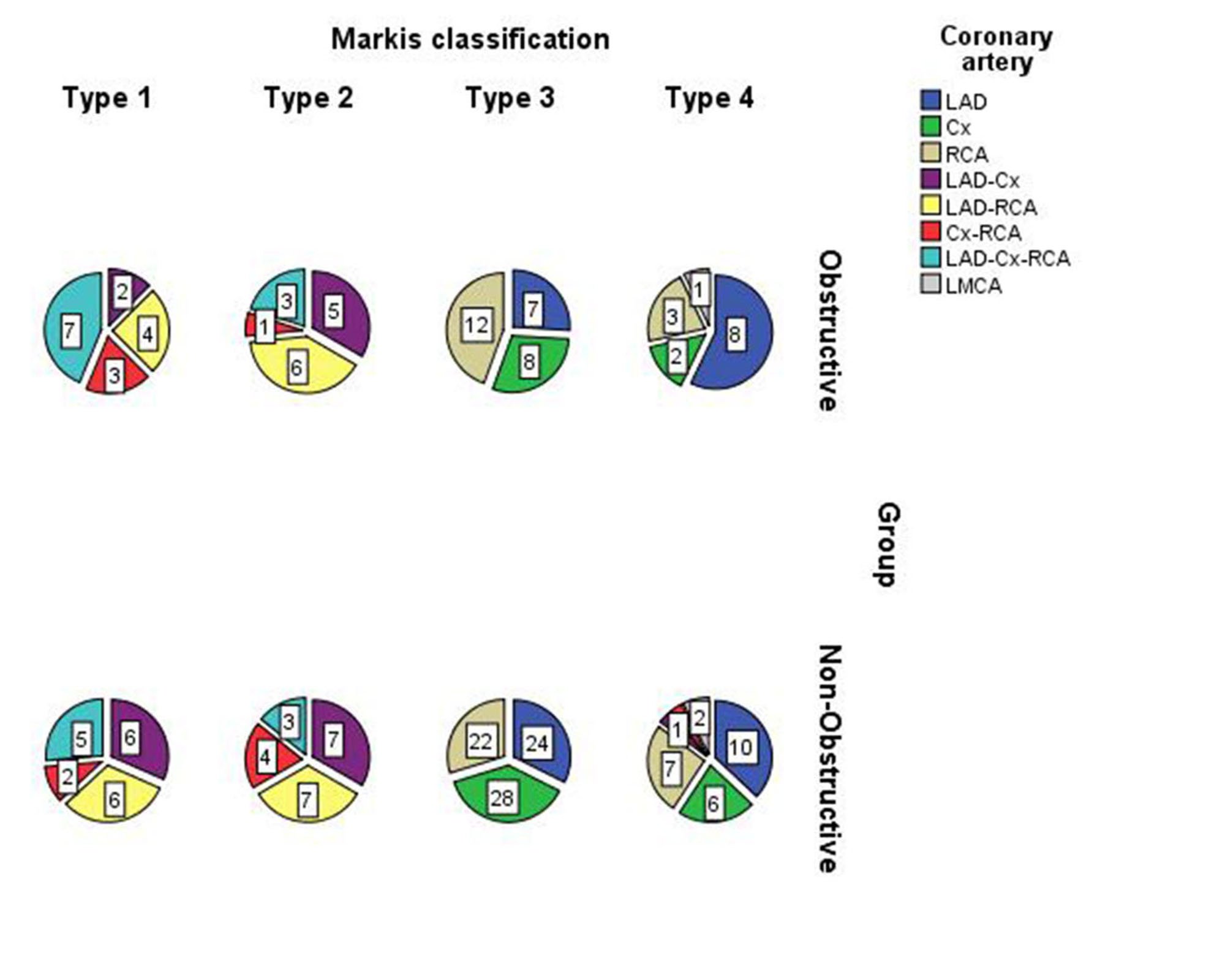
The incidence of obstructive CAE in patients with NSTE-ACS was equal in the left anterior descending coronary artery (20.8%) and right coronary artery (20.8%). According to the Markis classification, type 3 obstructive CAEs were most common in NSTE-ACS patients (Table 2).
Table 2. Angiographic characteristics of the study population

LAD: Left anterior descending artery; Cx: Circumflex artery; RCA: Right coronary artery; LMCA: Left main coronary artery.
Variables that were found to be significant in univariate regression analysis were included in the logistic regression analysis. AIP was considered as a predictor of obstructive CAE in newly diagnosed NSTE-ACS patients (Table 3).
Table 3. Analysis to identify the independet factors of obstructive coronary artery ectasia pattern

HTN: Hypertension; DM: Diabetes mellitus; COPD: Chronic obstructive pulmonary disease; CRP: C-reactive protein; AIP: Atherogenic Index of Plasma.
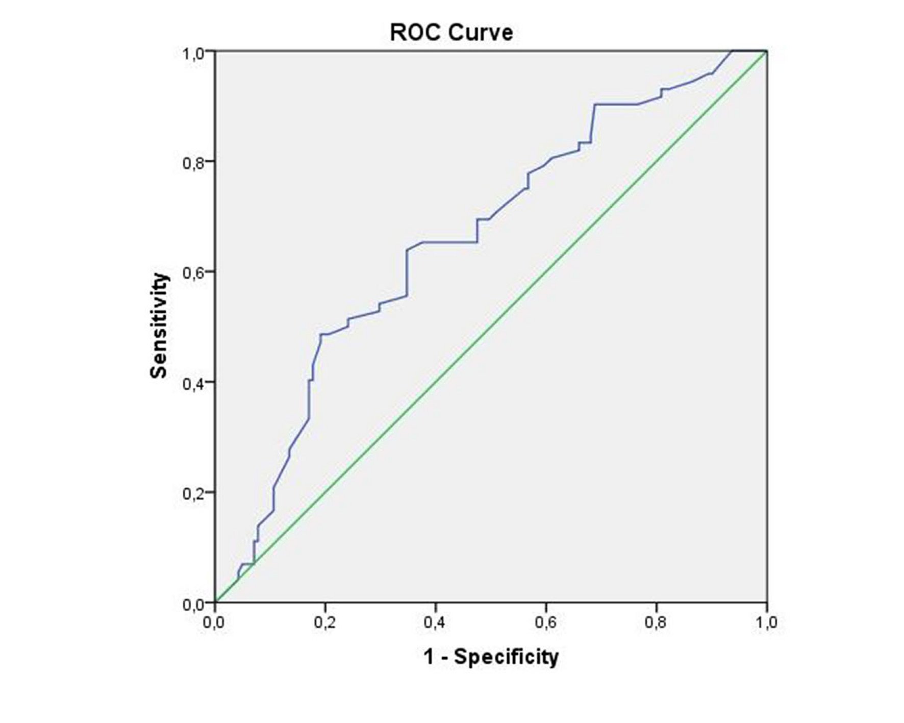
In the receiver operating characteristic analysis, AIP values above 0.33 showed 90% sensitivity and 68% specificity (area under the curve (AUC): 0.658, 95% CI: 0.581–0.734, p < 0.001) in terms of predicting obstructive CAE in NSTE-ACS patients (Figure 1). The distribution of the coronary arteries based on the Markis classification and the presence of obstruction is shown in Figure 2.

Figure 1. Receiver operator characteristic curve of the AIP to predict obstructive CAE in patients with NSTE-ACS (90% sensitivity and 68% specificity AUC: 0.658, p<0.001).
DISCUSSION
To the best of our knowledge, our study is the first of its kind to investigate the relationship between the presence of obstructive and non-obstructive ectasia and AIP in NSTE-ACS patients. This study concluded that the AIP value, which can be easily calculated, is associated with the presence of obstructive CAE and is a predictor of obstructive CAE risk.
CAEs are uncommon coronary disorders and their pathophysiological mechanism is unclear [15]. As the pathophysiological mechanisms are not fully understood, there are no specific treatment methods and guideline recommendations to prevent the progression of the disease. CAEs may present with anginal attacks or ACS similar to CVDs [16, 17]. Anginal attacks are usually caused by slow and turbulent flow in dilated CAs, and turbulent blood flow may trigger the activation of atherogenic genes in the long term, leading to endothelial dysfunction and subsequent undesirable clinical outcomes, such as MI [18].
Dyslipidemia is a significant risk factor for atherosclerosis [19]. Plasma HDL-C levels have an inverse relationship with the risk of atherosclerosis [20]. Other studies have demonstrated the relationship between increased TG and LDL-C values and atherosclerotic processes [21, 22]. Given this information, it would be reasonable to hypothesize that
CAEs, the pathogenesis of which is affected by atherosclerotic processes, are associated with dyslipidemia.
HTN, advanced age, obesity, smoking, and hypercholesterolemia are among the major risk factors for CVDs [23, 24]. The formulas derived from the plasma lipid profile have been considered as predictors for CVD risk in recent years. In study conducted on CVD risk prediction with TG/HDL-C or LDL-C/HDL-C ratios, it has been shown that combined formulas are superior to a single lipid marker [25]. The main goal of various clinical study was to obtain a well-established predictor of the CVD risk instead of using the classical ratio [26]. In addition, it has been demonstrated that AIP is superior to HDL-C, LDL-C, and TG values in predicting the risk of CAD [27]. Therefore, AIP values may be related to obstructive CAE. Indeed, in our study, AIP values were found to be significantly higher in the obstructive CAE group compared to those in the non-obstructive CAE group, whereas no difference were observed between the groups in HDL-C and LDL-C values. Moreover, we found that TG, an important parameter used in the calculation of AIP, was statistically and numerically significant in patients with obstructive CAE compared to those without. Hydrolysis of TGs by lipoprotein lipase yields TG-rich lipoprotein residues and fatty acids. Both lipolysis of fatty acids and lipoprotein residues have 4 times higher cholesterol transport capabilities [28]. Thus, it is likely that increased TG levels cause rapid progression of the atherosclerotic process and play an important role in the etiology of obstructive CAE.
The development of atherosclerotic plaque is associated with an increase in the small dense LDL-C (sdLDL-C) ratio. AIP is directly proportional to the increased number of sdLDL particles and inversely proportional to LDL-C particle size [29, 30]. In a previous study, the relationship between sdLDL and CAD has been demonstrated, and it has been shown that sdLDL plays a role in the atherosclerotic process [31]. Accordingly, increased AIP values in patients with obstructive CAE may be an indirect indicator of sdLDL values. In addition, the easy calculation of AIP may provide clinicians with important cell-related knowledge, which may ultimately aid in treatment planning.
CAEs associated with atherosclerotic processes may also be be associated with inflammatory parameters. Despite the exclusion of infectious diseases in our study, C-reactive protein (CRP), a simple indicator of inflammation, was statistically higher in patients with obstructive CAE. However, CRP values were not significantly different on multivariate logistic regression analysis in NSTE-ACS patients with obstructive ectasia. Atherosclerotic processes are important in obstructive and non-obstructive CAE, and AIP values can indirectly offer the clinician remarkable insights into atherosclerotic processes.
Our study had some limitations. First of all, it was a single-center, retrospective study. Secondly, the study design precludes the understanding of the efficacy of primary preventive medical therapy in patients with non-obstructive ectasia. Our results were obtained based on the current clinical and laboratory results. Multicenter and prospective studies are needed to support the results of the present study and to eliminate its limitations. The relationship between sdLDL values and AIP may be a topic of investigation in prospective studies to improve our understanding of the atherosclerotic process in obstructive CAE.
CONCLUSIONS
Considering the findings in literature and of our study, we hypothesized that obstructive CAE lesions in NSTE-ACS patients may be associated with AIP. In particular, our study showed that obstructive CAE was independently and significantly associated with AIP values above 0.33 compared to non-obstructive CAE. AIP may be a useful parameter for atherosclerotic risk management in patients with obstructive and non-obstructive CAE.


